Neutralization of White Spot Syndrome Virus by Monoclonal
Antibodies against Viral Envelope Proteins
Hsiu-Hui Shih(1)
(Manuscript received 12 May, 2004; accepted 10 June, 2004)
ABSTRACT: Two monoclonal antibodies (MAbs) recognizing envelope proteins of the white spot
syndrome virus (WSSV), 6E1 against VP28 and 3E8 against VP19, were applied to demonstrate their neutralizing ability to this virus by using both in vitro and in vivo assays. Mixtures of MAb 6E1 with virus filtrate were inoculated into the primary explant monolayer culture derived from the lymphoid Oka organs of Penaeus monodon. MAb was likely to neutralize the infectivity of virus to monolayer since cytopathic effects were apparently blocked in experiment group. WSSV was titrated using Blue-Cell ELISA and the neutralizing index was calculated to be 6.90 for 6EI and 5.83 for 3E8. Neutralized virus fluids injected intramuscularly into post larvae of P. monodon. The shrimp in the positive control, which were injected with WSSV only showed an increasing mortality and a 100% mortality was reached at day 34, whereas no shrimp died in the negative control. The mortality for 6E1 was 6.7% and for 3E8 was 13.3%. These results suggest that MAbs recognizing the WSSV envelope proteins could neutralize viral infectivity to both cultured cells and shrimp.
KEY WORDS: White spot syndrome virus, WSSV, MAbs, Neutralization, Primary culture.
INTRODUCTION
White spot syndrome is a major shrimp disease worldwide. The disease has also been found in other invertebrate aquatic organisms including crab and crayfish (Flegel, 1997). The causative agent is an enveloped, non-occluded and rod-shaped virus, shrimp white spot syndrome virus (WSSV) (Chou et al., 1995). The virus contains a 305 kb double-stranded circular DNA (Yang et al., 2001) and has been approved recently to be a representative of a new genus, Whispovirus, by the International Committee on Taxonomy of Viruses (ICTV) (Mayo, 2002). The virus particle consists of at least five major proteins: VP28, VP26, VP24, VP19 and VP15 (28, 26, 24, 19 and 15 kDa, respectively). VP28 and VP19 are associated with the viron envelope and VP26, VP24, and VP15 with the nucleocapsid (van Hulten et al., 2001). Although the protein profiles of WSSV have been revealed already, epitopes located on these proteins responsible for virus neutralization or playing roles in virus cell interaction and in the establishment of the systemic infection process were still obscure. Except that VP28 had been suggested to be likely to play a key role in the initial steps of the systemic WSSV infection of shrimp recently (van Hulten et al., 2001).
Since there is no established cell line, which can be infected with WSSV, the role of the envelope proteins VP19 or VP28 in virus neutralization is less well studied. Primary lymphoid cultures can be reproducibly established (Chen et al., 1986) and are infectable with WSSV (Shih, 2002), permitting an investigation into the neutralizing activity of antibodies independent of the effects of other defense mechanisms in vivo. In this study, panel monoclonal antibodies (MAbs) specific to two envelope proteins, VP28 and VP19 (Shih ___________________________________________________________________________
1. Department of Life Science and Institute of Zoology, National Taiwan University, Taipei 106, Taiwan. Tel: 886-2-33662504; Email: [email protected]
et al., 2001), are applied and their neutralizing ability for WSSV infectivity either in vitro or in vivo are evaluated.
MATERIALS AND METHODS
Virus and primary Oka lymphoid cell cultures
White spot syndrome virus used in this study was obtained from infected, farmed Penaeus
japonicus in Taiwan. A homogenate of the head soft tissues was prepared in TNE buffer (50
mM Tris, 100 mM NaCl, 1 mM EDTA, pH 7.4) after centrifugation and filtration (Shih, 2002). Primary cell cultures were prepared from Oka organs. Lymphoid Oka organs from
Penaeus monodon were harvested, pooled and processed for primary cell culture according to
procedures described previously (Chen et al., 1986). The explant cultures were grown in 2x Leibovitz's L15 medium (2xL15) supplemented with 20% fetal bovine serum (FBS), 100 IU/mL penicillin, 100 µg/mL streptomycin, 0.25 µg/mL amphotericin B and 10% shrimp muscle extract as reported before (Shih, 2002). Briefly following these protocols, confluent primary cultures of 24 h old were ready for use to titrate WSSV-containing fluids before and after neutralization by monoclonal antibodies.
Neutralization test of MAbs to WSSV in primary cultures
Two monoclonal antibodies recognizing viral envelope proteins produced in my lab (Shih
et al., 2001) were used in this study. MAb 6E1 against VP28 was IgG1 with a κ light chain, and MAb 3E8 against VP19 was IgM with a κ light chain too. Ascites, containing 6E1 or 3E8, or the mock medium (FBS-free L15) was mixed with an equal volume of ten-fold diluted WSSV filtrate and incubated at room temperature for 1 h with gentle shaking. To determine the titer of WSSV filtrate remained after neutralization, the mixtures were ten-fold diluted and inoculated into 24 h old primary monolayer cultured in 48-well plates. Virus adsorption was carried out for 1 h with gentle agitation at room temperature. The supernatant was replaced with 0.5 mL of fresh L15 medium containing 2% FBS in each well. The plates were incubated at 20℃for 3 days.
The TCID50 titer of each mixture was determined by observation of blue cell after the procession of the Blue-Cell ELISA described previously (Shih, 2002). The neutralization index (NI) was calculated by the difference between the titer of control and MAb treated samples, and expressed as log10 NI. All neutralization assays were performed in duplicate and reproduced twice by using two series of WSSV filtrates prepared independently.
In vivo neutralization assay
Post larvae of P. monodon at PL20 stage were injected intramuscularly in the fourth tail segment of the shrimp by a 29-gauge needle with 10 µL of each mixture described above. A negative (330 mM NaCl) and a positive control (WSSV only) were included. For each group 15 shrimp were used. The shrimp were subsequently cultured for a period of 35 days and the mortality was monitored daily.
The presence of WSSV in shrimp pre- and post-injection was tested by a PCR protocol (Wang et al., 1997) with DNA templates extracted from gill tissues using a GenomicPrep Cell and Tissue DNA Isolation Kit (Pharmacia Biotech, Sweden). The results of the analysis showed that the experimental batch of shrimp was WSSV-free before injection but that all became infected after injection.
RESULTS WSSV infectivity to primary lymphoid
cells could be neutralized by MAbs against its envelope proteins VP28 (Fig. 1). The morphology of monolayer cultured from the Oka lymphoid organ of P. monodon 3 days after seeding showed that the confluence was still remained. Besides any signs of deterioration of this culture, such as granularity around the nucleus, vacuolation within cytoplasm or monolayer detaching from the substrate were not observed (Fig. 1A). Mock medium could not neutralize WSSV infectivity and cytopathic effects appeared (Fig. 1B). On the contrary, neutra- lization was successively produced by MAb 6E1 against VP28 and the cytopathic effects was although not totally achieved but evidently delayed (Fig. 1C).
Results of the neutralization assays titrated by Blue-Cell ELISA methods showed those two MAbs possessed neutralizing activities. Neutralization index (log10 NI) of 6E1 was 6.90 and of 3E8 was 5.83, respectively (Table 1).
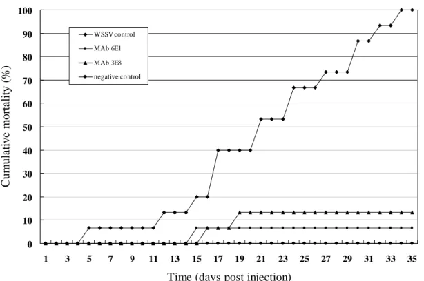
During in vivo neutralization assays, no shrimp died in the negative control injected only with 330 mM NaCl (Fig. 2). The shrimp in the positive control, which were injected with WSSV only showed an increasing mortality as the test proceeded and a 100% mortality was reached at day 34. WSSV could be neutralized by those two MAbs, for 6E1 a 6.7% mortality appeared at day 15 and lasted until the end of this assay. For 3E8, a 6.7% mortality appeared at day 16 and increased to 13.3% at day 20, then remained until the end of this experiment.
DISCUSSION
To demonstrate the role of the major WSSV envelope protein in WSSV infection, a specific polyclonal antiserum against VP28 was generated. And the VP28 antiserum was
Fig. 1. WSSV infectivity to primary lymphoid cells could be neutralized by MAb against its envelope proteins VP28. Neutralized mixtures of MAbs and WSSV-containing filtrate were inoculated into 24 h old primary monolayer and incubated for 3 days at 20℃. A: The morphology of monolayer cultured from the Oka lymphoid organ of P. monodon 3 days after seeding. B: Mock medium could not neutralize WSSV infectivity and cytopathic effects are showed. C: Neutralization produced by MAb 6E1 against VP28. Since the total neutralization was not achieved, the cells became ovoid but cytopathic effects was evidently delayed in (C). Scales = 50 µm in (A) and 30 µm in (B) & (C).
B A
Table 1. Neutralization index (log10 NI) of 2 MAbs against WSSV envelope proteins, VP28 and VP19.
MAb Epitopea NIb
6E1 VP28 6.90
3E8 VP19 5.83
a
As determined by Western blotting against WSSV (Shih et al., 2001).
b
The means of duplicate determinations and reproduced twice by using two series of inocula prepared independently are presented.
suggested to be able to neutralize WSSV infection of P. monodon in a concentration- dependent manner upon intramuscular injection (van Hulten et al., 2001). VP28 had been pronounced to locate in the spikes of the WSSV envelope and it was likely to play a key role in the initial steps of the systemic WSSV infection in shrimp (van Hulten et al., 2001). However, this observation did not prove the neutralizing activity of WSSV VP28 specific antibodies alone, because protective defense mechanisms might have coincidentally been induced and involved during virus elimination in shrimp. In contrast, the in vitro neutralization studies preformed with WSSV-infected primary shrimp lymphoid cells herein provide unequivocal evidence for the neutralization activity of the corresponding antibodies alone. In this study, the in vitro neutralization test was processed within 72 h to decrease the time for possible horizontal spread of virus, which may prevent the measurement of more effective neutralization.
Although the neutralizing activities of 3E8 and 6E1 were high (NI are 5.83 and 6.90, respectively), and mortalities could be extremely reduced (13.3% and 6.7%, respectively) but complete neutralization was never achieved. A constant residual infectivity was observed under several assay conditions. The observation of only partial neutralization is consistent with related studies which showed that for virtually all viruses a small fraction escapes neutralization even when a large excess of neutralizing antibodies is used (Burton et al., 2000).
Binding to epitopes located on VP19, the other envelope protein of WSSV, with MAb 3E8 could reduce the infectivity of this virus in vitro and in vivo as revealed for the first time in this study. But researches concerning the WSSV envelope proteins have been merely focused on VP28 so far. Polyclonal or monoclonal antibodies specific to VP28 had been generated against different geographical virus isolates (Liu et al., 2002; Shih et al., 2001; van Hulten et al., 2001) to detection, identification and localization of the WSSV. In addition, a gene (vp28) encoding VP28 had been cloned and expressed either in Escherichia coli (Zhang
et al., 2002) or in insect cell (van Hulten et al., 2000b). A computer-assisted analysis which
revealed a striking similarity between WSSV VP24, VP26 and VP28 at the amino acid and nucleotide sequence level, but still neglected VP19 (van Hulten et al., 2000a). Further research to reveal the distribution of VP19 on the viral envelope and its possible role in WSSV infection will be required. Such information might be useful to realize the mechanisms of neutralization and to control the spreading of this virus.
ACKNOWLEDGEMENTS
I would like to thank L. F. Tan for processing the primary culture, and B. C. Owi for the injection and culture of shrimp. This work was supported by the Fisheries Administration, the Council of Agriculture, under Grant 90AS-1.4.5-FA-F3(11).
Fig. 2. Neutralization assay of WSSV infection in P. monodon using monoclonal antibodies 6E1 against VP28 or 3E8 against VP19.
LITERATURE CITED
Burton, D. R., R. A. Williamson and P. W. H. I. Parren. 2000. Antibody and virus: binding and neutralization. Virology 270: 1-3.
Chen, S.-N., S.-C. Chi, G.-H. Kou and I.-C. Liao. 1986. Cell culture from tissues of grass prawn, Penaeus monodon. Fish Pathol. 24: 161-166.
Chou, H.-Y., C.-Y. Huang, C.-H. wang, H.-C. Chiang and C.-F. Lo. 1995. Pathogenicity of a baculovirus infection causing white spot syndrome in cultured penaeid shrimp in Taiwan. Diseases Aquat. Organisms 23: 165-173.
Flegel, T. W. 1997. Major viral diseases of the black tiger prawn (Penaeus monodon) in Thailand. World J. Microbiol. and Biotechnol. 13: 433-442.
Liu, W., Y.-T. Wang, D.-S. Tian, Z.-C. Yin and J. Kwang. 2002. Detection of white spot syndrome virus (WSSV) of shrimp by means of monoclonal antibodies (MAbs) specific to an envelope protein (28 kDa). Diseases Aquat. Organisms 49: 11-18.
Mayo, M. A. 2002. A summary of taxonomic changes recently approved by ICTV. Arch. Virol. 147: 1655-1656.
Shih, H.-H. 2002. Detection and titration of white spot syndrome virus using a Blue-Cell ELISA. J. Fish Diseases 25: 185-189.
Shih, H.-H., C.-S. Wang, L.-F. Tan and S.-N. Chen. 2001. Characterization and application of monoclonal antibodies against white spot syndrome virus. J. Fish Diseases 24: 143-150. van Hulten, M. C. W., R. W. Goldbach and J. M. Vlak. 2000a. Three functionally diverged
major structural proteins of white spot syndrome virus evolved by gene duplication. J. General Virol. 81: 2525-2529.
van Hulten, M. C. W., M. Westenberg, S. D. Goodall and J. M. Vlak. 2000b. Identification of 0 10 20 30 40 50 60 70 80 90 100 1 3 5 7 9 11 13 15 17 19 21 23 25 27 29 31 33 35
Time (days post injection)
C u m u la ti ve mor ta lity ( % ) WSSV control MAb 6E1 MAb 3E8 negative control
two major virion protein genes of white spot syndrome virus of shrimp. Virology 266: 227-236.
van Hulten, M. C. W., J. Witteveldt, M. Snippe and J. M. Vlak. 2001. White spot syndrome virus envelope protein VP28 is involved in the systemic infection of shrimp. Virology 285: 228-233.
Wang, C.-S., Y.-J. Tsai, G.-H. Kou and S.-N. Chen. 1997. Detection of white spot disease virus infection in wild-caught greasy back shrimp, Metapenaeus ensis (de Haan) in Taiwan. Fish Pathol. 32: 35-41.
Yang, F., J. He, X. Lin, Q. Li, D. Pan, X. Zhang and X. Xu. 2001. Complete genome sequence of the shrimp white spot bacilliform virus. J. Virol. 75: 11811-11820.
Zhang, X., C. Huang, X. Xu and C.-L. Hew 2002. Identification and localization of a prawn white spot syndrome virus gene that encodes an envelope protein. J. General Virol. 83: 1069-1074.