Contents lists available atScienceDirect
Journal of Chromatography A
j o u r n a l h o m e p a g e :w w w . e l s e v i e r . c o m / l o c a t e / c h r o m a1-Butyl-3-methylimidazolium-based ionic liquids and an anionic surfactant:
Excellent background electrolyte modifiers for the analysis of benzodiazepines
through capillary electrophoresis
Hsiu-Li Su, Wan-Chun Kao, Kuan-Wen Lin, Cheng-yu Lee, You-Zung Hsieh
∗Department of Applied Chemistry, National Chiao Tung University, 1001 Ta Hsueh Road, Hsinchu 300, Taiwan
a r t i c l e i n f o
Article history: Received 9 July 2009
Received in revised form 16 February 2010 Accepted 24 February 2010
Available online 3 March 2010 Keywords: 1-Butyl-3-methylimidazolium bis(trifluoromethanesulfonyl)imide Ionic liquids Benzodiazepine Capillary electrophoresis
a b s t r a c t
In this study, we found that adding 1-butyl-3-methylimidazolium-based ionic liquids (ILs) and sodium dodecyl sulfate (SDS) as modifiers in the background electrolyte (BGE) for capillary elec-trophoresis enhanced the separation of benzodiazepines. In particular, 1-butyl-3-methylimidazolium bis(trifluoromethanesulfonyl)imide ([BMIM][NTf2]) was the best IL additive for the separation system because its anionic moiety interacted favorably with the benzodiazepines. We added SDS because of its known effect on the separation of hydrophobic analytes. We optimized the separation conditions in terms of the concentrations of the IL, SDS, and organic solvent, the pH, and the BGE’s ionic strength. The optimal BGE, containing 170 mM [BMIM][NTf2] and 10 mM SDS, provided baseline separation, high efficiency, and satisfactory peak shapes for the benzodiazepines. The separation mechanism was based on heteroassociation between the anionic moiety of the IL and the benzodiazepines, with SDS improv-ing the resolution of the separation. The limits of detection for the seven analytes ranged from 2.74 to 4.42g/mL. We subjected a urine sample to off-line solid phase extraction (SPE) prior to the analysis of its benzodiazepine content. Our experimental results reveal that the combination of [BMIM][NTf2] and SDS provides adequate separation efficiency for its application to CE analyses of benzodiazepines after SPE concentration.
© 2010 Elsevier B.V. All rights reserved.
1. Introduction
Ionic liquids (ILs) are compounds comprising organic cations and inorganic or organic anions that have melting points below 100◦C [1]. Because the choice of the anionic and cationic com-ponents affects the properties of the ILs, they can be regarded as designer solvents[2]. ILs exhibit many interesting properties, including excellent solvation over a wide range of viscosities and temperatures[3], negligible vapor pressure, and non-volatility. ILs have been used in analytical separations[1,4]as (i) the station-ary phases of gas chromatography (GC) columns[5,6], on account of their low volatility and thermal stability, and (ii) as stationary phases for liquid chromatography (LC) columns[7,8], because their non-molecular characteristics result in unusual selectivities. There-fore, the use of ILs provides GC and LC methods with additional variables for successful separation and analysis.
Much effort has been exerted recently in the application of ILs to capillary electrophoresis (CE). For example, ILs have been used
∗ Corresponding author. Tel.: +886 3 5731785; fax: +886 3 5723764. E-mail address:[email protected](Y.-Z. Hsieh).
as support coatings covalently bonded to the inner walls of capil-laries[4]; when cationic imidazolium-based ILs were applied, the electroosmotic flow (EOF) of the capillary was reversed relative to that of the uncoated capillary. Qin and Li[9]used this technique to weaken the interactions between the analytes and the capillary wall to determine metal or ammonium ions. ILs have also been used as dynamic wall coatings in aqueous CE[1]. Qi et al.[10]used ILs in the BGE to analyze the anthraquinones present in Chinese herbs; in such cases, the ILs play dual roles: as EOF modifiers and as species that associate with the analytes. ILs have also been applied to non-aqueous CE, where dynamic coating of the capillary wall is greatly decreased and/or inexistent[1,11]. The resulting improved resolution has been attributed to heteroassociation between the ILs and the analytes. Vaher et al. [12] separated polyphenolic compounds by applying 1-alkyl-3-methylimidazolium-based ILs possessing various counter anions. ILs have also been used as modifiers to improve separations performed through micellar electrokinetic chromatography (MEKC) [1]. Mwongela et al.[3] employed imidazolium-based ILs in polymeric surfactant systems to analyze both achiral and chiral compounds. The addition of the ILs appears to increase the ionic strength and decrease the EOF, thereby influencing the analytes’ migration times. Tian et al. added 0021-9673/$ – see front matter © 2010 Elsevier B.V. All rights reserved.
Fig. 1. Structures of benzodiazepines and [BMIM][NTf2]. *Values of pKaare those of the conjugate acids of the benzodiazepines.
1-butyl-3-methylimidazolium tetrafluoroborate ([BMIM][BF4]) as
a modifier for the MEKC separation of lignans. The BMIM+cation
appears to modify the sodium dodecyl sulfate (SDS) micelles and, thereby, change the degree of solute partitioning[13].
Benzodiazepines are sedative, hypnotic, and anxiolytic drugs. Because insomnia, anxiety, and depression occur frequently in modern society, the rate of benzodiazepine consumption is increas-ing. Overdoses of these drugs can cause acute symptoms; therefore, it is desirable to monitor their concentrations in the body[14]. CE is a suitable method for separating benzodiazepines because of its rapidity, efficiency, and selectivity. Although SDS micelles have been employed previously to determine benzodiazepines [15–17], the resolution of this approach is poor when simulta-neously analyzing several benzodiazepines. To obtain the highest degree of resolution of benzodiazepines, we investigated the effects of adding ILs and SDS as modifiers in CE separations. Among the known ILs, imidazolium-based ILs have been used most com-monly in CE [18–21] because their cheaper prices or simpler syntheses have made them as the first choice for analytical scien-tists. In particular, 1-butyl-3-methylimidazolium–based ILs have been used most often in both aqueous and non-aqueous CE. Hence, in this study, we selected 1-butyl-3-methylimidazolium bis(trifluoromethanesulfonyl)imide ([BMIM][NTf2]) as a modifier
to analyze a mixture of benzodiazepines, including diazepam, clorazepate, bromazepam, nitrazepam, alprazolam, flunitrazepam, and lorazepam (Fig. 1). We optimized the separation conditions in
terms of the concentrations of the IL, SDS, and organic solvent, the pH, and the ionic strength. In addition, we determined the effects that the interactions of the ILs and SDS with the benzodiazepines had on their CE separation. We then used the developed method to analyze benzodizepines in spiked urine samples after solid phase extraction (SPE).
2. Materials and methods 2.1. Chemicals and reagents
All reagents and chemicals were of analytical grade. Lorazepam, bromazepam, clorazepate, nitrazepam, diazepam, alprazolam, and flunitrazepam standards were obtained from Sigma–Aldrich (St. Louis, MO, USA). 1-Methylimidazole (99%), 1-bromohexadecane (C16H33Br, 99%), and 1-methylpyrrolidine
(98%) were purchased from Acros Organics (Geel, Belgium). Lithium bis(trifluoromethanesulfonyl)imide (LiNTf2, 97%) was purchased
from Alfa Aesar (Ward Hill, MA, USA). Disodium hydrogenphos-phate (Na2HPO4), sodium dihydrogenphosphate (NaH2PO4), and
sodium hydroxide (NaOH) were purchased from Fluka (Buchs, Switzerland). SDS was purchased from J.T. Baker (Phillipsburg, NJ). Methanol was purchased from Echo Chemical (Miaoli, Taiwan). Ethyl acetate was purchased from Grand Chemical (Bangkok, Thai-land). Citric acid was obtained from Merck (Darmstadt, Germany). Dichloromethane was obtained from Tedia (Fairfield, OH, USA).
Deionized water was purified through a Millipore Milli-Q water system (Milford, MA, USA).
2.2. Apparatus
A Beckman P/ACE MDQ CE system (Fullerton, CA, USA) was used to effect the separations. A diode array detector was employed for detection. Separations were performed in a 50-cm (effective length: 40 cm)× 50-m I.D. fused-silica capillary (Polymicro Technologies, Phoenix, AZ, USA), which was assembled in cartridge format. A per-sonal computer running 32 Karat software controlled the P/ACE instrument and allowed data analysis. Samples (80g/mL) were pressure-injected at 3.45 kPa for 3 s. The detection wavelength was set at 230 nm. The separations were performed under a positive applied potential (20 kV). Prior to use, the separation capillary was preconditioned (at 138 kPa) sequentially with MeOH (10 min), 1 M HCl (10 min), deionized water (2 min), 1 M NaOH (10 min), and then deionized water again (2 min).
2.3. Synthesis of ILs
[BMIM][Cl] was prepared using a slight modification of a lit-erature procedure[22]. 1-Methylimidazole (0.15 mol) was reacted with 1-chlorobutane (0.15 mol) for 48 h at 80◦C. The resulting salt was washed twice with ethyl acetate to yield a yellow liquid. After drying under vacuum for 2 days, [BMIM][Cl] was obtained as a highly viscous liquid.
[BMIM][NTf2] was synthesized through the reaction of
[BMIM][Cl] (0.035 mol) and LiNTf2(0.035 mol) in acetone (50 mL) at
room temperature for 6 h. After removing the solvent under rotary evaporation, the residue was dissolved in CH2Cl2 (100 mL) and
washed with water (3× 25 mL) and then the solvent was removed under rotary evaporation[23]. [BMIM][Cl] and [BMIM][NTf2] were
analyzed qualitatively using1H NMR spectroscopy.
2.4. Preparing standards and urine samples
Stock standard solutions (1 mg/mL) of the seven benzodi-azepines were prepared in MeOH and refrigerated at 4◦C. Prior to analysis, each stock solution was diluted to 80g/mL with MeOH and water (1:1, v/v) to form the working solution. The [BMIM][NTf2] BGEs were prepared at concentrations ranging from
100 to 240 mM in 10–30 mM phosphate buffer containing 50–60% MeOH and 5–20 mM SDS. Between runs, the capillary was flushed (at 138 kPa) sequentially with MeOH (1.5 min), water (1.5 min), 0.1 M NaOH (1 min), water (1.5 min), and the BGE (5 min). Cal-ibration curves were obtained after preparing solutions of the standards individually at 10, 20, 50, 100, and 150g/mL. Experi-ments were performed four times at each concentration. Samples of human urine were collected and frozen at−20◦C; when required
for an assay, they were thawed and warmed to room tempera-ture.
2.5. Solid phase extraction
Oasis®MCX column-type cartridges were obtained from Waters
(Milford, MA, USA). The cartridges (3 mL/60 mg) were first con-ditioned with methanol (2 mL) and H2O (2 mL). The loading
sample was a drug-free urine sample (2 mL) spiked with seven benzodiazepines (1000g/mL, 25 L) and citric acid/disodium hydrogenphosphate buffer (1 M, 100L); the pH was 5.0. The con-centration of benzodiazepines in the final solution was 11g/mL. The column was washed sequentially with 0.1 M HCl (0.5 mL) and methanol (0.5 mL). The elution solution was a mixture of dichloromethane, isopropyl alcohol, and sat. ammonium hydroxide (78:20:2, v/v/v); the mixture was dried under a stream of nitrogen
gas[24]. For assaying, the residue was dissolved in a mixture of methanol (125L) and water (125 L).
3. Results and discussion
In preliminary experiments, we observed poor separation effi-ciency of the seven benzodiazepines when using SDS surfactant as the sole modifier. For example, when using 5 mM phosphate buffer (pH 8.5) containing 65 mM SDS and 55% MeOH, the analysis required a long time (ca. 40 min) to achieve separation. In corre-sponding experiments using the ILs, we first investigated the effect of the cationic moiety of the ILs by comparing the performance of [BMIM][Cl] and [EMIM][Cl] as modifiers. Our results revealed that [BMIM][Cl] separated the benzodiazepines with better reso-lution. Next, to study the effect of the anionic moiety of the ILs on the separation of the benzodiazepines, we compared the perfor-mance of [BMIM][Cl], [BMIM][PF6], and [BMIM][NTf2] as additives.
We found that [BMIM][NTf2] had a dramatic effect on the
sepa-ration of these analytes. To achieve satisfactory sepasepa-ration using [BMIM][NTf2] in the BGE with SDS as a modifier, we investigated
the combined effects of the concentrations of [BMIM][NTf2], SDS,
and organic solvent, the pH of the phosphate buffer, and the ionic strength on the migration time and selectivity of the benzodi-azepines.
Fig. 2. Electropherograms of the seven benzodiazepines recorded at [BMIM][NTf2]
concentrations of (A) 100, (B) 170, and (C) 240 mM. Conditions: 20 mM phos-phate buffer (pH 7.0) containing 55% MeOH; sample concentration, 80g/mL in MeOH/water (1:1, v/v); separation voltage, 20 kV; detection at 230 nm. Peak iden-tification: (*) system peak; (1) lorazepam; (2) bromazepam; (3) clorazepate; (4) nitrazepam; (5) diazepam; (6) alprazolam; (7) flunitrazepam.
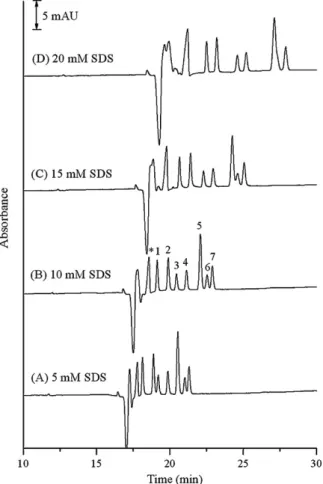
Fig. 3. Electropherograms of the seven benzodiazepines recorded at SDS
concen-trations of (A) 5, (B) 10, (C) 15, and (D) 20 mM. Conditions: 20 mM phosphate buffer (pH 7.0) containing 170 mM [BMIM][NTf2] and 55% MeOH; sample concentration,
80g/mL in MeOH/water (1:1, v/v).
3.1. Effect of [BMIM][NTf2] concentration
In aqueous CE, the capillary wall is coated and the direction of the EOF is reversed after adding imidazolium ions to the BGE. In contrast, the degree of dynamic coating by imidazolium ions is decreased in non-aqueous CE[1,7]. Because we used a high content of organic solvent in this study, we first investigated the influence of the IL on the direction of the EOF, applying reverse polarity volt-age and a BGE containing 170 mM [BMIM][NTf2] and 55% MeOH.
Neither the EOF marker nor the analyte signal was observed when the voltage was set at−20 kV. After increasing the concentration of [BMIM][NTf2] to 240 mM, we still did not observe any signals
for the analytes within 60 min. When we applied a separation voltage of +20 kV, however, the signals for the analytes emerged. These results indicate that adding [BMIM][NTf2] to the BGE did not
lead to a significant degree of coating of the capillary wall. We suspect that the direction of the EOF was not influenced by the added imidazolium ions in the BGE because the BGE contained an organic solvent content of greater than 50%. Thus, the main role of [BMIM][NTf2] was that of an association agent, not as an EOF
modifier.
Fig. 2 displays the electropherograms obtained when using [BMIM][NTf2] in the BGE at concentrations ranging from 100 to
240 mM—in the absence of SDS—at a constant buffer pH (pH 7.0) and a constant concentration of organic solvent. The reso-lution of the signals for bromazepam, clorazepate, nitrazepam, diazepam, alprazolam, and flunitrazepam increased upon increas-ing the [BMIM][NTf2] concentration, but the signal for lorazepam
remained overlapped with the system peak. The mobility and
selectivity of the analyte changed significantly upon changing the concentration of [BMIM][NTf2]. At 170 mM [BMIM][NTf2], we
observed the best separation of the seven analytes; therefore, we employed this concentration in our subsequent experi-ments.
3.2. Effects of the concentrations of SDS and organic solvent Fig. 3reveals the effect of the SDS concentration (5–20 mM) on the separation of the analytes in the BGE containing 170 mM [BMIM][NTf2]. An increase in the SDS concentration improved the
resolution of the signals for lorazepam, bromazepam, clorazepate, and nitrazepam. For diazepam, alprazolam, and flunitrazepam, 10 mM SDS provided the optimal resolution; when the SDS concen-tration exceeded 15 mM, the signals for diazepam and alprazolam migrated together. The improved separation efficiencies for the BGEs containing 5–10 mM SDS were probably due to synergistic effects between the ILs and SDS. Overall, the best results were obtained when using 10 mM SDS in the BGE.
We added organic solvent in this system to increase the solubility of [BMIM][NTf2] in the BGE ([BMIM][NTf2] is only
slightly soluble in water). In a preliminary experiment, we attempted to separate the benzodiazepines through non-aqueous CE using MeOH/ACN mixtures [50–100% (v/v)] containing 170 mM [BMIM][NTf2] (pH 5.0), but we could not obtain satisfactory
separa-tion. Because [BMIM][NTf2] is quite soluble in MeOH, we employed
MeOH at concentrations greater than 50% to improve the solubility of the IL in the BGE for CE. When we increased the concentra-tion of MeOH in the BGE from 50 to 60% (v/v), the separaconcentra-tion time increased (Fig. 4). We suspect that the decrease in the EOF was due
Fig. 4. Electropherograms of the seven benzodiazepines recorded at MeOH
concen-trations of (A) 50, (B) 55, and (C) 60%. Conditions: 20 mM phosphate buffer (pH 7.0) containing 170 mM [BMIM][NTf2] and 10 mM SDS.
to the decreased dielectric constant, zeta potential, and viscosity of the BGE. Because micelles are difficult to form in BGEs contain-ing more than 50% MeOH, it appears that the analytes’ enhanced separation was a result of solvophobic interactions with individual surfactant molecules. Because adding 55% MeOH to the BGE pro-vided the optimal resolution and a stable baseline, we employed this concentration in our subsequent experiments.
3.3. Effects of buffer pH and electrolyte concentration
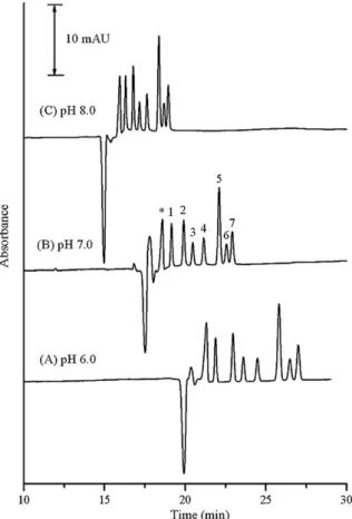
Changing the BGE’s pH affects both the EOF and the charge of the analyte.Fig. 5reveals the effect of the pH (pH 6.0–8.0) of the phosphate buffer on the analytes’ migrations, while maintaining the other separation conditions constant. The EOF increased upon increasing the pH from 6.0 to 8.0. Because the analytes possessed no net charge in the BGE within this pH range, the main effect on their migration times was caused by a variation of the EOF. At pH 8.0, the resolution of diazepam, alprazolam, and flunitrazepam was poor; at pH 6.0, the analysis time was ca. 28 min. Therefore, the optimal conditions for the separation of these analytes involved a pH of 7.0. Fig. 6displays the effect of the phosphate concentration in the BGE on the analytes’ separation. Although the addition of the IL also contributed to the electrolyte’s ionic strength[23], we kept its concentration constant in this study. The EOF decreased upon increasing the phosphate concentration, presumably due to nar-rowing of the double-layer thickness at the capillary wall. At 10 mM phosphate, the EOF was too fast to completely resolve the peaks for diazepam, alprazolam, and flunitrazepam. At 30 mM phosphate, the analysis time was extended to 27 min. Hence, the best resolution for all of the analytes within a reasonable analysis time occurred when we added 20 mM phosphate to the BGE.
Fig. 5. Effect of pH on CE analysis. Conditions: 20 mM phosphate buffer containing
170 mM [BMIM][NTf2], 55% MeOH, and 10 mM SDS. Inset: effect of pH on the EOF.
Fig. 6. Effect of phosphate concentration on CE analysis. Conditions: phosphate
buffer (pH 7.0) containing 170 mM [BMIM][NTf2], 55% MeOH, and 10 mM SDS. Inset:
effect of phosphate concentration on the EOF.
3.4. Mechanism of separation using [BMIM][NTf2] and SDS
Although the analytes possessed no net charge in the BGE at pH 7.0, their signals appeared after that of the EOF marker. This phe-nomenon suggested that the association of the benzodiazepines with the free anions was the cause of their separation. In addi-tion, the migration order of the analytes was the same for the BGEs containing either [BMIM][NTf2] or [BMIM][NTf2]/SDS, but it
dif-fered from that obtained when the BGE containing SDS only. These experimental results suggest that [BMIM][NTf2] was the dominant
agent in the separation mechanism, with the anionic moiety of [BMIM][NTf2] involved in heteroassociation interactions with the
analytes and SDS improving the resolution through hydrophobic, electrostatic, and/or hydrogen bonding interactions. Thus, the dif-ferent degrees of association of the individual benzodiazepines allowed their mixture to be separated adequately.
3.5. Calibration curve and detection limits
Table 1lists the ranges of linearity, limits of detection (LODs), relative standard deviations (RSDs) of the migration times and peak areas, and peak efficiencies for the analytes under the optimized separation conditions for a BGE containing 170 mM [BMIM][NTf2] and 10 mM SDS. The benzodiazepines were
com-pletely resolved within 23 min. The calibration curve was linear from 10 to 150g/mL and the coefficients of determination (r2)
were all greater than 0.9924. The LODs [signal-to-noise (S/N) ratio = 3] of the seven analytes ranged from 2.74 to 4.42g/mL. The theoretical plate numbers (per meter) ranged from 2.64× 10⁵ to
Table 1
Calibration curves, coefficients of determination (r2), limits of detection (LODs), relative standard deviations (RSDs), and theoretical plate numbers for benzodiazepines
separated through CE.
Lorazepam Bromazepam Clorazepate Nitrazepam Diazepam Alprazolam Flunitrazepam Calibration curvea y = 163x–759 y = 177x–1295 y = 104x–400 y = 157x–1294 y = 211x–1851 y = 107x–613 y = 128x–1110 Coefficient of determination 0.9982 0.9973 0.9976 0.9924 0.9960 0.9975 0.9969
LOD (S/N = 3;g/mL) 4.23 4.42 4.23 3.63 2.74 3.17 3.63
LOQ (S/N = 10;g/mL) 14.1 14.7 14.1 12.1 9.14 10.6 12.1
RSD (%; n = 5)
(a) Migration time (min) 0.18 0.23 0.18 0.16 0.15 0.15 0.16
(b) Peak area 5.30 2.43 4.68 6.39 4.85 7.07 9.59
Theoretical plates (N, per meter)b 3.52× 10⁵ 2.79× 10⁵ 2.95× 10⁵ 2.75× 10⁵ 2.64× 10⁵ 2.75× 10⁵ 2.83× 10⁵ aCalibration line (10–150g/mL): peak area (arbitrary units) = slope × concentration (g/mL) + y-intercept.
bN = 5.54(t
R/W1/2)2; tR, migration time; W1/2, width at half peak height; in 20 mM phosphate buffer (pH 7.0) containing 170 mM [BMIM][NTf2], 10 mM SDS, and 55% MeOH.
1.06× 10⁵to 1.41× 10⁵]. The RSDs of the migration times for the
seven analytes were all less than 0.23% under the optimized separa-tion condisepara-tions. These results imply that combining ILs with SDS for the analysis of benzodiazepines provides satisfactory repeatability. 3.6. Analyzing spiked urine samples
We employed SPE prior to performing CE separation of the ben-zodiazepines in a urine sample to eliminate interference from the urine sample and to concentrate the analytes.Fig. 7reveals that the presence of the seven analytes in the spiked urine sample was dis-cernable without interference from any unknown compounds. The
Fig. 7. CE electropherogram, obtained using [BMIM][NTf2] and SDS, of a urine
sam-ple spiked with the seven benzodiazepines. Analysis was performed under the optimized conditions. Other conditions were the same as those used to obtainFig. 2.
analytes were confirmed by their migration times and absorbance spectra. Under the optimized SPE conditions, the recoveries were 50.1% for lorazepam, 88.2% for bromazepam, 81.8% for clorazepate, 74.7% for nitrazepam, 78.0% for diazepam, 68.5% for alprazolam, and 72.6% for flunitrazepam; the repeatabilities (RSD; n = 4) of the extractions were 2.3, 5.4, 5.4, 9.8, 9.7, 2.8 and 5.1%, respectively. 4. Concluding remarks
In this study, we used CE to separate a mixture of seven benzo-diazepines within 23 min after adding an IL to the BGE and using SDS as a modifier. The addition of [BMIM][NTf2] and SDS
pro-vided high selectivity and improved the resolution of the seven analytes of interest, presumably because the different degrees of association of the individual benzodiazepines allowed their mix-ture to be separated satisfactorily, relative to the results obtained using the IL or SDS alone. [BMIM][NTf2] appeared to play a
dom-inant role during the separation process. The anionic moiety of [BMIM][NTf2] behaved as a heteroassociation site for the
benzo-diazepines; added SDS improved the resolution of the separation. The optimal BGE for separation of the benzodiazepines comprised 170 mM [BMIM][NTf2] at pH 7.0, 20 mM phosphate buffer, 10 mM
SDS, and 55% MeOH. We also applied this method, in conjunction with SPE, to the successful analysis of the seven benzodiazepines in a spiked sample of human urine. Our results suggest that ILs can contribute significantly to CE-based separations.
Acknowledgment
This study was supported by a grant (NSC 98-2113-M-009-016-MY3) from the National Science Council, Taiwan.
References
[1] A. Berthod, M.J. Ruiz-Ángel, S. Carda-Broch, J. Chromatogr. A 1184 (2008) 6. [2] J.L. Anderson, D.W. Armstrong, G.-T. Wei, Anal. Chem. 78 (2006) 2893. [3] S.M. Mwongela, A. Numan, N.L. Gill, R.A. Agbaria, I.M. Warner, Anal. Chem. 75
(2003) 6089.
[4] S.A. Shamsi, N.D. Danielson, J. Sep. Sci. 30 (2007) 1729. [5] J.L. Anderson, D.W. Armstrong, Anal. Chem. 75 (2003) 4851. [6] J.L. Anderson, D.W. Armstrong, Anal. Chem. 77 (2005) 6453. [7] H. Qiu, S. Jiang, X. Liu, L. Zhao, J. Chromatogr. A 1116 (2006) 46. [8] Q. Wang, G.A. Baker, S.N. Baker, L.A. Colón, Analyst 131 (2006) 1000. [9] W. Qin, S.F.Y. Li, J. Chromatogr. A 1048 (2004) 253.
[10] S. Qi, S. Cui, X. Chen, Z. Hu, J. Chromatogr. A 1059 (2004) 191.
[11] M. López-Pastor, B.M. Simonet, B. Lendl, M. Valcárcel, Electrophoresis 29 (2008) 94.
[12] M. Vaher, M. Koel, M. Kaljurand, J. Chromatogr. A 979 (2002) 27. [13] K. Tian, S. Qi, Y. Cheng, X. Chen, Z. Hu, J. Chromatogr. A 1078 (2005) 181. [14] R.C. Baselt, Disposition of Toxic Drugs and Chemicals in Man, 4th edition,
Chem-ical Toxicology Institute, Foster City, CA, 1995. [15] M. Tomita, T. Okuyama, J. Chromatogr. B 678 (1996) 331. [16] M.F. Renou-Gonnord, K. David, J. Chromatogr. A 735 (1996) 249. [17] Y. Suzuki, H. Arakawa, M. Maeda, Biomed. Chromatogr. 18 (2004) 150.
[18] T.-F. Jiang, Y.-L. Gu, B. Liang, J.-B. Li, Y.-P. Shi, Q.-Y. Ou, Anal. Chim. Acta 479 (2003) 249.
[19] L. Yu, W. Qin, S.F.Y. Li, Anal. Chim. Acta 547 (2005) 165. [20] M.-E. Yue, Y.-P. Shi, J. Sep. Sci. 29 (2006) 272.
[21] Y. Franc¸ois, A. Varenne, E. Juillerat, A.-C. Servais, P. Chiap, P. Gareil, J. Chro-matogr. A 1138 (2007) 268.
[22] S. Carda-Broch, A. Berthod, D.W. Armstrong, Anal. Bioanal. Chem. 375 (2003) 191.
[23] P. Lozano, T. De Diego, D. Carrié, M. Vaultier, J.L. Iborra, Biotechnol. Lett. 23 (2001) 1529.
[24] C.-W. Huang, H.-P. Jen, R.-D. Wang, Y.-Z. Hsieh, J. Chromatogr. A 1110 (2006) 240.
![Fig. 1. Structures of benzodiazepines and [BMIM][NTf 2 ]. *Values of pK a are those of the conjugate acids of the benzodiazepines.](https://thumb-ap.123doks.com/thumbv2/9libinfo/7490550.115014/2.892.191.688.81.648/fig-structures-benzodiazepines-bmim-values-conjugate-acids-benzodiazepines.webp)
![Fig. 2. Electropherograms of the seven benzodiazepines recorded at [BMIM][NTf 2 ]](https://thumb-ap.123doks.com/thumbv2/9libinfo/7490550.115014/3.892.479.835.489.1027/fig-electropherograms-seven-benzodiazepines-recorded-bmim-ntf.webp)
![Fig. 7. CE electropherogram, obtained using [BMIM][NTf 2 ] and SDS, of a urine sam-](https://thumb-ap.123doks.com/thumbv2/9libinfo/7490550.115014/6.892.75.410.535.1065/fig-electropherogram-obtained-using-bmim-ntf-sds-urine.webp)
