Chiral liquid crystals as biosensing platforms
Mon-Juan Lee
a,*, Yu-Chien Sung
b, Yu-Cheng Hsiao
b, and Wei Lee
b,**
a
Department of Bioscience Technology, Chang Jung Christian University, Guiren Dist., Tainan
71101, Taiwan
b
Institute of Imaging and Biomedical Photonics, College of Photonics, National Chiao Tung
University, Guiren Dist., Tainan 71150, Taiwan
ABSTRACT
The texture observation has long been the core technique in liquid crystal (LC)-based bioassays. Its working principle stems from the dark-to-bright texture change induced by the interruption of the initially homeotropic alignment in nematic bulks or from the radial-to-bipolar configuration transition in LC droplets in the presence of biomolecules. One of the drawbacks of this observational scheme, which requires a polarizing optical microscope, is the difficulty in quantitative analysis. In this invited paper, we report on our recent development of alternative optical biosensing techniques based on cholesteric LCs (CLCs) without the use of a polarizer. The increase in structural order in a vertically anchored CLC cell in the quasi-planar state provides a means to allow detection and quantification of the concentration of biomolecules immobilized on the interface between the mesophase and the surfactant DMOAP for LC vertical alignment.
Keywords: Liquid crystals, Cholesteric liquid crystals, Transmission spectroscopy, Biophotonics, Biosensors
1. INTRODUCTION
Studies have established that liquid crystal (LC)-based biosensors are powerful tools for the detection of biological binding events at solid surfaces fabricated with surface-immobilized biomolecules or self-assembled monolayers (SAMs), which enables semi-quantitation by observing the change in the optical texture of LCs.1–6 In particular, various LC-based
immunosensors are developed in which LCs or LC droplets serve as the sensing element for the detection of immunotargets such as microbial immunocomplexes, hepatitis B antibody, or cardiac biomarker.7–11
Recently, we reported an LC-based optical immunosensor for the detection of cancer biomarker CA125, which is a mucin-like glycoprotein overexpressed in several types of cancer, including ovarian cancer and breast cancer.12–14 A
eutectic nematic LC with a wide nematic range (30–95 C) and large birefringence (Δn = 0.333) was applied to improve the sensitivity of the LC-based immunosensor,13,14 whose detection limit was further lowered through surface
modification.12 These techniques generally rely on disruption of LC alignment by biomolecules, and the consequent
change in optical texture of LCs, to determine the relative amount of biomolecules.
To establish absolute quantitative methods for LC-based biosensing, the unique interfacial orientation in association with the Bragg reflection of chiral nematic or cholesteric LCs (CLCs) was exploited. CLCs are extensively applied in reflective polarizers, color filters, and reflective displays. The helical structure of CLCs selectively reflects certain wavelengths of light according to the handedness as the CLC chirality, and the surface-induced ordering transitions of CLCs can be measured spectroscopically. We demonstrated a quantitative, color-indicating protein assay method based on vertically anchored CLC (VAC), in which the helical pitch of CLCs changed with the concentrations of bovine serum albumin (BSA), thus altering the transmission spectrum and enabling the color-indicating capability of CLC-based biosensors (Fig. 1). As a result, a detection limit of 1 fg/ml BSA was achieved with this VAC biosensing approach.15 In
this study, we attempt to develop a quantitative VAC immunosensor for the detection of CA125 (Fig. 2). Both the optical textures and transmission spectra of CLC in the presence of various concentrations of CA125 captured by immobilized anti-CA125 antibodies were analyzed so that quantitative strategies for the VAC immunosensor can be derived.
*[email protected]; phone +886 (0)6 278-5123 ext. 3212; fax +886 (0)6 278-5010 **[email protected]; phone +886 (0)6 303-2121 ext. 57826; fax +886 (0)6 303-2535
Z
Ibl# hICq-..\y/d.o:r
..r\.allir
.y//i.,/:\%/e,i
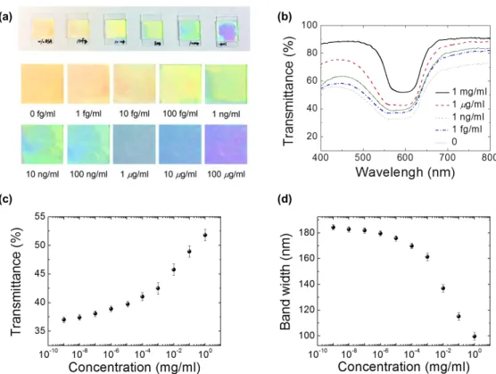
IrAbell \a\i.al U.1/rAVl1 b.IWl.11 \\li.11 IIyQÁ\\Nr1p+1 OyIlell AIM 40U0 Figure 1. Quantitation of BSA by VAC biosensor. (a) Representative VAC cells of ~5 μm in thickness featuring the color-indicating capability of the VAC biosensor at different BSA concentrations. (b) Transmission spectra of the VAC cells prepared with various concentrations of BSA. (c) Correlation of minimum transmittance and (d) bandwidth of Bragg’s reflection in the transmission spectra to BSA concentration. (Adopted from Ref. 15.)
Figure 2. Schematic of the CLC structure in a VAC immunosensor. The configuration of CLC in the vicinity of the DMOAP−CLC interface tends to be directed by the DMOAP alignment layer to be in the focal conic (FC) state, while others in the bulk confined in the cell remain in the intrinsic planar (P) state. When CA125 is bound specifically to anti-CA125 antibodies immobilized on the DMOAP-coated glass substrate, the fraction of the CLC in the FC state reduces because the CA125 immunocomplex weakens the vertical anchoring strength of DMOAP, thus altering the transmission of light by CLC in the VAC cell. (a) Severe disruptions of the self-assembled planar configuration near the substrates caused by the DMOAP aligning layers. (b) The weak blocking effect on the vertical anchoring strength in the presence of a low immunocomplex population immobilized on one of the substrates. (c) Effective shielding by immunocomplex that reduces DMOAP’s vertical aligning efficacy.
2. METHODOLOGY
2.1 Materials
LCD-grade glass slides (15 mm × 20 mm) were obtained from Mesostate (Taiwan). The silane surfactant dimethyl-octadecyl[3-(trimethoxysilyl)propyl]ammonium chloride (DMOAP) and the protein BSA (66 kDa) were purchased from Acros Organics and Sigma–Aldrich, respectively. HDN, a eutectic nematic mixture of LCs (∆n = 0.333 at a wavelength of 589.3 nm and temperature of 20 C), was provided by Jiangsu Hecheng Display Technology Co., Ltd. CLC was prepared by mixing the LC host E44 (Daily-Polymer Corporation) uniformly with the chiral dopant S5011 (Merck) at a concentration of 2.31 wt%. The resulting CLC exhibits a characteristic photonic bandgap in the visible transmission spectrum due to its intrinsic periodically helical structure. Recombinant human CA125/MUC16 was a product of R&D Systems, while anti-CA125 mouse antibodies were manufactured by Santa Cruz Biotechnology.
2.2 Preparation of DMOAP-coated glass slides
LCD-grade glass slides were immersed successively in aqueous solutions of detergent, deionized (DI) water, and 99.5% ethanol, accompanied by sonication for 15 min. The slides were dried under a stream of nitrogen and then baked at 74 °C for 15 min. The cleaned glass slides were immersed in an aqueous solution of 1% (v/v) DMOAP and reacted for 15 min. After rinsing with copious amount of DI water, the glass slides were dried under a stream of nitrogen and heated at 85 °C for 15 min, followed by mechanical buffing for three times with a rubbing machine.
2.3 Immobilization of anti-CA125 antibodies and formation of CA125 immunocomplex
Aqueous solutions of the CA125 antigen or anti-CA125 antibody of the desired concentrations were prepared by diluting in DI water. Anti-CA125 antibody was immobilized by dispensing a 200-l droplet of anti-CA125 antibody (at 0.1
g/ml) on a coated glass surface, which was allowed to dry at 35 °C for 3 h. The entire surface of the DMOAP-coated glass slide with immobilized anti-CA125 antibody was then reacted with 10 l of 0.001, 0.01, 0.1 or 1 g/ml CA125 at 7 °C for 1.5 h by dispensing the CA125 solution and covering the glass slide with a cover glass. After removal of the cover glass, the glass slide was rinsed twice with DI water and dried on a hot plate at 30 °C for 2 min to remove excess water.
2.4 Assembly of the LC cell
Unlike our previous study of VAC biosensing of BSA where a cell is composed of two identical glass substrates (covered with the protein of the same concentration),15here each LC optical cell was assembled with an intact
DMOAP-coated glass substrate and a glass slide covered by the CA125 antigen–antibody immunocomplex. 3-m ball spacers were used to separate the two substrates, which were then sealed incompletely with AB glue and dried at room temperature for 2 h. The empty cells were filled with HDN as references or CLC at room temperature by capillary action.
2.5 Optical texture observation and transmission spectrometry
The optical textures of the LC cells were examined under an Olympus BX51 polarized optical microscope equipped with crossed polarizers in the transmission mode. The transmission spectra of the CLC cells were acquired with an Ocean Optics HR2000+ high-resolution fiber-optic spectrometer.
3. RESULTS AND DISCUSSION
3.1 Optical textures of HDN and CLC in the presence of various concentrations of CA125
Anti-CA125 antibodies immobilized on DMOAP alignment layer was reacted with various concentrations of the CA125 antigen and detected with HDN or CLC. As indicated in our previous studies,12,14 the concentration of the anti-CA125
antibody was adjusted to ensure that in the absence of CA125, the antibodies alone have no effect on the homeotropic alignment of HDN, which was aligned perpendicularly to the glass surface by DMOAP, and its optical texture was completely dark (Fig. 3(a)). In the presence of CA125, which binds specifically to anti-CA125 antibodies, the CA125 antigen–antibody immunocomplex resulted in significant disruption of the alignment of LCs, and the optical texture of
Figure 3. Optical textures of HDN (top) and CLC (bottom) in the presence of various concentrations of CA125. A 200-μl droplet of 0.1 μg/ml anti-CA125 antibody was dispensed on a DMOAP-coated glass slide, followed by drying and reaction on the entire surface with (a,f) 0, (b,g) 0.001, (c,h) 0.01, (d,i) 0.1 and (e,j) 1 μg/ml CA125. HDN (a–e) or CLC (f–j) was filled in the space between two glass substrates described above to form a nematic or CLC cell, respectively. The optical textures were examined under an Olympus BX51 polarized optical microscope equipped with crossed polarizers in the transmission mode. The red dashed circle indicates the perimeter of the droplet of anti-CA125 antibody.
HDN therefore changed from dark to bright (Fig.3(b)–(e)). In contrast, the change in birefringence texture of CLC with increasing CA125 concentration was not as significant as that of HDN (Fig.3(f)–(j)). However, the appearance of the CLC cells with anti-CA125 antibodies was different from that without immobilized antibody as a result of the intrinsic optical activity of CLCs.
3.2 Transmission spectra of vertically anchored CLC in the presence of various concentrations of CA125
Our previous study demonstrated that the configuration of the vertically anchored CLC was sensitive to the amount of immobilized BSA on the alignment layer, which, in turn, affected the reflection and transmission of light, leading to the color-indicating feature of the VAC biosensor.15 As shown in Fig. 1(a), a distinct color was reflected from the VAC cell
at each BSA concentration. When the VAC cells were analyzed by transmission spectrometry, we observed that the overall transmittance increased, while the reflection bandwidth decreased with increasing BSA concentration (Fig.1(b)). Correlation of the minimum transmittance and bandwidth at half maximum of the Bragg reflection with BSA concentration can therefore be utilized in quantitation (Fig.1(c), (d)).
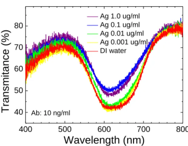
In this study, the VAC optical cell with CA125 antigen–antibody immunocomplex atop one of the two substrates was subjected to transmission spectroscopic measurement. The overall transmittance of the VAC immunosensor was the lowest when the immobilized anti-CA125 antibodies were reacted only with DI water (Fig. 4). This is attributable to the effective scattering caused by the disruption of the helically planar (P) structure or generation of the focal conic (FC) domains near the substrates. In the presence of CA125, the overall transmittance increased gradually with increasing CA125 concentration, indicating that the effective volume of CLC in the P state varied sensitively with the amount of CA125. The working principle for the quantitative VAC biosensor can be explained by alteration in the configuration of CLC by CA125 (Fig.2). In the absence of CA125, most CLC molecules at the interfaces of DMOAP and CLC are directed by DMOAP to align themselves vertically to the glass substrate. CLC molecules close to the DMOAP−CLC interfaces are in the scattering state, most likely to be in the FC state, while others in the bulk away from the interfaces remain in their intrinsic alignment, namely the P state. As the concentration of CA125 increased, the increasing amount of the larger CA125 immunocomplex thus formed weakened the vertical anchoring strength of DMOAP. Owing to the shielding effect on the strong vertical anchoring provided by the aligning surfactant DMOAP near the substrate, more CLC molecules took the P structure, giving rise to a reduced extent of light scattering and, hence, an increase in total transmittance. Consequently, the minimal transmittance of the VAC cell can be correlated with CA125 concentration to develop and optimize an algorithm for the quantitation of CA125. Figure 4 indicates that no significant change in distorted Bragg’s reflection or overall spectral profile took place in various conditions of CA125 concentrations, presumably due to the shielding effect to be only one-sided (i.e., limited) as well as the relatively thin cell gap (~3 μm) to restrain the total volume of CLC molecules in the P state. According to this preliminary finding, the desired color-
400 500 600 700 800 40 50 60 70 80
Tra
n
s
mita
n
c
e
(
%)
Wavelength (nm)
Ag 1.0 ug/ml Ag 0.1 ug/ml Ag 0.01 ug/ml Ag 0.001 ug/ml DI water Ab: 10 ng/mlFigure 4. Transmission spectra of the VAC immunosensor in the presence of various concentrations of CA125. The VAC cells shown in Fig. 3(f)−(j) were subjected to transmission spectroscopy measurement to obtain the transmission spectra.
indication feature observed in our previous CLC-based BSA sensing seems to be hardly realized in the present work toward CA125 immunosensing. Further studies along this line will be rigorously pursued in this laboratory for improvement including retaining the color-indicating function in the VAC platform.
4. CONCLUDING REMARKS
A quantitative VAC immunosensor for determining the concentration of cancer biomarker CA125 was developed in this study. Cancer screening is an important procedure in cancer prevention and early diagnosis. Traditional label-based immunoassays are time-consuming and rely on fluorescence- or enzyme-labeled antibodies for detection, which render the procedure complex and costly. Development of label-free immunodetection techniques such as the VAC immunosensor provides a possibility to simplify and lower the cost of the cancer screening procedure. In addition, the color-indicating properties of VAC biosensing may contribute to the development of point-of-care devices, eliminating the use of expensive detection instruments. By exploiting the intrinsic optical activity of CLC as well as transmission spectroscopy, the VAC immunosensor provides a new approach to the quantitation of CA125 as well as other protein targets.
ACKNOWLEDGMENTS
This study is financial supported by the Ministry of Science and Technology, Taiwan, under grant Nos. 104-2112-M-009-008-MY3 and 101-2314-B-309-001-MY3.
REFERENCES
[1] Kim, S. R., Shah, R. R. and Abbott, N. L., “Orientations of liquid crystals on mechanically rubbed films of bovine serum albumin: a possible substrate for biomolecular assays based on liquid crystals,” Anal. Chem. 72(19), 4646–4653 (2000).
[2] Skaife, J. J. and Abbott, N. L., “Quantitative interpretation of the optical textures of liquid crystals caused by specific binding of immunoglobulins to surface-bound antigens,” Langmuir 16, 3529–3536 (2000).
[3] Skaife, J. J., Brake, J. M. and Abbott, N. L., “Influence of nanometer-scale topography of surfaces on the orientational response of liquid crystals to proteins specifically bound to surface-immobilized receptors,” Langmuir 17, 5448–5457 (2001).
[4] Chen, C. H. and Yang, K. L., “Oligopeptide immobilization strategy for improving stability and sensitivity of liquid-crystal protease assays,” Sensors and Actuators B 204, 734–740 (2014).
[5] Bi, X., Lai, S. L. and Yang, K. L., “Liquid crystal multiplexed protease assays reporting enzymatic activities as optical bar charts,” Anal. Chem. 81(13), 5503–5509 (2009).
[6] Hartono, D., Xue, C.-Y., Yang, K.-L. and Yung, L.-Y. L., “Decorating Liquid Crystal Surfaces with Proteins for Real-Time Detection of Specific Protein–Protein Binding,” Adv. Funct. Mater. 19, 3574–3579 (2009). [7] Helfinstine, S. L., Lavrentovich, O. D. and Woolverton, C. J., “Lyotropic liquid crystal as a real-time detector of
microbial immune complexes,” Lett. Appl. Microbiol. 43(1), 27–32 (2006).
[8] Alino, V. J., Pang, J. and Yang, K. L., “Liquid crystal droplets as a hosting and sensing platform for developing immunoassays,” Langmuir 27(19), 11784–11789 (2011).
[9] Chen, C. H. and Yang, K. L., “Liquid crystal-based immunoassays for detecting hepatitis B antibody,” Anal. Biochem. 421(1), 321–323 (2012).
[10] Zapp, E., Westphal, E., Gallardo, H., de Souza, B. and Cruz Vieira, I., “Liquid crystal and gold nanoparticles applied to electrochemical immunosensor for cardiac biomarker,” Biosens. Bioelectron. 59, 127–133 (2014). [11] Tan, L. N. and Abbott, N. L., “Dynamic anchoring transitions at aqueous-liquid crystal interfaces induced by
specific and non-specific binding of vesicles to proteins,” J. Colloid Interface Sci. 449, 452–461 (2015). [12] Su, H. W., Lee, M. J. and Lee, W., “Surface modification of alignment layer by ultraviolet irradiation to
dramatically improve the detection limit of liquid-crystal-based immunoassay for the cancer biomarker CA125,” J. Biomed. Opt. 20(5), 57004 (2015).
[13] Sun, S. H., Lee, M. J., Lee, Y. H., Lee, W., Song, X. and Chen, C. Y., “Immunoassays for the cancer biomarker CA125 based on a large-birefringence nematic liquid-crystal mixture,” Biomed. Opt. Express 6(1), 245–256 (2015).
[14] Su, H. W., Lee, Y. H., Lee, M. J., Hsu, Y. C. and Lee, W., “Label-free immunodetection of the cancer biomarker CA125 using high-Deltan liquid crystals,” J. Biomed. Opt. 19(7), 077006 (2014).
[15] Hsiao, Y. C., Sung, Y. C., Lee, M. J. and Lee, W., “Highly sensitive color-indicating and quantitative biosensor based on cholesteric liquid crystal,” Biomed. Opt. Express 6(12), 5033–5038 (2015).