行政院國家科學委員會補助專題研究計畫
□ 成 果 報 告
□期中進度報告
肌腱與韌帶的組織工程-以間質幹細胞、生物反應爐、與力學
刺激之統合研究: 第一年成果報告
計畫類別:V 個別型計畫 □ 整合型計畫
計畫編號:
NSC91- 2314- B- 039- 023
執行期間:
90 年 8 月 1 日至 91 年 7 月 31 日
計畫主持人:
曾國峰
共同主持人:
計畫參與人員:許晉榮,許弘昌,張景明。
成果報告類型(依經費核定清單規定繳交):V 精簡報告 □完整報告
本成果報告包括以下應繳交之附件:
□赴國外出差或研習心得報告一份
□赴大陸地區出差或研習心得報告一份
□出席國際學術會議心得報告及發表之論文各一份
□國際合作研究計畫國外研究報告書一份
處理方式:除產學合作研究計畫、提升產業技術及人才培育研究計畫、
列管計畫及下列情形者外,得立即公開查詢
□涉及專利或其他智慧財產權,□一年□二年後可公開查詢
執行單位:
中國醫藥學院中醫學系
中 華 民 國
九十一年
十二月
二十六日
行政院國家科學委員會專題研究計畫成果報告
肌腱與韌帶的組織工程-以間質幹細胞、生物反應爐、與力學刺激
之統合研究: 第一年成果報告
Tissue Engineer ing of Tendons and Ligaments – An Appr oach using
Mesenchymal Stem Cells, Bior eactor s, and Mechanical Stimulation
計畫編號:NSC91-2314-B-039-023
執行期間:90 年 8 月 1 日至 91 年 7 月 31 日
主持人:曾國峰 中國醫藥學院中醫學系
一、中文摘要 韌帶與肌腱是人體肌肉骨骼系統中兩種非 常重要的組織,它們的主要功能在於力量 的傳導、引向與拮抗等。在臨床領域中, 韌帶與肌腱的傷害相當常見,而臨床上吾 人也有許多治療的方式。 以韌帶受傷而言,直接修補斷裂的韌帶 通常造成不佳之結果,所以臨床上我們通 常需要以肌腱或其他人造物來增強修補或 是與以重建,但修補或重建後之結果似乎 也不是很令人滿意。肌腱受傷,包括有撕 裂傷,斷裂或是慢性的受傷,也是非常常 見。臨床上僅有急性的肌腱撕裂傷或是斷 裂的修補才有較佳之結果。慢性或是未治 療之肌腱斷裂在修補後之結果常是留有許 多後遺症。由於有這些不甚理想之結果, 以組織工程之方式來製造新的韌帶或是肌 腱似乎是一個最佳的選擇,因為所製造出 的新韌帶或肌腱將是自體產生的,沒有免 疫排斥問題。 在 此 計 畫 中 我 們 提 出 了 一 個 兩 年 計 畫,運用多學門的結合來製造新韌帶以供 臨床使用。此一計畫主要是延續吾人過去 兩年以間質母細胞來修補填充巨大骨骼 缺損之研究,將之延伸至韌帶與肌腱。吾 人的假說是間質母細胞在主要伸長力學 刺 激 下 會 被 導 引 入 韌 帶 與 肌 腱 此 一 Lineage 的路線。吾人利用間質幹細胞培 養技術,生物培養反應爐,以 PGA 聚合物 組成組織架構,以及不同的 Tensile 力學 刺激來進行此一研究計畫。此一成果報告 敘述第一年計畫之成果,主要在於生物培 養反應爐之製造與 PGA 聚合物組織架構的 組成。此外對於有關骨質疏鬆與骨髓內所 取得之間質幹細胞之數目與繁衍分化能 力也有許多成果報告。 關鍵詞:間質幹細胞,力學,細胞分化及增 生,骨頭,生物力學。 英文摘要AbstractLigaments and tendons are two very important tissues of the musculolskeletal system. Their primary functions are force
transmission, direction, and resistance.
Injuries to the ligaments and tendons are quite frequent in clinical settings, and there are many options in treatment. For ligament injuries, direct repair often yield poor results.
Thus augmentation with graft or
reconstruction with tendons is required. Tendon injuries, including severance and chronic tear/rupture, are also common. Only acute repair of the severed tendons has good clinical results. Chronic and neglected tears of the tendons are often with
unsatisfactory results following repair.
Tissue engineering of the ligaments and tendons appears to be an excellent choice for repair and regeneration of new ligaments and tendons for clinical use in that the newly engineered tissues will be autologous with no immune response and reaction. We are proposing a two-year research project in which a multi-discipline approach will be used to engineer new ligaments. Following
our previous experience with bone
marrow-derived mesenchymal stem cells (MSCs) in filling and repairing bone defects, we are extending the study into ligaments and tendons. The hypothesis is that with
principal tensile mechanical stimulation, the MSCs will be directed into the lineage toward formation of ligaments and tendons. An integrated approach using MSC-based
techniques, tissue bioreactors, tissue
scaffolds using PGA polymers, and different tensile stimulation is proposed. This proposal is formed into a two-year project. In this progress report, we forwarded the results of designing, manufacturing and fabrication of the tissue bioreactor for the use of the second-year project. We also reported the relationship between the numbers of MSC derived from the bone marrow as well as their differentiation and proliferative capacity. Their relationship to the occurrence of osteoporosis is also forwarded.
Keywords: Mesenchymal stem cell, Bio-Mechanics,
Differentiation and Proliferation, Bone, Cartilage,
二、緣由與目的 (Introduction & Purpose)
Tendons and ligaments are two very important structures of the musculoskeletal system, with their main functions being force transmission, direction/re-direction and force
resistance. Proper function of the
locomotion as well as refined movement of the humans and animals requires integral structure and proper function of the tendons and ligaments.
Ligaments are short bands of tough but
flexible fibrous connective tissue that bind the bones of the body together and support the organs in place. Anatomically ligaments are composed of fibers arranged in a spirally wound pattern. Depending on the location and the function, ligaments are found in the forms of cords, bands or in bundles. As tendons, a hierarchical structure is found in ligaments. Also like tendons, a crimp appearance is found in ligaments (Figure 1).
Biochemically, ligaments have similar
compositions as tendons, with over 90% of type I collagen and less than 10% of type III
collagen fibers.2 The mechanical functions of
ligaments are several folds: (1) maintain normal joint kinematics; (2) provide passive joint stability; and (3) take part in normal
joint proprioception.2,3 The mechanical
properties of ligaments depend on several factors including age, location, strain rate, and its viscoelastic behavior. For example, the human anterior cruciate ligament (ACL) has a typical failure strength of 50 to 150 MPa and a failure strain ranging from 12% to
15%, depending on the testing conditions.3
Injury to the tendons and ligaments is not uncommon in humans. In the United States only, at least 120,000 patients per year
undergo tendon or ligament repairs.4 The
number of anterior cruciate ligament (ACL) reconstruction using autografts or allografts was estimated to be about 30,000 per year in
the US.4,5 The incidence of tendon injuries,
owing to degeneration, avulsion, and
severance/laceration are also high.
Similarly, the incidence of tendon and ligament injuries in Taiwan was also high, with an estimated number of 1,200 ACL reconstruction procedures performed yearly.
To date, repair of acute tendon laceration or severance provides some marginally satisfactory results. Repair of chronic or neglected tendon rupture, however, often results in re-rupture or severe impairment of
the injured limb’s function. Reconstruction
of severely damaged tendons has thus taken three directions: the use of devices to prevent scar formation, the implantation of implants to enhance pseudosheath formation before tendon grafting, and the use of artificial
tendon substitutes to bridge tendon grafts.4, 6
To achieve these goals, tendon substitutes,
including Teflon®, Gor-Tex®, nylon, Dacron®,
silicon and others, were used. All but silicon tubes result in disastrous and
detrimental results in tendon repair.6
Numerous surgical techniques were also devised to enhance repair of injured tendon. Despite these therapeutic options, the results
for chronic tendon repair are often
un-satisfactory both for surgeons and
patients.6
Ligament injuries are the most common injuries to joints in general and to the knee in particular, accounting for 25% to 40% of all
knee injuries in most studies.3, 4 Due to the
poor results of repairing torn ligaments, reconstruction of the ACL with either
augmentation or primary substitution is the
mainstay therapeutic option at present.7
Although reconstruction of the ACL using
patellar tendon autograft provides a
satisfactory short-term outcome, long-term results are often less satisfactory with complications such as re-rupture, loosening
and ligamentation of the tendon.7
With the high incidence of tendon and ligament injuries as well as poor long-term clinical results after repair or reconstruction, the need for bioengineered tendons and ligaments is important and crucial. Within the last few years, attempts were made to discover new biocompatible materials for ligament and tendon replacement. Newly regenerated tendons and ligaments are probably the best choice for reconstruction of tendon and ligament injuries, in that they are autologous and that, giving appropriate stimulation, they will probably possess the
same biochemical and biomechanical
functions as the original tissues.
Over the past few years, several
approaches are proposed to produce
tissue-engineered ligaments and tendons both
in vitro and in vivo.6, 12-15 However, the
development of these approaches always involves the use of biocompatible and, preferably, biodegradable materials that can (1) provide mechanical resistance, and (2) can be colonized and reorganized by living
cells in vitro or in situ post-grafting.
Collagen fiber matrices are the most commonly used biomaterial for tissue engineering of tendons and ligaments. With its high tensile strength (30-60 MPa) and
small diameter, cross-linked collagen
prostheses appeared to be a good biomaterial for reconstruction of ACL, at least in rabbits. However, the absence of living cells in the prostheses make it a basic requirement that the biocompatible collagen prostheses need to be replaced and remodeled in vivo by
fibroblasts and other stem cells.12, 13 One
approach using cultured fibroblasts
incorporated into collagen matrices are yet another option for tissue engineering of
tendons and ligaments.6, 14, 15 The major
advantage of co-culture of fibroblasts with collagen matrices is that, with cultivation of
the fibroblasts the collagen matrices will be able to produce collagen for the regeneration of new tendons and ligaments. The newly bioengineered tissues, however, lacked the strengths and characteristic histological pattern of normal ligaments or tendons. Furthermore, the insertions of ligaments and tendons to bone remained a weak point
during reconstruction and repair.6, 14, 15
Several factors may have contributed to these disadvantages. First, the biomaterials used were probably not the most suitable ones for tissue engineering of ligaments and tendons. Secondly, co-culture of fibroblasts may have less than optimal effects in producing tendons/ligaments and their insertions to bone. Thirdly, the culture conditions were probably not optimal in regenerating new tissues. And finally, proper mechanical
stimulation was absent for excellent
regeneration and remodeling of tendons and ligaments.
The hypothesis of this proposal is that tissue engineering of tendons and ligaments require a proper integration of mesenchymal stem cell-based technology, tissue reactor cultivation, proper mechanical stimulation and appropriate biomaterials. We proposed to use marrow-derived mesenchymal stem cells as the cells to differentiate and proliferate cells responsible for production and fabricating new tissues, PLA and PGA based matrices as the scaffolds, tissue reactor techniques as the cultivation reservoir, and finally simultaneous mechanical stimulation to engineer new tendons and ligaments. A two-year integrated project proposal is forwarded to investigate the possible tissue engineering of tendons and ligaments.
Bioreactors are closed system that can
continuously supply nutrients and
automatically control tissue culture
parameters according to the changing needs
of the growing tissue construct.20-22
Numerous investigators have successfully cultured and bioengineered cartilage explants
in bioreactors.20-22 The major advantages of
bioreactors are that they can continuously supply nutrients and apply mechanical or other biophysical stimulation at the same
time. With appropriate mechanical stimulation, for example shearing, tissue constructs grown in tissue reactors are found
to be able to resemble natural cartilage.20, 21
Our hypothesis is that even with good tissue culture techniques, excellent bioengineered tendons and ligaments will not be formed without proper mechanical stimulation. For a tendon or ligament to be properly regenerated and remodeled to be with specific histological patterns and with
mechanical integrity, continuous or
intermittent tensile and/or torsional stress is
necessary.23, 24
For the tissue constructs to be able to
transform into new ligaments, a
biodegradable matrix scaffold is also necessary. Among numerous biodegradable materials, including type I collagen, gelatin, chitosan, PGA, PLA, PCL, used in
orthopaedics and research settings,
Poly-Glycolic Acid (PGA), Poly-Lactic Acid (PLA), and their co-polymers appear to be a good choice for tissue scaffold for tissue engineering of ligaments and tendons. PGA has a high melting point and low solubility in organic solvents. Due to its hydrophilic nature, surgical sutures made from PGA
polymers (e.g. Vicryl) tend to lose their
mechanical strength more rapidly, typically
over a period of 4 to 6 weeks
post-implantation.27, 28 PLA is more
hydrophobic than PGA, hence a lower backbone breakdown rate than PGA. The period for sutures made from PLA polymers
range from 8 to 12 weeks.27, 28 A pilot study
from our laboratory showed that it took at least two to four weeks to have the MSCs generate enough cells and extra-cellular matrices for the newly engineered ligaments. Therefore, we’ll be using both PGA polymers and PLA-PGA polymers to serve as the scaffold for tissue engineering of the ligaments.
In the principal investigator’s previous
articles,23, 24 the relationship between skeletal
tissue differentiation/proliferation and
mechanical stimulation was proposed
according to Carter’s mechanobiology view
(Figure 3).26 Our previous results in mice
and rabbits have shown that with principal
compression strain history, skeletal tissue was directed into the lineage of bone, either cancellous or cortical bone, while, with
shearing stress, cartilage (at least
fibrocartilage) was formed in mice.23, 24
These results provide some valid
verifications that Carter’s view of
mechanobiology was basically correct, at least in terms of bone and cartilage differentiation and proliferation. In this proposal, we are furthering our previous studies and extending into ligaments and
tendons (i.e. fibrous tissue in Figure 3). As
proposed by Carter et al. and revised in our
previous articles, our hypothesis is that for proper differentiation and proliferation of tendons and ligaments to occur, a principal tensile strain (or stress) history must be present during the differentiation and proliferation processes. The pattern of tensile strain history needs to be validated further in this study.
Thus in this proposal, we proposed to use a combination of MSC separation, isolation and cultivation, bioreactor tissue culture system and appropriate mechanical stimulation to engineer new ligaments and tendons for possible clinical application. The global hypotheses are: (1) Bone marrow mesenchymal stem cells have the potential to differentiate into tenocytes and fibroblasts that are responsible for regenerating new tendons and ligaments; (2) A proper scaffold, embedded with MSC’s, when subjected to intermittent tensile stimulation, will be engineered into a normal tendon and/or ligaments; (3) Custom-made bioreactors will be able to greatly facilitate the tissue engineering of the tendons and ligaments. The purposes of this proposal are thus: (1) further evaluate the possibility of directing MSCs into the tendon/ligament lineage; (2) test the possibility of using bioreactor and mechanical stimulation simultaneously to engineer new tendons and ligaments; (3) test the theory of mechanobiology as forwarded
by Pauwels25 and Carter.26
三、研究方法 (Material and Methods)
The purpose of this first-year project is first to manufacture custom-made bioreactors
specifically designed for the purpose of the study, to evaluate the culture conditions and flow rates of the culture medium for optimal culture of the tissue constructs in the bioreactors, and to investigate the best materials and scaffold constructs used for tissue constructs.
(1) Isolation and cultivation of Human
Mesenchymal Stem Cells (hMSCs) 1, 2, 3:
Human subjects will be derived from at least 12 young male individuals who suffer from long bone fractures requiring operation. Before operation and harvest of the bone marrow, informed consent will be obtained from the patients. Withdrawal of about 20 to 30 ml of bone marrow from the anterior
superior iliac spine will be done
simultaneously at the time of the operation under either spinal or general anesthesia. At the time of withdrawal of bone marrow, 2 ml of heparin were added into the syringe to prevent clotting. Further isolation of the adherent MSCs will be performed according
to the protocols stated previously.3, 4 Briefly,
the isolation of the hMSCs involves (1) adding 2 ml of PBS solution into 10 ml of heparinized bone marrow, (2) centrifuging the mixed solution at 3000 rpm for ten minutes under 25 degrees Celsius, and (3) removing the supernatants with pipets. Then mix the remaining solution with another 5 ml of PBS solution, add 5 ml of Percoll (1.073 g/ml, 95%) via pipets carefully toward the bottom of the test tubes, and
centrifuge once again at 25°C at 3000 rpm
for ten minutes. Remove the interface layer to another test tube, add another 5 ml of PBS
solution, mix thoroughly, centrifuge at 25°C
at 3000 rpm for ten minutes, and then remove the supernatant. The remaining solution was then added with 5 ml of DMEM medium containing 10% fetal bovine serum. Cell density was then counted and the cells
diluted to the density of 106/ml, ready for
tissue construct culture.
The isolated MSCs will be further culture expanded by changing DMEM medium with 10% FBS every other day, and the cells will
be culture-expanded to a density of 107/ml.
The cells are then ready for tissue culture in the bioreactor.
(2) Design and Fabrication of Tissue Culture BioReactors:
Bioreactors can provide mixing which significantly improved the yield and spatial uniformity of cell seeding, and increased the rates of cell proliferation and tissue regeneration. They have been used in culture of the cartilage growth and the results were better than the traditional culture
methods.5-7 Based on the design used by
Freed and Vynjak-Novakovic,5 We have
modified and re-designed a custom-made bioreactor which will serve as the culture bioreactor for this current study. The major difference between Freed’s design and our
design lies in the fact that a
tensile-conpression actuator was attached to the bioreactor, which will provide a tensile strain on the tissue constructs in the bioreactor (Figure 1). Culture media can be infused into the bioreactor via the inlet and exit via the outlet, and the flow rate can be adjusted. The bioreactor is also a rotating wheel which can provide some shearing stress (which will be analyzed using fluid dynamics principles). Every bioreactor can harvest two to four tissue constructs at the same time. The bioreactors will be made of stainless steel with influx and outflow ports. The diameter of the bioreactor is set to be 2.5 centimeters and the length is about 8 cm.
Figure 1. Draft Design for the custom-made bioreactor
(3) Tissue Culture Constructs:
For the tissue constructs to be able to
transform into new ligaments, a
biodegradable matrix scaffold is necessary. Among numerous biodegradable materials, including type I collagen, gelatin, chitosan, PGA, PLA, PCL, used in orthopaedics and research settings, poly(glycolic acid) (PGA),
Poly(Lactic Acid) (PLA), and their
co-polymers appear to be a good choice for a
tissue scaffold for tissue engineering of ligaments and tendons. PGA has a high melting point and low solubility in organic solvents. Due to its hydrophilic nature, surgical sutures made from PGA polymers tend to lose their mechanical strength more rapidly, typically over a period of 4 to 6
weeks post-implantation.8,9 PLA is more
hydrophobic than PGA, hence a lower backbone breakdown rate than PGA. The period for sutures made from PLA polymers
range from 8 to 12 weeks.8, 9 A pilot study
from our laboratory showed that it took at least two weeks to have the MSCs generate enough cells and extra-cellular matrices for the newly engineered ligaments. Therefore, we’ll be using both PGA polymers and
PLA-PGA polymers (e.g. Vicryl) to serve as
the scaffold for tissue engineering of the ligaments.
There are numerous methods of fabricating polymer scaffolds: fiber bonding, solvent casting, particulate leaching, membrane
lamination, and melt molding.8 For a polymer
that can be used as a scaffold for tissue engineering purpose, the polymer has to be biocompatible, biodegradable, and with a certain amount of mechanical integrity to
withstand mechanical stimulation. Vicryl
sutures have been used extensively in surgery to close wound and repair numerous tissues. It has a failure stress of about 50 to 100 MPa and an ultimate strain of about 2% at initial
implantation. Degradation of the Vicryl to
a failure stress less than 30 MPa takes about four to six weeks in vivo, and complete
resorption of the material takes
approximately three months. With its initial mechanical strength and long degradation
time, Vicryl is a suitable material for the
scaffold used in this study. Two patterns of ligament scaffolds will be fabricated. The first one is made with the technique of melt molding to produce a 3-dimentional ACL PGA polymer construct (8x8x35 mm in size) with a porosity of about 96%, a bulk density
of about 50 g/cm3, and a strength of about
100 MPa. The constructs will be sterilized with ethylene oxide. An outside contractor will manufacture this scaffold construct. The second scaffold construct will be made
directly using Vicryl sutures. We will
weave 1-O Vicryl sutures into a
3-dimentional construct with the shape similar to human ACL (8x8x35 mm in size). The second construct will be sterilized by ethylene oxide and saved for further use.
(4) Tissue Culture Conditions in the
Bioreactors:
Prior to cell seeding, the scaffolds are pre-wetted in culture medium, clamped at both ends to specially designed soft tissue grips, positioned with forceps to zero tensile strain ( no tensile strain will be applied onto the scaffolds), and fixed onto the graspers in the bioreactors (Figure 1). The bioreactors will be filled with 150 ml of culture medium
and placed in a humidified 370C/5% CO2
incubator for 8-12 hours prior to cell inoculation. The scaffolds are then
inoculated with the previously
culture-expanded MSCs. The bioreactors are to be rotated around their center of rotation at a speed of 20 rpm. Continual supply of the culture medium will be given via the fluid influx inlet at a flow rate of 2 to
3 ml/min.5 The scaffolds are then sampled at
timed intervals of 3, 7, 14, and 21 days for further analysis.
(5) Histological Analysis:
Cultured scaffold constructs are to be fixed with 70% alcohol solution, and then embedded in glycol methecrylate (GMA).
The 4-µm thick longitudinal sections are
stained with H-E or trichrome de Masson methods. Histomorphometric methods will
be used to examine the scaffold’s
microscopic pattern. Parameters analyzed will be gross histology, relative cellularity, presence of Type I and Type III collagen fibers via in situ hybridization.
四、結果 (RESULTS)
(1) Isolation and cultivation of Human Mesenchymal Stem Cells (hMSCs): The isolation, separation and culture-expansion of the hMSCs have gone well as planned. Batches of bone marrows were harvested during surgical procedures from a total of 30 patients over the past year. Human study protocols were followed rigidly and all patients were given consent forms. The bone marrow harvest sites were from the posterior superior iliac spine (PSIS) and from the proximal femur during spinal operations and total hip replacements. Through a series of investigations, we found that the yield of hMSCs was dependent on the patients’ age and the site of bone marrow harvest. Bone marrows harvested from the iliac crests had better MSC yield than from the proximal femur or from the tibia. The younger the patient is, the better the culture yield (Figure 2). Mono-nucleated cells (e.g. MSCs) were found at a frequency of 0.01 to 0.0001% of multinucleated cells in the human bone marrow. The number of mono-nucleated cells decreased gradually with age. The negative trend was statistically significant (p<0.01). y = 0.0009x2- 0.1284x + 5.8692 R2= 0.769 0.00 2.00 4.00 6.00 8.00 0 20 40 60 80 100 Age N umb er o f M N C s ( 10^ 6) /ml B M
Figure 2. Number of MSCs vs. Age
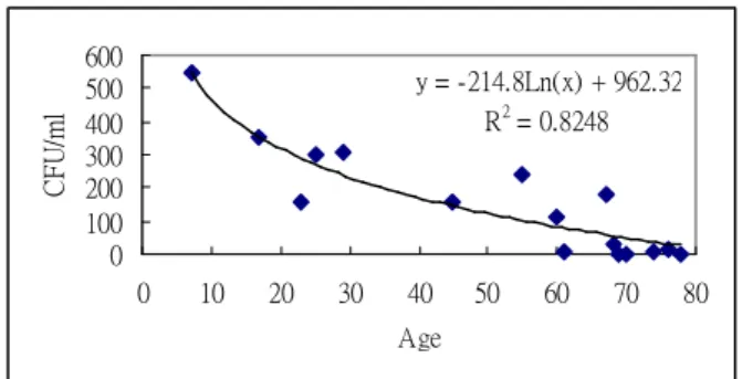
(2). The proliferative and differentiation capacity of the MSCs decreased with Age and Implications in osteoporosis treatment: Mean Colony Forming Unit (CFU) number per ml of bone marrow also decreased significantly with age in the current study (p<0.05). A significant linear negative relationship between the CFU number per ml of BM and age was noted (Figure 3).
y = -214.8Ln(x) + 962.32 R2= 0.8248 0 100 200 300 400 500 600 0 10 20 30 40 50 60 70 80 Age C F U /m l
Figure 3. CFU/ml BW vs. Age
Of all the 30 patients, 20 of them had measurements of axial bone mineral density (BMD) using Dual X-ray Absortiometry (DXA). The BMD data confirmed previous reports that axial skeleton BMD peaks at around 35 years of age and gradually decreased with age (Figure 4).
y = 6E-05x3- 0.0089x2+ 0.3832x - 1.1464 R2= 0.8672 0 1 2 3 4 5 0 20 40 60 80 100 Age B M D ( g/ cm^ 2)
Figure 4. L-spine BMD vs. Age
Further characterization of the number of CFU with axial skeleton BMD revealed an interesting relationship. A statistically significant positive relationship (p<0.05) between the number of mean CFU per ml of bone marrow and bone mineral density of the L-spine was noted (Figure 5)
. y = 0.0059x + 1.6345 R2= 0.5997 0 1 2 3 4 5 0 100 200 300 400 Mean CFU/ml Bone Marrow
B M D ( g/ cm^ 2) Figure 5. BMD vs. CFU/ml BW
(3) Design and Fabrication of Tissue Culture BioReactors: The designs and fabrication of the tissue culture bioreactors for the current study have shifted greatly due
to a numerous reasons. The initial design draft was shown on Figure 6. The main problems were with the issue of providing culture medium inflow and outflow as well as administering intermittent tensile force on the tissue scaffolds at the same time. Connector leakage and breakage were frequently encountered. These problems
were solved slowly but effectively.
Currently, we have fabricated three sets of bioreactors, named as Bio-Tendon, which will enable us to provide continuous flow of medium as well as intermittent, or continuous tensile mechanical stimulation on the tissue scaffolds (Figure 7). Two magnetic actuators were put on the ends of the Bio-Tendon and they will provide both rotational and tensile movements on the tissue scaffold, which will be mounted in the center. Continuous flow of culture medium is to be administered through the in and out vents on the Bio-Tendon.
Figure 6. Draft of the Bioreactor
Figure 7. Completed Bio-Tendon Bioreactor
(4) Culture scaffolds and Primary Results: For further detailed studies, we’ve used polyethylene (PE) and Polyglycolic acid (PGA) as the scaffolds for the tissue engineering of tendons. Primary results using the Bio-Tendon along with intermittent tensile strain of 0 to 1% have resulted in
promising results. Excellent cell seeding was found both in the PE and PGA scaffolds, and with about 14 days of culture and tensile stimulation in the Bio-Tendon, large amounts of type-I collagen fibers were found in the scaffolds (Figures 8 & 9). Currently all other studies are underway and are ready for the second-year study.
Figure 8. H&E Stain of a representative engineered scaffold histological section.
(Tensile strain 0~1% for 14 days, X400)
Figure 9. Crystal Violet Stain of a representative engineered scaffold histological section (Tensile strain 0~1% for 14 days, X400)
五、討論 (Discussion)
The results showed that the two-year project was right on schedule with successful fabrication of the bioreactors for the tissue engineering of tendons and ligaments. A specially designed bioreactor, named as Bio-Tendon, was fabricated and put to test. The Bio-Tendon showed good capability to both provide culture medium for cell expansion and differentiation as well as subject the tissue scaffolds to intermittent tensile mechanical stimulation. With continual medium supply and intermittent tensile stimulation, the scaffolds (either from PE or PGA) would gradually turn into constructs with great amount of type-I collagen. With no further ossification of the type-I collagen fiber, the scaffolds appeared
to be with tendon and ligament
characteristics. Further characterization of the engineered constructs biochemically, biomechanically and histologically, will be necessary in the near future. Currently, all these studies are under way in the second-year project.
As a side-project of the year-one study, we’ve found that the number of MSCs decreased gradually with age. The number of MNCs (a fairly good presentation of MSCs) was present at a percentage of 0.01~0.0001% per PMNs in the human bone marrow, and the number of MNCs decreased gradually with age. Therefore, there is a decrease in MSC frequency in human bone marrow as we age. The MSC’s capacity to proliferate into colony-forming units also decreased significant with age. Furthermore, a positive correlation between the L-spine BMD and the number of CFU per ml of bone marrow was also noted. Hence, we have hypothesized that there are age-related decrease in the number of MSC in human bone marrow and there are also impairments in the regulation of bone cell production. Although the underlying mechanisms to the findings are still unknown, numerous factors, such as gene, growth factors, leptons, and others, are thought to be important in the etiology of osteoporosis. It is possible that the decrease in the number of bone marrow MSCs and their subsequent differentiation
into the bone cell lineage play a very significant role in the pathogenesis of osteoporosis. Inverse relationship between
the differentiation of adipocytic and
osteogenic cells in bone marrow had been forwarded by several investigators (Beresford
et al., 1992; Rogers et al., 1995; Jaiswal et al., 2000). Our current results further
support their findings.
From these results, it is highly possible that the occurrence of osteoporosis has to do
with the degrading proliferative and
differentiation capacity of the MSCs in the bone marrow. Therefore, it might be possible that we can simply inject solutions loaded with massive amount of mesenchymal stem cells and other growth factors into the elderly to help rejuvenate the MSCs, increased their bone formation capacity, and treat osteoporosis-related diseases.
The results showed that the number of mononucleated cells, e.g. MSC’s, decreased with age. The MSC’s capacity to differentiate into osteoblasts also decreased with age. The hypothesis that there are age-related decrease in the number of MSC’s and impairments in the regulation of bone cell production was verified. Furthermore, the potential of the MSC’s to differentiate into the bone cell lineage was also impaired. It’s possible that the decrease in the number of bone marrow MSC’s and their subsequent differentiation into the bone cell lineage play a very significant role in the pathogenesis of osteoporosis. Further studies are warranted to investigate the possibility of using culture-expanded mesenchymal stem cells to treat osteoporosis.
六、Refer ences
1. Caplan AI. The mesengenic process.
Clinics in Plastic Surgery, 21(3): 429-435, 1994.
2. Wakitani S, Goto T, Pineda SJ, Young
RG, Mansour JM, Caplan AI, and
Goldberg VM. Mesenchymal
cell-based repair of large, full-thickness defects of articular cartilage. J Bone Join Surg, 76-A (4): 579-592, 1994.
Tissue transformation into bone in vivo.
A potential practical application.
JAMA, 266(14): 1953-1955, 1991.
4. Lazarus HM, Haynesworth SE, Gerson
SL, Rosenthal NS, and Caplan AI. Ex vivo expansion and subsequent infusion of human bone marrow-derived stromal
progenitor cells (mesenchymal
progenitor cells): implications for
therapeutic use. Bone Marrow
Transplantation, 16: 557-564, 1995.
5. Connolly J, Griese R, Lippiello L, et al.
Development of an osteogenic
bone-marrow preparation. J Bone Join Surg, 71-A: 684-691, 1989.
6. Khouri RK, Tark KC, and Shaw WW.
Prefabrication of flaps using an
arteriovenous bundle and angiogenesis factors. Surg Forum, 39: 597-599, 1988.
7. Haynesworth SE, Goshima J, and
Goldberg VM. Characterization of cells with osteogenic potential from human marrow. Bone, 13: 81-88, 1992.
8. Langer R, Vacanti JP. Tissue
engineering. Science, 260:920-926, 1993.
9. Langer R. Vacanti JP. Artificial organs.
Scientific American, 273(3):130-3, 1995. 10. Nerem RM, Sambanis A. Tissue
Engineering: from biology to biological substitutes. Tissue Engineering, 1:3-12, 1995.