©2010 Taipei Medical University
C A S E R E P O R T
J Exp Clin Med 2010;2(2):87–891. Case Report
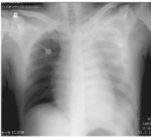
The patient, a 41-year-old man, was involved in an au-tomobile crash. He was not wearing a seat belt and the airbag exploded. A witness called 911 immediately, and the emergency medical team arrived in 2 minutes. On extraction, the patient was unconscious. Initial evalua-tion showed blood pressure of 69/45 mmHg and Glasgow coma scale (GCS) score of E1V1M1. He was brought to a local hospital, where his blood pressure was 65/20 mmHg, pulse rate was 113/min and respiration rate was 18/min. After fluid resuscitation with 2000 mL of normal saline and 4 units of packed red blood cells, his blood pres-sure improved to 104/50 mmHg. Chest film (Figure 1) showed massive hemothorax on the left side. A chest
tube was inserted and the patient was transferred to our hospital, which is a tertiary referral center. The ground transport time was about 45 minutes.
On arrival to our hospital, his blood pressure was 70/50 mmHg and pulse rate was 106/min. The patient was pale and drowsy with GCS score of E2M4V1. There was no open wound or deformity over his trunk or limbs. Both pupils were of equal size (3 mm) and reactive. The jugular veins were engorged and the trachea was slightly deviated to the right. Breath sounds were decreased over the left hemithorax and heart sounds were muffled, but fluid drainage through the chest tube was only 200 mL. Bedside ultrasonography (Figure 2) showed marked fluid accumulation in the pericardial sac with signs of cardiac tamponade. Pericardiocentesis was performed and 20 mL
Cardiac rupture after blunt injury of the chest is uncommon and usually fatal. The inci-dence of biatrial cardiac rupture is rarer and more likely to be lethal. We report a case of biatrial multiple lacerations following blunt chest injury. At referral, the chest film showed left side massive hemothorax. A chest tube was inserted and primary resuscitation was carried out. The patient’s vital signs became stable and he was transferred to our hospital for further management. The time from the injury occurring and the patient arriving at our hospital was 117 minutes. Unexplained hypotension was noted on arrival and bedside ultrasonography revealed cardiac tamponade. After emergent pericardiocentesis, the patient’s hemodynamics stabilized. Emergency surgery was performed and the patient was discharged without any sequelae. We successfully managed this case through a high index of suspicion, aggressive investigation, and prompt exploration.
Received: Oct 10, 2009 Revised: Feb 3, 2010 Accepted: Feb 6, 2010
KEY WORDS:
atrial rupture; blunt cardiac rupture; cardiac tamponade; pericardiocentesis
Survival of a Patient With Blunt Traumatic
Biatrial Rupture
Che-Wei Lin
1,2, Wai-Mau Choi
3, Wen-Han Chang
3, Cheng-Hsin Lin
4,
Cheuk-Sing Choy
1*
1Department of Emergency Medicine, Taipei Medical University Hospital, Taipei, Taiwan 2Department of Emergency Medicine, Tungs’ Taichung MetroHarbor Hospital, Taichung, Taiwan 3Department of Emergency Medicine, Mackay Memorial Hospital, Taipei, Taiwan
4Department of Surgery, Chi Mei Medical Center, Tainan, Taiwan
*Corresponding author. Department of Emergency Medicine, Taipei Medical University Hospital, 252, Wu-Hsing Street, Sinyi District, Taipei 11042, Taiwan.
88 C.W. Lin et al of blood was drained (Figure 3). Cardiac performance then improved, and the blood pressure rose to 104/ 59 mmHg, with a pulse rate of 90/min.
The patient was sent to the operating room for emer-gency surgery. A computed tomography scan was first performed and showed biatrial cardiac rupture with massive hemopericardium and hemothorax. Moreover, malposition of the chest tube was noted. In the operat-ing room, cardiac arrest was noted after sternotomy and pericardiotomy. Primary closure of the lacerations was done immediately without cardiopulmonary by-pass, and the heart resumed spontaneous beating after resuscitation. Multiple lacerations in both atria, includ-ing two in the right and four in the left were noted. The lacerations were 2–5 mm in length. Although the pre-operative condition was grave, the patient recovered uneventfully after surgery, and was discharged 2 weeks later without neurologic or cardiovascular sequelae.
Figure 1 Massive hemothorax on the left side. There is no
obvious mediastinum widening and no rib fractures.
A B
Figure 2 Bedside ultrasonography: (A) precardiocentesis diastolic phase; (B) precardiocentesis systolic phase. The arrows
indi-cate fluid collection in the pericardial sac. Chambers collapse is noted. Pericardial effusion is around 1.5 cm.
A B
Figure 3 Bedside ultrasonography: (A) postcardiocentesis diastolic phase; (B) postcardiocentesis systolic phase. The amount of
Traumatic biatrial rupture 89
2. Discussion
Cardiac rupture is the most serious complication of blunt chest injury. It was found in 0.3–2.0% of patients who suffered blunt trauma.1,2 Among cardiac injuries, the incidence of atrial rupture is 44%; biatrial tear is rare, accounting for only 4% of the total.3 Mortality from atrial tear is approximately 75%.3 Our case had multiple lacer-ations in both atria, which is extremely rare.
Cardiac tamponade is the leading cause of death in cardiac rupture.4 Because of myocardial tearing, blood or blood clots accumulate in the pericardium, prevent-ing the heart from fillprevent-ing with blood. Consequently, the heart is constricted and cardiac output is decreased. If the pericardium is defective, then tamponade pressure can be partially relieved.4 Our case had multiple peri-cardial tear injuries, which probably explains why he reached our hospital alive.
The initial diagnosis was massive hemothorax, and he was transferred to our hospital for further management. However, his blood pressure dropped to 70/50 mmHg on arrival and the drainage volume through the chest tube was incompatible with other signs. In line with ATLS (Advanced Trauma Life Support) guidelines, and in light of the unexplained hypotension, our proper next course of action was aggressive reinvestigation. Tube malposition, obstruction and further injury were suspected. We resurveyed the patient and bedside ul-trasonography showed signs of cardiac tamponade. Urgent pericardiocentesis was done to improve hem-odynamic status. The pericardial tear had caused hemo-thorax, which had resulted in misdiagnosis at the local hospital. It is of paramount importance to always have a high index of suspicion for further injury, even when the initial diagnosis and treatment have been completed.
Pericardiocentesis can improve the hemodynamics in patients with heart rupture.5 Especially in trauma cases, even small changes in pericardial fluid can compromise cardiac output. Immediate pericardiocentesis can de-crease the pressure in the pericardial sac and stabilize the hemodynamic status, thus earning more time for definitive surgery. There are two major approaches for pericardiocentesis. The subxiphoid puncture site is the traditionally recommended and preferred route for the
evacuation of fluids. However, studies of echocardio-graphically guided pericardiocentesis have found that the intercostal space near the heart apex, as opposed to the subxiphoid area, may provide more direct and eas-ier access to fluids in the pericardial sac. One drawback to the latter approach is possibly a higher incidence of pneumothorax, especially in patients with chronic lung disease. Echocardiography is therefore highly recom-mended when a patient’s condition permits sufficient time to determine the optimal approach to pericardio-centesis.6 Emergent thoracotomy in the emergency room is clearly indicated when cardiac arrest occurs during resuscitation of a trauma patient, as massive injuries to the cardiopulmonary system are the most frequently encountered causes requiring cardiac massage.
It is now recommended that bedside ultrasonogra-phy be performed in trauma patients. In hypotensive states, typical tamponade signs like Beck’s triad are dif-ficult to recognize. Ultrasonography represents an ad-vanced technique for detecting potentially fatal cardiac tamponade.
In conclusion, we have reported a case of multiple biatrial laceration following blunt chest injury. This con-dition is rare and its mortality is high. Our successful management was due to a high index of suspicion, aggressive investigation and early exploration.
References
1. Brathwaite CE, Rodriguez A, Turney SZ, Dunham CM, Cowley R. Blunt traumatic cardiac rupture. A 5-year experience. Ann Surg 1990;212:701–4.
2. Pulley SC, Nirula R. Survival of an elderly patient with blunt trau-matic cardiac rupture. J Trauma 2007;63:E119–20.
3. Fulda G, Brathwaite CE, Rodriguez A, Turney SZ, Dunham CM, Cowley RA. Blunt traumatic rupture of the heart and pericardium: a ten-year experience (1979–1989). J Trauma 1991;31:167–72. 4. Hendel PN, Grant AF. Blunt traumatic rupture of the heart.
Successful repair of simultaneous rupture of the right atrium and left ventricle. J Thorac Cardiovasc Surg 1981;81:574–6. 5. Lu LH, Choi WM, Wu HR, Liu HC, Chiu WT, Tsai SH. Blunt cardiac
rupture with prehospital pulseless electrical activity: a rare successful experience. J Trauma 2005;59:1489–91.
6. Harper RJ. Chapter 16—Pericardiocentesis. In: Roberts JR, Hedges JR. Clinical Procedures in Emergency Medicine, 5th ed. Philadelphia: Saunders, 2010:287–307.