Hsiang-Lin Tsai Jan-Sing Hsieh Fang-Jung Yu Deng-Chyang Wu Fang-Ming Chen Che-Jen Huang Yu-Sheng Huang Tsung-Jen Huang Jaw-Yung Wang Accepted: 5 January 2006 Published online: 20 April 2006 # Springer-Verlag 2006
Perforated colonic cancer presenting
as intra-abdominal abscess
Abstract Background and aims: The various presentations of carcino-ma of the colon are well known. Abscess formation occurs in 0.3 to 0.4% and is the second most common complication of perforated lesions. Perforation and penetration of adja-cent organs with intra-abdominal abscess formation as the initial pre-sentation is uncommon. Materials and methods: A retrospective analy-sis was made between January 1998 and December 2003 at the Kaohsiung Medical University Hospital. Six colon cancer patients with intra-ab-dominal abscess as the initial presen-tation were enrolled into this study. Among them, two were men and four were women. Results: During the 6-year period, there were 756 patients with colonic carcinoma but only six of those patients (0.79%) presented with abscess formation as the initial finding. The initial pre-operative diagnosis was ruptured colonic
di-verticulitis with abscess formation in three patients, and the other three patients were as follows: one ruptured appendicitis with abscess, one right subcutaneous inguinal abscess, and one omphalitis with abdominal wall abscess. Subsequent colonoscopy was performed in two patients, and colon cancer was recognized. The most common associated symptoms/signs were palpable abdominal mass, ab-dominal pain, and anemia. All of them underwent a one-stage surgical procedure, and adjuvant chemothera-py was given. One patient died of peritoneal carcinomatosis and liver metastases 1 year post-operatively. The other five patients are still alive. Conclusions: It is difficult to make an accurate diagnosis of abscess for-mation as the first evidence of colonic carcinoma pre-operatively. The one-stage resection of the lesion seems to be an acceptable treatment. For pa-tients with intra-abdominal abscess, clinicians should be aware of this differential because it is easily ig-nored pre-operatively.
Keywords Perforated colonic carcinoma . Abscess formation . One-stage surgical resection
Introduction
Cancer of the colon does not always present with the familiar symptoms of rectal bleeding, anemia, change of
bowel habits, or abdominal pain [1]. Less common
manifestations include perforation and abscess formation, which are usually intra-peritoneal but may also be located in the extra-peritoneal spaces. The incidence of colonic
perforation ranges from 2.6 to 10% [2,3], including cases
of free perforation into the peritoneal cavity and those H.-L. Tsai
Division of General Surgery, Department of Surgery,
E-Da Hospital/I-Shou University, No. 1, E-Da Road, Jiau-Shu Tsuen, Yan-Chau Shiang,
Kaohsiung 824, Taiwan
H.-L. Tsai . J.-S. Hsieh . F.-M. Chen . C.-J. Huang . Y.-S. Huang .
T.-J. Huang . J.-Y. Wang
Department of Surgery, Faculty of Medicine, College of Medicine, Kaohsiung Medical University, Kaohsiung 807, Taiwan
J.-S. Hsieh . F.-M. Chen . C.-J. Huang . Y.-S. Huang . T.-J. Huang .
J.-Y. Wang (*)
Division of Gastrointestinal and General Surgery, Department of Surgery, Kaohsiung Medical University Chung-Ho Memorial Hospital, No. 100, Tzyou 1st Road, Kaohsiung 807, Taiwan
e-mail: [email protected] Tel.: +886-7-3122805
Fax: +886-7-3114679 F.-J. Yu . D.-C. Wu
Department of Medicine, Faculty of Medicine, College of Medicine, Kaohsiung Medical University, Kaohsiung 807, Taiwan
F.-J. Yu . D.-C. Wu
Division of Gastrointestinal Medicine, Department of Internal Medicine, Kaohsiung Medical University Chung-Ho Memorial Hospital, No. 100, Tzyou 1st Road, Kaohsiung 807, Taiwan
T able 1 Reported cases of abscesses as the presenting sign in carcinoma of the colon Patient ’s number 1 2 3 4 5 6 Age/sex 78/male 68/female 76/female 43/female 71/female 46/male Location of abdominal mass LUQ RUQ RLQ LUQ Right groin Periumbilical area Size of abdominal abscess (cm 2 ) 5×4 12×12 5×5 5×6 5×6 6×7 Abdominal CT Y es Y es Y es Y es No No Colonoscopy Y es N o N o N o Y es No Initial pre-operative diagnosis Diverticulitis rupture with abscess Diverticulitis rupture with abscess Appendicitis rupture with abscess Diverticulitis rupture with abscess Right inguinal subcutaneous abscess Omphalitis with abdominal wall abscess Date of sur gery Jan-31-1998 Feb-26-1998 Oct-1 1-1998 Jan-31-2001 Aug-6-2001 Dec-4-2003 One-stage sur gery Y es Y es Y es Y es Y es Y es Operative method Extended right hemicolectomy Extended right hemicolectomy Right hemicolectomy Extended right hemico-lectomy Right hemicolectomy Extended right hemicolectomy Location of the tumor T ransverse colon T ransverse colon Appendix T ransverse colon Cecum T ransverse colon Histology Adenocarcinoma, PD Adenocarcinoma, MD Adenocarcinoma, MD Adenocarcinoma, MD Adenocarcinoma, PD Mucinous carcinoma, PD TNM T3 No Mo T3 No Mo T3 N1 Mo T3 No Mo T4 N2 Mo T4 No Mo Stage II II III II III II Distant metastases No No No No No No T ype of abscess Intraperitoneal abscess Intraperitoneal abscess Intraperitoneal abscess Intraperitoneal abscess Retroperitoneal abscess with abdominal wall involved Intraperitoneal abscess with abdominal wall involved Site of perforation Proximal to tumor Proximal to tumor Proximal to tumor Proximal to tumor T umor itself T umor itself Culturing materials of abscess None done Schwanella putr efaciens None done Escherichia coli Escherichia coli Escherichia coli + Bacte-roides fragilis Complications None None W ound infection None None Anastomotic leakage, enterocutaneous fistula Results Alive and well 6 years after sur gery Alive and well 6 years after sur gery Alive and well 6 years after sur gery Alive and well 3 years after sur gery Died 1 year after sur gery due to carcinomatosis and liver metastases Alive and well 1 year after sur gery CT Computed tomography , PD poorly dif ferentiated, MD moderately differentiated
where the tumor has perforated locally resulting in abscess or fistula formation. In a previous series, abscess formation occurred in 0.3 to 0.4% of colonic carcinoma and is the second most common complication of perforated lesions
[4–8].
Kelley et al. [9] reported that perforation is the most
lethal complication of colorectal carcinoma, with a fourfold operative mortality and a 5-year survival that is one fourth that of the overall population. Perforation and penetration of adjacent organs, with abscess formation as the initial presentation, is uncommon. Perforation may occur a distance from the tumor and may be due to the necrosis of stercoral ulcers or increased pressure proximal to an obstructing lesion. Penetration of the wall and the adjacent tissue by the neoplasm is of prime importance in the
pathogenesis of adjacent abscess formation [1].
Most of the reported cases of perforated colonic carcinoma presenting as an abscess were not diagnosed pre-operatively. In our data, four of six cases were not appropriately diagnosed pre-operatively even if most of them were evaluated under the initial evaluation of abdominal computed tomography (CT). We report herein six cases of intra-abdominal abscess resulting from perforating carcinoma of the colon and their clinical outcomes.
Materials and methods
Between January 1998 and December 2003, 756 patients with colonic malignancy admitted to Department of Surgery of the Kaohsiung Medical University Hospital were reviewed. Of the 756 patients, six patients with abscess formation as the initial presentation of perforated colonic carcinoma were included in our study. All of the medical records of these six patients were reviewed. Characteristics and data, which included commonly associated symptoms/signs, location and size of the palpable abdominal mass, initial diagnosis, location of the colon cancer, histology of the tumor, tumor stage, distant metastases, type of abscess, operative complica-tions, and clinical results were analyzed. Clinical stages and pathological features of the primary tumors were defined according to the criteria of the American Joint
Commission on Cancer [10].
Abdominal CT scan was performed pre-operatively for the diagnosis except for two patients. Two of the six patients who received colonoscopy was subsequently diagnosed as with colonic cancer after CT-guided percu-taneous drainage or incision and drainage under the initial tentative diagnosis of diverticulitis rupture with abscess formation and inguinal subcutaneous abscess, respectively. All of the six patients underwent a one-stage surgical resection of the lesion, with three who were diagnosed with intra-abdominal abscess undergoing surgery within 24 h of admission. Culture samples from abscess materials
obtained by surgical method were available for four patients (66.7%). Cultures were made for aerobes, anaer-obes, mycobacteria, and fungi examination. Finally, all six patients were contacted, evaluated, and treated with
5-fluorouracil (5-FU) (450 mg/m2) plus leucovorin (LV)
(200 mg/m2) administered weekly for 6 weeks in an
8-week cycle regularly, with an average follow-up of 55 months (range 12 to 87 months).
Results
Of the six patients, two were men and four were women. Their ages ranged from 43 to 78 years, with an average of
63.7 years. Their demography is shown in Table 1. The
most frequent pre-operative diagnosis was ruptured colonic diverticulitis with abscess formation (three out of six, 50%). The duration of hospitalization was from 15 to 52 days, with an average of 30.2 days. All presented with a palpable abdominal mass and four had a diagnosis of intra-abdominal mass confirmed by intra-abdominal CT before
surgery (Fig. 1). However, the other two patients had no
abdominal CT done because the initial impression was either subcutaneous or abdominal wall abscess. Both patients were subsequently found to have perforated colonic cancer and intra-abdominal abscess with abdom-inal wall involvement post-operatively.
Transverse colon was the most frequent site of the perforated colonic cancer with abscess formation in this study (four out of six, 66.7%), and two cases were found to have perforation on the tumor itself and four on the site proximal to the tumor. Four of six were TNM stage II (66.7%) and two were TNM stage III (33.3%). None had distant metastasis. Intra-peritoneal abscess (five out of six,
Fig. 1 A 68-year-old female patient was diagnosed with intra-abdominal abscess formation. Contrast-enhanced CT scan shows a heterogeneous mass (arrowheads) located at the anterior intraperi-toneal space. Postoperatively, she was a case of perforated transverse colonic carcinoma with abscess formation
83.3%) was the most frequent type of abscess formation as the initial presentation and only one patient had a retroperitoneal abscess in our series. The most commonly isolated organism by culture was Escherichia coli (three out four, 75%).
The most common symptoms/signs in our cases were abdominal mass (100%), abdominal pain (100%), and anemia (100%). However, the frequent symptom of bowel habit change in most colon cancer patients was not present in our patients. The associated symptoms and signs are
summarized in Table2.
All of the six patients underwent a one-stage surgery, which seemed to be an acceptable procedure for the patients. Surgical complications included wound infection (one out of six, 16.7%) and anastomotic leakage with entero-cutaneous fistula (one out of six; 16.7%). All were regularly followed-up post-operatively in our hospital, with an average follow-up of 55 months. Five of the six are still alive.
Discussion
The presentation of colorectal cancer can be quite variable
and depends on the site of the lesion [11, 12]. This
variability is well known and approximately one third of patients with colorectal carcinoma will have a major complication of the disease. The most common initial complication is bowel obstruction, occurring in 8 to 21% of
patients [9,13,14], with some authors reporting complete
obstruction that range from 8 to 40% [15, 16]. Seventy
percent of obstructing lesions is found in the left colon, coinciding with the predominance of left-side lesions. Perforation and penetration of adjacent organs with abscess formation as the initial presentation of the carcinoma is uncommon, and the incidence of perforation in a previous
large series is 2.6–7.8% [5,7,12]. This includes cases of
free perforation into the peritoneal cavity and those where the tumor had perforated locally, resulting in abscess or fistula formation. Of our patients, two were found to have
perforation on the tumor itself and four on the site proximal to the tumor.
Abscess formation occurs in 0.3 to 0.4% of colonic carcinoma and is the second most common complication of
perforative lesions [4, 7]. The abscess may remain
localized in the paracolic area, may extend as a flank abscess, or may track along various tissue planes and
present as an abscess at another site, e.g., in the thigh [17]
or subcutaneously on the trunk [18]. However, most of the
abscesses occur in the intra-abdominal cavity, especially intra-peritoneally, although fistula formation is usually uncommon. In our study, intra-peritoneal abscess is present in five of the six patients (83.3%), which is consistent with previous reports.
Cross-sectional imaging techniques, such as ultrasound images and abdominal CT, have become the most common
techniques for diagnosing intra-abdominal abscesses [19].
With the advent of abdominal computed tomography, it has been possible to identify and accurately determine the
location of an intra-abdominal abscess before surgery [20].
However, most of the reported cases of perforated colonic carcinoma presenting as an abscess did not have an accurate diagnosis pre-operatively. In our series, most of them were diagnosed as ruptured diverticulitis with abscess formation (three out of six, 50%); but unlike diverticular abscesses, pericolic abscesses caused by carcinomas are
rarely small or mobile [21]. General or localized
extra-colonic spread of infection is more frequently encountered
in neoplasm than in inflammatory conditions [1]. CT will
frequently be used, as in our cases, and will show the nature, location, and extent of the inflammatory compo-nent. However, the source of the inflammation may not be apparent. With recent advances of imaging techniques, the diagnosis of intra-abdominal abscess has improved. In our reported cases, the lack of an accurate pre-operative diagnosis by abdominal CT may be due to the larger intra-abdominal abscess in these patients.
Pertinent to this discussion are abscesses which usually develop intra-peritoneally in the pericolic region. This is
thought to be related to the efficient “walling off ” and
localization by the primary inflammatory process, while tumor penetration is said to be facilitated by the out-stripping of its blood supply by rapid growth and subsequent necrosis. In our patients, perforation occurred at the tumor site due to the penetration of the carcinoma through the bowel wall in two of six cases. In most reported cases of colonic carcinoma, perforation is into the perito-neal cavity, which is similar with our observation. When this colonic perforation occurs, the findings can be mistaken for diverticulitis, particularly when it occurs on the right side or in the elderly in Asia.
Stainland et al. [11] mentioned that the presentation of
colorectal cancer can be quite varied and depends on the site of the lesion. Right-sided colonic lesions frequently present with symptoms of abdominal pain, palpable mass, or anemia. In this series, all of the six patients had
right-Table 2 The associated symptoms/signs
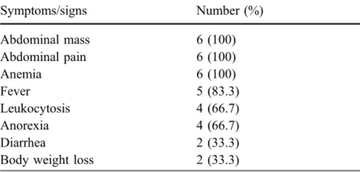
Symptoms/signs Number (%) Abdominal mass 6 (100) Abdominal pain 6 (100) Anemia 6 (100) Fever 5 (83.3) Leukocytosis 4 (66.7) Anorexia 4 (66.7) Diarrhea 2 (33.3)
sided colonic cancer and, therefore, presented with the aforementioned symptoms. The anterior abdominal wall is one of the most common presenting points of such
abscesses [21]. In our analysis, one case involved the
anterior abdominal wall due to a perforated transverse colonic carcinoma with an abscess formation. We also have a retro-peritoneal abscess resulting from a perforated cecal carcinoma involving the anterior abdominal wall. Extra-peritoneal perforation with subsequent abscess formation in the subcutaneous tissue occurs very infrequently.
Survival was worse for patients with obstructive or
perforated cancer [2, 9], and adjuvant chemotherapy was
highly recommended for these patients [22]. All of our six
patients were administrated with 5-FU/LV chemotherapy after the resection of tumors. Despite the previously
unfavorable results of colonic cancer with perforation, the clinical outcomes of our cases were considerably acceptable after the one-stage operation. The favorable prognosis may be due to the fact that our cases were TNM stage II (66.7%) and III (33.3%). The surgical complica-tions included one wound infection (16.7%) and one anastomotic leakage with entero-cutaneous fistula (16.7%). Fortunately, they recovered uneventfully after an exclu-sively conservative treatment.
In conclusion, a one-stage surgical procedure seems feasible for these cases and the clinical outcomes are generally favorable. However, the long-term survival of patients with perforated colonic carcinoma with abscess remains to be elucidated.
References
1. Dean DT, Howard MP (1983) Retro-peritoneal abscess: a presentation of colon carcinoma. Gastrointest Radiol 8:177–181
2. Chen HS, Shen-Chen SM (2000) Ob-struction and perforation in colorectal adenocarcinoma: an analysis of prog-nosis and current trends. Surgery 127:370–376
3. Mandava N, Kumar S, Pizzi WF, Aprile IJ (1996) Perforated colorectal carci-noma. Am J Surg 172:236–238 4. Donaldson GA (1958) The
manage-ment of perforative carcinoma of the colon. N Engl J Med 258:201–207 5. Miller LD, Boruchow IB, Fitts WT
(1986) An analysis of 284 patients with perforative carcinoma of the colon. Surg Gynecol Obstet 123:1212–1218 6. Devitt JE, Roth-Moyo LA, Brown FN
(1970) Perforation complicating adenocarcinoma of colon and rectum. Can J Surg 13:9–12
7. Welch JP, Donaldson GA (1974) Per-forative carcinoma of colon and rectum. Ann Surg 180:734–740
8. Kobayashi H, Sakurai Y, Shoji M, Nakamura Y, Suganuma M, Imazu H, Hasegawa S, Matsubara T, Ochiai M, and Funabiki T (2001) Psoas abscess and cellulitis of the right gluteal region resulting from carcinoma of the rectum. J Gastroenterol 36:623–628
9. Kelley WE, Brown PW, Lawrence W (1981) Penetrating, obstructing and perforating carcinomas of the colon and rectum. Arch Surg 116:381–384 10. Greene FL, Page DL, Fleming ID, Fritz
AG et al (2001) In: AJCC cancer staging handbook. Springer, Berlin Heidelberg New York, pp 111–118 11. Stainland JR, Ditchburn J, Dombal FT
(1967) Clinical presentation of disease of the large bowel. A detailed study of 642 patients. Gastroenterology 70:22–28
12. Andaz S, Heald RJ (1993) Abdominal wall abscess—an unusual primary pre-sentation of a transverse colonic carci-noma. Postgrad Med J 69:826–828 13. Ohman U (1982) Prognosis in patients
with obstructing colorectal carcinoma. Am J Surg 143:742–747
14. Rovito PF, Verazin G, Prorok I (1990) Obstructing carcinoma of the cecum. J Surg Oncol 45:177–179
15. Kronborg O, Baker O, Sprecjler M (1975) Acute obstruction in cancer of the colon and rectum. Dis Colon Rectum 18:22–27
16. Stower MJ, Hardcastle JD (1986) The results of 1115 patients with colorectal cancer treated over an 8-year period in a single hospital. Eur J Surg Oncol 11:119–123
17. Cooke RV (1956) Advanced carcinoma of the colon with emphasis on the inflammatory factor. Ann R Coll Surg Engl 18:46–61
18. Shucksmith HS (1963) Subcutaneous abscess as the first evidence of carci-noma of the colon. Br J Surg 50:514–515
19. Gupta H, Dupuy DE (1997) Advances in imaging of the acute abdomen. Surg Clin North Am 77:1245–1263 20. Haaga JR, Havrilla TR (1997) CT
detection and aspiration of abdominal abscesses. Am J Roentgenol 128: 465–474
21. John PW (1976) Unusual abscesses in perforating colorectal cancer. Am J Surg 131:270–274
22. Sugarbaker PH, Gianola FJ, Speyer JC, Wesley R et al (1984) Prospective, randomized trial of intravenous versus intraperitoneal 5-fluorouracil in patients with advanced primary colon or rectal cancer. Surgery 1985;98:414–422