between pagetoid migrations of atypical melanocytes in superficial spreading melanoma from real Paget’s cells especially in the presence of pigmentation.
Pigmentation may be seen as a deposit of melanin in basal keratinocytes, throughout the epidermis and even within some Paget’s cells. It may be also demon-strated by the presence of numerous melanophages in the dermis. But, morphological and cytological criteria must always be completed by immunohistochemical studies to have an accurate diagnosis. Indeed, Paget’s disease stains for epithelial markers especially for cytokeratin. Cytokeratin 7 has recently been shown more sensitive for discriminating pigmented mammary Paget’s disease from malignant melanoma. C-erbB and hormonal receptors may also be positive, whereas PS100 and HMB45 are always negative.
Pigmented mammary Paget’s disease should be considered in the differential diagnosis of pigmented lesions of the nipple and the areola. Differential diagnosis with malignant melanoma relies on immunohistochemical studies.
Pigmented Mammary Paget’s Disease Presenting as an
Enlarged Areola
Chao-Chin Wang, MD,* Kuo-Hsien Wang, MD,* Chieh-Jui Cheng, MD,
and Ju-Hsin Chen, MD
àDepartments of *Dermatology and
Pathology, Taipei Medical University Hospital, Taipei, Taiwan;
and
àDepartment of Dermatology, Taipei Medical University-Wan Fang Hospital, Taipei, Taiwan
A
66-year-old Taiwanese woman presented herself with a 6-month asymptomatic enlargement of right areola. She did not rub, nor scratch but applied povidone-iodine topically on the lesion (Fig. 1a). Phys-ical examination showed an enlarged areola on the right breast with mottled hyperpigmentation and Figure 3. Intraductual carcinoma infiltrating the dermis(Hematox-ylin-Eosin stain· 400).
Figure 4. Cytokeratin positive (·400).
Address correspondence and reprint requests to: Ju-Hsin Chen, MD, Department of Dermatology, Taipei Medical University-Wan Fang Hospital, No.111, Sec. 3, Singlong Rd., Wunshan District, Taipei City 116, Taiwan, or e-mail: dermahsin007@yahoo.com.tw
DOI: 10.1111/j.1524-4741.2009.00751.x ª2009 Wiley Periodicals, Inc., 1075-122X/09
The Breast Journal, Volume 15 Number 4, 2009 421–423
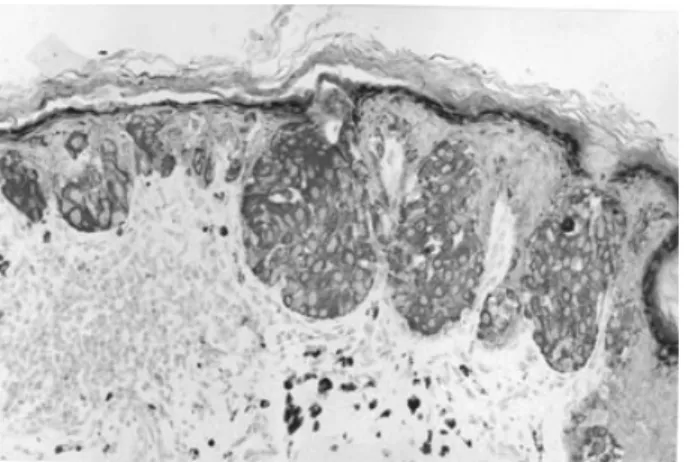
minimal scaling (Fig. 1b). She did not have nipple retraction, palpable breast mass or lymphadenopathy. The finding of incisional skin biopsy from the left lower quadrant of the right areola showed nests of large epithelioid cells in the epidermis and some of them contained melanin pigments (Fig. 2a). A moder-ate lymphocytic infiltrmoder-ate and some melanophages were seen in the papillary dermis (Fig. 2b).
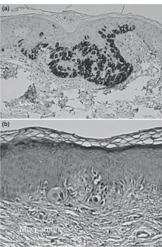
Immunohistochemically, the tumor cells were posi-tive for cytokeratin-7 (CK-7) (Fig. 3a) but did not react with HMB-45, S-100, carcinoembryonic antigen (CEA), and CK-20. Histochemical stain with mucicar-mine was focally positive (Fig. 3b). These findings were compatible with the diagnosis of pigmented mammary Paget’s disease (MPD). Further image stud-ies including mammography and ultrasonography
showed no underlying abnormality in the right breast. But she declined any further treatment and has been lost to follow-up since.
Pigmented MPD is a rare clinicopathological variant of MPD. The disease has hyperpigmented patches or plaques involving the areola and nipple, simulating mel-anoma and pigmented epidermotropic metastases from breast carcinoma clinically and histopathologically. The lesion in our patient had a subtle hyperpigmented plaque that looked no more than an ‘‘enlarged areola.’’ The histological picture of pigmented MPD consists of clusters of large epithelioid cells (Paget cells) within the epidermis; some cases show proliferations of dendritic melanocytes as solitary units along the dermo-epider-mal junction while others show abundant melanin within the cytoplasm of the Paget cells without increased melanocytes. The Paget cells in MPD have a characteristic immunohistochemical profile that is
(a)
(b)
Figure 1. (a) Enlargement of right areola. (b) Close-up view of the right side areola shows mottled hyperpigmentation with minimal scaling.
(a)
(b)
Figure 2. (a) Clusters of large epithelioid cells are seen in the epidermis. Some of the neoplastic cells contain pigments inside (H&E, 100·). (b) These large cells have pale, abundant cytoplasm and hyperchromatic nuclei. Focal vacuolization of epidermal basal cells, moderate lymphocytic infiltrates and some melanophages are noted in the papillary dermis (H&E, 400·).
uniformly positive for CK-7 and epithelial membrane antigen (EMA), variably positive for CEA, and is nega-tive for HMB-45 and S-100 protein. Its differential diagnoses from malignant melanoma in situ, pagetoid Bowen’s disease, and Toker cell hyperplasia can be difficult but the histochemical and the immunohisto-chemical profiles are often useful.
Melanoma cells have strongly positive staining with melanocyte markers such as S-100 protein, HMB-45, and Melan-A, whereas they react negatively with cytokeratins, CEA, and EMA. Pagetoid Bowen’s disease stains negatively with mucin. It is pancyto-keratin positive but is usually CK-7 negative. Toker cell hyperplasia has increased nonneoplastic Toker cells, which are mainly in small groups and may have small tubular structures within the cellular aggregates. These cells are smaller than the Paget cells in MPD and do not have hyperchromatic nuclei or cytological aty-pia. Moreover, they are negative for mucin and PAS.
In summary, we report the first case of pigmented MPD presenting as an enlarged areola. Based on the information in this report, we suggest that physicians should consider the differential diagnosis of pigmented MPD if the patient has any pigmented change of the nipple–areola complex.
Primary Leiomyosarcoma of the Breast
Bengu Cobanoglu, MD,* Muge Sezer, MD,* Pervin Karabulut, MD,*
Sirin Ozer, MD,* and Ayse Murat, MD
*Department of Pathology, University of Firat, Elazig, Turkey;
Department of Radiology, University
of Firat, Elazig, Turkey
A
64-year-old female patient referred to the hospi-tal for the presence of a firm lump in her leftbreast. It has been present for 5 months. There was no other breast complaint and no family history of breast cancer.
On physical examination, an ill-defined area was palpable in the upper outer quadrant of the left breast. There was no other breast complaint and no family history of breast cancer.
An ultrasound of the left breast revealed hypoechoic lesion. There was no microcalcification. Mammography
(a)
(b)
Figure 3. Immunohistochemical pictures of the lesion. (a) Positive staining with CK-7; (100·). (b) Reactive with mucicarmine; (400·).
Address correspondence and reprint requests to: Bengu Cobanoglu, MD, Department of Pathology, University of Firat, Medical Faculty, Elazig 23119, Turkey, or e-mail: bengus_2005@yahoo.com, bengus_2006@yahoo. com.
DOI: 10.1111/j.1524-4741.2009.00752.x ª2009 Wiley Periodicals, Inc., 1075-122X/09
The Breast Journal, Volume 15 Number 4, 2009 423–425