Photoluminescence of Zn Se x Te 1 x Zn Te multiple-quantum-well structures grown
by molecular-beam epitaxy
Y. T. Shih, Y. L. Tsai, C. T. Yuan, C. Y. Chen, C. S. Yang, and W. C. Chou
Citation: Journal of Applied Physics 96, 7267 (2004); doi: 10.1063/1.1818712
View online: http://dx.doi.org/10.1063/1.1818712
View Table of Contents: http://scitation.aip.org/content/aip/journal/jap/96/12?ver=pdfcov Published by the AIP Publishing
Articles you may be interested in
Growth of Zn x , Cd ( 1 x ) Se Zn x Cd y Mg ( 1 x y ) Se – In P quantum cascade structures for emission in the 3 – 5 m range
J. Vac. Sci. Technol. B 28, C3G24 (2010); 10.1116/1.3276438
Time-resolved photoluminescence of isoelectronic traps in Zn Se 1 x Te x semiconductor alloys Appl. Phys. Lett. 93, 241909 (2008); 10.1063/1.3054162
Effects of alloying and localized electronic states on the resonant Raman spectra of Zn 1 x Mg x O nanocrystals Appl. Phys. Lett. 91, 091901 (2007); 10.1063/1.2775813
Stability characteristics of the low-temperature ferrimagnetism in the Mn 2 x Zn x Sb system Low Temp. Phys. 33, 70 (2007); 10.1063/1.2409637
Exciton–phonon coupled states in CdTe / Cd 1x Zn x Te quantum dots J. Appl. Phys. 93, 2906 (2003); 10.1063/1.1540740
Photoluminescence of ZnSe
xTe
1−x/ ZnTe multiple-quantum-well structures
grown by molecular-beam epitaxy
Y. T. Shiha)
Institute of Photonics, National Changhua University of Education, Changhua, Taiwan 50007, Republic of China
Y. L. Tsai
Institute of Applied Physics, Chung Yuan Christian University, Chungli, Taiwan 32023, Republic of China
C. T. Yuan, C. Y. Chen, C. S. Yang, and W. C. Chou
Institute of Electrophysics, National Chiao Tung University, Hsinchu, Taiwan 30050, Republic of China
(Received 21 May 2004; accepted 23 September 2004)
This work investigates photoluminescence (PL) spectra from ZnSexTe1−x/ ZnTe multiple-quantum-well structures grown on GaAs(001) substrates by molecular-beam epitaxy. The PL data reveal that the band alignment of the ZnSexTe1−x/ ZnTe system is type II. The thermal activation energy for quenching the PL intensity was determined from the temperature-dependent PL spectra. The activation energy was found to increase initially and then decrease as the thickness of the ZnSexTe1−xlayers decreases from 7 to 3 nm. The temperature-dependent broadening of the
PL linewidth was also investigated. The LO-phonon scattering was found to be the dominant broadening mechanism. © 2004 American Institute of Physics.[DOI: 10.1063/1.1818712]
I. INTRODUCTION
The wide-band-gap II-VI semiconductors ZnSe and ZnTe and their mixed crystals ZnSexTe1−x have attracted
much interest because of their great potential for use as pos-sible blue/green light-emitter materials. During the past sev-eral years, remarkable advances in molecular-beam epitaxy
(MBE) have enabled the growth of ZnSexTe1−xepilayers and
ZnSexTe1−x/ ZnTe heterostructures. Many efforts have been
made to characterize these structures optically. Brasil and co-workers1,2 reported the optical characterization of ZnSexTe1−xepilayers grown by MBE. Rajakarunanayake and
co-workers3,4investigated the photoluminescence(PL) spec-tra from ZnSexTe1−x/ ZnTe superlattices. They found a type-II band alignment with a large valence-band offset. Yang et al.5 investigated the PL spectra from ZnSexTe1−x
epilayers. The peaks of the Te-bound excitons and excitons bound to the Te clusters have been identified. Baranovskii et
al.6 investigated the PL spectra from ZnSexTe1−x/ ZnTe
single-quantum wells. The dominant line broadening mecha-nism was attributed to the compositional disorder. On the other hand, a high-brightness green light-emitting diode, us-ing ZnSeTe as the active layer, has been reported for the realization of blue/green light-emitting devices.7
In this work, ZnSexTe1−x/ ZnTe multiple-quantum-well (MQW) structures were grown by molecular-beam epitaxy
on GaAs(001) substrates. PL was measured to characterize the structures. The thermal activation energy was determined from the temperature-dependent PL spectra. The variation of the activation energy with the thickness of ZnSexTe1−xlayers
and the temperature-dependent broadening of the PL line-widths were investigated.
II. EXPERIMENTS
ZnSexTe1−x/ ZnTe (x=0.22 and 0.54) MQW structures
were grown on GaAs(001) substrates, using a Veeco Applied EPI 620 MBE system. The GaAs substrates were indium mounted on a molybdenum block. Oxides on the surfaces of the substrates were removed by heating the substrates to 650 ° C until clear reflection high-energy electron diffraction patterns were observed. Veeco Applied EPI 40 cc low-temperature effusion cells were employed to evaporate the elemental solid sources Zn, Se, and Te. Before the ZnSexTe1−x/ ZnTe multiple-quantum wells were grown, a
ZnSe layer of 76 Å for samples A–C共x=0.54兲 and D–F 共x = 0.22兲 or 2m for samples G–I共x=0.22兲, and a ZnTe layer of 360 nm were grown as buffer layers. The ZnSe epilayer was grown as a buffer layer because a recent report8 demon-strated that the crystallinity of a ZnTe epilayer grown on a GaAs substrate can be remarkably improved using a ZnSe buffer layer, and that the strain of a ZnTe layer with a ZnSe buffer layer was less than that without a ZnSe buffer layer.
During the growth of the ZnSe0.54Te0.46/ ZnTe
共ZnSe0.22Te0.78/ ZnTe兲 multiple-quantum wells, the cell
tem-peratures of Zn, Te, and Se were 270(300), 300 (325), and 155共160兲 °C, respectively, while the temperature of the sub-strate was fixed at 300 ° C. Ten periods of ZnSexTe1−x/ ZnTe
quantum wells were grown in each sample. The thickness of the ZnTe layers was fixed at 200 Å, whereas the thickness of the ZnSexTe1−xlayers varied from 30 to 70 Å. After the ten periods of the ZnSexTe1−x/ ZnTe quantum wells had been
deposited, a ZnTe layer of 300 Å was grown as a cap layer. The thicknesses of the epilayers were estimated from the growth rate. Energy dispersive x-ray spectroscopy was em-ployed to determine the Se concentration, x, in the ZnSexTe1−x epilayers. Table I presents relevant sample
pa-rameters.
a)Author to whom correspondence should be addressed; electronic mail:
0021-8979/2004/96(12)/7267/5/$22.00 7267 © 2004 American Institute of Physics
Photoluminescence was measured to characterize the samples optically. The samples were loaded onto the cold finger of a closed-cycle refrigerator to maintain their tem-peratures from 10 to 300 K. A 351.0-nm beam at an aver-age power of 10 mW from an argon-ion laser was used to excite the PL spectra. A SPEX 1403 double-grating spec-trometer, equipped with a thermoelectric-cooled photomulti-plier tube, was employed to analyze the spectra.
III. RESULTS AND DISCUSSION
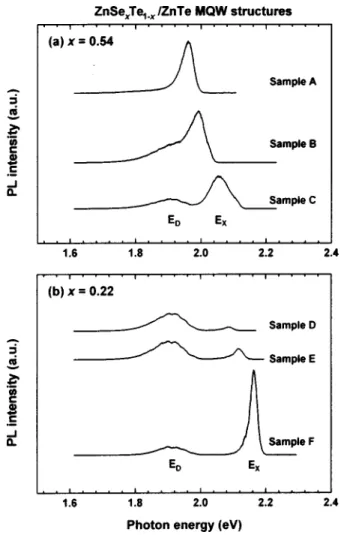
Figure 1 presents the low-temperature PL spectra ob-tained from ZnSexTe1−x/ ZnTe MQW structures. These PL
spectra include two types of emission peaks. As the thickness of the ZnSexTe1−x layers decreases, the emission peaks at
higher energies (labeled EX) exhibit a large blueshift, while those at lower energies(labeled ED) remain in the same po-sition. The EX emission peaks are associated with the inter-band excitonic transition. The composition-dependent energy gap of the MBE-grown ZnSexTe1−xepilayers at the low tem-perature can be expressed as1
Eg共x兲 = xEZnSe+共1 − x兲EZnTe− bx共1 − x兲, 共1兲
where EZnSe= 2.820 eV and EZnTe= 2.392 eV are the
energy-band gaps for ZnSe and ZnTe, respectively, and b = 1.507 eV is the bowing parameter. Accordingly, the emis-sion energies of the EX peaks in Fig. 1 are less than the energy gaps of either ZnTe or ZnSexTe1−x. This result reveals
that the EXtransitions in Fig. 1 are type-II transitions. Figure 2 schematically depicts the band alignment of the ZnSexTe1−x/ ZnTe system. In this type-II system, electrons
are confined in the ZnSexTe1−x layers while holes are con-fined in the ZnTe layers. Therefore, the lowest energy inter-band transition (labeled EX) is spatially indirect. The large blueshift of the EXemission peaks is attributed to the quan-tum confinement of electrons. As the thickness of the ZnSexTe1−x layers decreases, the strong quantum
confine-ment increases the subband energy Ee in the ZnSexTe1−x
quantum well and increases the interband transition energies. The ED emission peaks in Fig. 1 do not shift as the thickness of the ZnSexTe1−x layers decreases and so can be
assigned to optical transitions related to interface defects. PL measurements were made for samples G–I to examine the origin of the ED peaks. Figure 3 presents the results. The
growth parameters of samples G–I were similar to those of samples D–F, except that the buffer layers of samples G–I were thicker. The thicker buffer layers reduce the effect of the interface defects on the PL spectra. Therefore, Fig. 3 does not exhibit the EDpeaks.
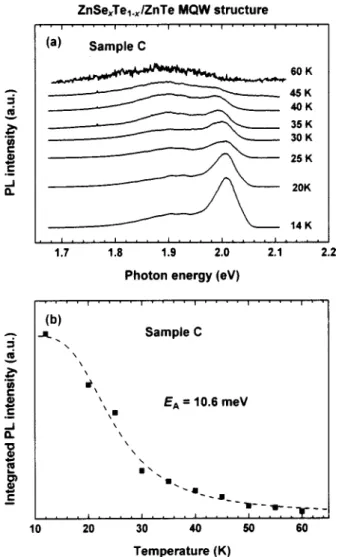
The temperature dependence of the PL spectra was also investigated. Figure 4(a) presents the temperature-dependent PL spectra from sample C. The temperature dependence TABLE I. Sample parameters of ZnSexTe1−x/ ZnTe multiple-well structures.
Sample Se concentration x of ZnSexTe1−x layers Thickness of buffer layer ZnSe/ ZnTe (nm) Thickness of layers ZnSexTe1−x/ ZnTe (nm) A 0.54 7.6/ 360 7 / 20 B 0.54 7.6/ 360 5 / 20 C 0.54 7.6/ 360 3 / 20 D 0.22 7.6/ 360 7 / 20 E 0.22 7.6/ 360 5 / 20 F 0.22 7.6/ 360 3 / 20 G 0.22 2000/ 360 7 / 20 H 0.22 2000/ 360 5 / 20 I 0.22 2000/ 360 3 / 20
FIG. 1. Photoluminescence spectra of ZnSexTe1−x/ ZnTe
multiple-quantum-well structures.
FIG. 2. Band alignment of ZnSexTe1−x/ ZnTe system.
7268 J. Appl. Phys., Vol. 96, No. 12, 15 December 2004 Shihet al.
from the PL spectra of the other samples reveals similar op-tical characteristics. As the temperature increases, the PL in-tensity drops rapidly and the interband exciton transition peaks are redshifted and broadened. The redshift is caused by the decrease in the band-gap energy as the temperature in-creases. Additionally, the small binding energy of the exciton in a type-II band alignment system is such that at higher temperature, the thermal energy is comparable to the binding energy of the exciton, and the exciton-phonon interaction is considerable, resulting in a weaker and broader PL peak.
The temperature dependence of the integrated PL inten-sity 共IPL兲 of an exciton emission peak could be expressed
as9,10
IPL共T兲 =
I0
1 + A exp共− EA/kBT兲
, 共2兲
where T is the temperature, kBis the Boltzmann constant, I0
is the integrated PL intensity near 0 K, A is a constant, and
EAis the thermal activation energy. EA is responsible for the quenching of PL intensity in the temperature-dependent PL spectra. Figure 4(b) presents the measured IPLfor sample C as a function of temperature. As the temperature increases to 50– 60 K, an abrupt drop of over one order of magnitude in the integrated PL intensity is observed. These measured val-ues of integrated PL intensity were fitted using Eq. (2) and the thermal activation energy EAof the MQW structures was obtained. Usually, EA is obtained simply from the slope of the Arrhenius plot of ln共IPL兲 versus 1/T at the
high-temperature limit. However, such a method might cause a large error because it relies entirely on data measured at high temperature. Figure 4(b) clearly shows that the theoretical curve is satisfactorily consistent with the experimental data. Similar results were obtained for the other samples.
Figure 5 presents the obtained thermal activation ener-gies EA of samples A–C共x=0.54兲 and D–F 共x=0.22兲. EAis plotted as a function of the thickness of the ZnSexTe1−x
lay-ers. The thickness of the ZnSexTe1−x layers decreases from
7 to 3 nm, and EA tends to increase initially and then de-crease.
The temperature-induced quenching of luminescence in the MQW structures proceeds mainly by two mechanisms: thermal emission of(at least one type of) charge carriers out of confined quantum-well states into barrier states11and ther-mal dissociation of excitons into free-electron-hole pairs.10 Electrons are confined in ZnSexTe1−x layers, whereas holes
are confined in ZnTe layers so samples with wider ZnSexTe1−x layers exhibit weaker wave-function overlap,
which results in small exciton binding energy. Therefore, the second quenching mechanism dominates. The primary means of quenching PL intensity is the thermal dissociation of excitons into free-electron-hole pairs. After the excitons have been broken apart, electrons are free to diffuse into the barrier, and the PL is quenched. The thermal activation en-ergy EAfor this process is determined from the exciton bind-ing energy and decreases as the thickness of the ZnSexTe1−x layers increases.
However, samples with narrower ZnSexTe1−xlayers have
narrower wells and therefore increased subband energy Ecof the electrons (Fig. 2). The structures herein are common-cation systems, so the conduction-band offset is small and leads to small delocalization energy Ve− Ec, facilitating the escape of the electrons from the quantum wells. Therefore, FIG. 4. (a) Temperature-dependent photoluminescence spectra of sample C of ZnSexTe1−x/ ZnTe multiple-quantum-well structures.(b) Variation of
in-tegrated PL intensity with temperature of sample C of ZnSexTe1−x/ ZnTe
multiple-quantum-well structures. FIG. 3. Photoluminescence spectra of samples G–1 of ZnSexTe1−x/ ZnTe
multiple-quantum-well structures.
the first quenching mechanism dominates. The main cause of the quenching of the PL intensity is the thermal emission of electrons into barrier states. The activation energy EAcan be regarded as the delocalization energy of electrons and de-creases as the thickness of the ZnSexTe1−xlayers decreases.
The temperature-dependent broadening of exciton emis-sion peaks of the ZnSexTe1−x/ ZnTe MQW structures was also investigated. Figure 6 presents the linewidths [half width at half maximum(HWHM)] of the PL peaks as func-tions of temperature. The luminescence linewidths gradually increase with temperature. Scattering processes with acoustic phonons, LO phonons, and ionized impurities are consid-ered, and the temperature-dependent luminescence linewidth of excitons in quantum wells thus expressed as12
⌫共T兲 = ⌫0+⌫LAT +
⌫LO
exp共បLO/kBT兲 + 1
+⌫impexp共− 具Eb典/kBT兲, 共3兲
where⌫0is the inhomogeneous broadening,⌫LAis the coef-ficient of the exciton–acoustic-phonon interaction,⌫LOis the exciton–LO-phonon coupling constant, បLO is the LO-phonon energy, ⌫imp is a factor of proportionality that ac-counts for the density of the impurities, and 具Eb典 is the im-purity binding energy averaged over all possible locations of the impurities.
The measured PL linewidths were fitted by Eq.(3). The solid curves in Fig. 6 represent the theoretical results. They agree well with the experimental data. Table II presents the fitted values of⌫0,⌫LA,⌫LO, andបLO. In the fitting
proce-dure, the contribution of the impurity scattering process to linewidth broadening can be ignored, so⌫imp= 0. Therefore, the concentrations of impurities in the samples herein were very low. Figure 6 and Table II together reveal that LO-phonon scattering is the mechanism that dominates the broadening of the linewidth as the temperature increases. FIG. 5. Variation of activation energy with thickness of ZnSexTe1−xlayers in
ZnSexTe1−x/ ZnTe multiple-quantum-well structures.
FIG. 6. Variation of PL linewidth with temperature of ZnSexTe1−x/ ZnTe
multiple-quantum-well structures.
TABLE II. Parameters obtained by fitting Eq.(3) to the HWHM VS T data in Fig. 6. Sample Parameters ⌫0 (meV) ⌫ LA 共10−2 meV/K兲 (meV)⌫LO ប LO (meV) A 23.37 8.02 38.56 10.80 B 22.71 11.27 86.06 17.08 C 22.59 6.01 22.92 8.27 D 10.94 3.23 15.21 6.29 E 9.91 1.47 27.46 7.18 F 9.88 0.18 31.61 7.73
7270 J. Appl. Phys., Vol. 96, No. 12, 15 December 2004 Shihet al.
IV. SUMMARY
In summary, ZnSexTe1−x/ ZnTe (x=0.22 and 0.54)
multiple-quantum-well structures were grown on GaAs(001) substrates using a Veeco Applied EPI 620 molecular-beam epitaxy system. Photoluminescence spectra were measured to characterize optically the MQW structures. The emission peaks from the interband transition were blueshifted as the thickness of the ZnSexTe1−x layers decreased. The PL data
reveal that the band alignment of ZnSexTe1−x/ ZnTe system
is type II.
The temperature dependence of the PL spectra was in-vestigated. As the temperature increased, the PL peaks be-came weaker and broader. The thermal activation energy that is responsible for quenching the PL intensity was determined from the plots of integrated PL intensity versus temperature. The activation energy tended to increase initially and then decrease as the thickness of the ZnSexTe1−xlayers decreased from 7 to 3 nm. For samples with wider ZnSexTe1−xlayers, the primary mechanism of quenching the PL intensity is ther-mal dissociation of excitons into free-electron-hole pairs. For samples with narrower ZnSexTe1−x layers, the dominant quenching mechanism is thermal emission of electrons into barrier states. The temperature-dependent broadening of PL linewidth was analyzed in terms of exciton–acoustic-phonon
and exciton–LO-phonon scattering processes. LO-phonon scattering was found to be the dominant broadening mecha-nism.
ACKNOWLEDGMENT
The authors would like to thank the National Science Council of the Republic of China for financially supporting this research under Contract Nos. NSC-92-2112-M-018-014, NSC-93-2112-M-018-005, and NSC-92-2112-M-009-041.
1
M. J. S. P. Brasil, R. E. Nahory, F. S. Turco-Sandroff, H. L. Gilchrist, and R. J. Martin, Appl. Phys. Lett. 58, 2509(1991).
2
F. S. Turco-Sandroff, R. E. Nahory, M. J. S. P. Brasil, R. J. Martin, and H. L. Gilchrist, Appl. Phys. Lett. 58, 1611(1991).
3
Y. Rajakarunanayake, M. C. Phillips, J. O. McCaldin, D. H. Chow, D. A. Collins, and T. C. McGill, Proc. SPIE 1285, 142(1990).
4
M. C. Phillips, Y. Rajakarunanayake, J. O. McCaldin, D. H. Chow, D. A. Collins, and T. C. McGill, Proc. SPIE 1285, 152(1990).
5
C. S. Yang, D. Y. Hong, C. Y. Lin, W. C. Chou, C. S. Ro, W. Y. Uen, W. H. Lan, and S. L. Tu, J. Appl. Phys. 83, 2555(1998).
6
S. D. Baranovskii, U. Doerr, P. Thomas, A. Naumov, and W. Gebhardt, Phys. Rev. B 48, 17149(1993).
7
D. B. Eason et al., Appl. Phys. Lett. 66, 115(1995).
8
T. W. Kim and H. I. Lee, Mater. Res. Bull. 37, 1763(2002).
9
J. D. Lambkin, D. J. Dunstan, K. P. Homewood, L. K. Howard, and M. T. Emeny, Appl. Phys. Lett. 57, 1986(1990).
10
I. Y. Gerlovin, Y. K. Dolgikh, V. V. Ovsyankin, Y. P. Efimov, I. V. Ignat’ev, and E. E. Novitskaya, Phys. Solid State 40, 1041(1998).
11
S. Weber, W. Limmer, K. Thonke, R. Sauer, K. Panzlaff, G. Bacher, H. P. Meier, and P. Roentgen, Phys. Rev. B 52, 14739(1995).
12
J. Lee, E. S. Koteles, and M. O. Vassell, Phys. Rev. B 33, 5512(1986).