Photobiological Sciences
PAPER
Cite this:Photochem. Photobiol. Sci., 2014, 13, 254
Received 9th August 2013, Accepted 2nd October 2013 DOI: 10.1039/c3pp50276g www.rsc.org/pps
Laser trapping-induced crystallization of
L
-phenylalanine through its high-concentration
domain formation
†
Ken-ichi Yuyama,
aChi-Shiun Wu,
aTeruki Sugiyama*
band Hiroshi Masuhara*
aWe present the laser trapping-induced crystallization of L-phenylalanine through high-concentration domain formation in H2O and D2O solutions which is achieved by focusing a continuous-wave (CW)
near-infrared laser beam at the solution surface. Upon laser irradiation into the H2O solution, laser
trap-ping of the liquid-like clusters increases the local concentration, accompanying laser heating, and a single plate-like crystal is eventually prepared at the focal spot. On the other hand, in the D2O solution, a lot of
the monohydrate needle-like crystals are observed, not at the focal spot where the concentration is high enough to trigger crystal nucleation, but in the 0.5–1.5 mm range from the focal spot. The dynamics and mechanism of the amazing crystallization behaviour induced by laser trapping are discussed from the viewpoints of the concentration increase due to laser heating depending on solvent, the large high-con-centration domain formation by laser trapping of liquid-like clusters, and the orientational disorder of molecules/clusters at the domain edge.
Introduction
Since the invention of the Ruby laser in 1960, lasers have been contributing greatly to the development of modern chemistry as a light source for spectroscopic/imaging measurements, micro/nano fabrication as well as for inducing photochemical reactions. In addition to these applications based on light absorption, lasers have also enabled us to utilize the photo-mechanical force so-called radiation pressure. Radiation pressure of a focused laser beam has attracted much attention and has been widely employed as optical tweezers for trapping and manipulating micrometre-sized objects in many research fields of physics, optics, and biology.1Over the past decades, the study on laser trapping in solution has progressed with the size reduction of the target objects from micrometres to nano-metres, and many groups including us have elucidated the trapping dynamics of nanoparticles,2,3 polymers,4 quantum dots,5–7 proteins,8,9 DNA,10 and so on. For nanoparticles in solution, laser irradiation generally provides its assembly
confined within the focal volume,2,3which is never expanded to the outside of the focal spot. On the other hand, laser trap-ping phenomena in molecular systems are strongly affected by their chemical properties and mutual intermolecular–cluster interactions. We have demonstrated that the laser trapping-induced assembly of protein and amino acid clusters is not confined in the focal volume, but is expanded to its outside due to intermolecular–cluster interactions, heat transfer, and convection flow.11,12 In the case of glycine, the large, dense domain formed by laser trapping was stable for longer than 1 min even without laser irradiation.12These advances strongly suggest the promising development of laser trapping studies as molecular photoscience.
In 2007, we for the first time succeeded in triggering crystal nucleation by applying the laser trapping technique to the solution surface of a supersaturated glycine–D2O solution, and
have called this phenomenon“laser trapping crystallization”.13 Since then, laser trapping crystallization has been applied to some amino acids such as glycine14,15andL-alanine,16and we also found that the polymorph depends strongly upon the laser power and polarization. Moreover, most recently we have succeeded in demonstrating the laser trapping crystallization of oneL-phenylalanine (L-Phe) plate-like crystal in unsaturated H2O solution, where the trapping site shifts from the focal
spot to the edge of the growing crystal.17For experiments on laser trapping crystallization, choosing a solvent is also critical because local temperature elevation by laser irradiation depends on the overtone vibrational mode of the solvent and †This article is dedicated to late Professor Nicholas John Turro for his
pioneer-ing research on modern molecular photochemistry by which many scientists including us have been stimulated.
a
Department of Applied Chemistry and Institute of Molecular Science, National Chiao Tung University, Hsinchu, Taiwan. E-mail: [email protected]; Fax: +886-3-572-3764; Tel: +886-3-571-2121 ext. 56595
b
Instrument Technology Research Center, National Applied Research Laboratories, Hsinchu, Taiwan. E-mail: [email protected]; Fax: +886-3-577-3947; Tel: +886-3-577-9911 ext. 556
Published on 07 October 2013. Downloaded by National Chiao Tung University on 29/04/2014 00:32:18.
View Article Online
the laser heating often inhibits crystal nucleation. For example, in the laser trapping crystallization of glycine–D2O solution,
crystallization was achieved at the focal spot,13,14 whereas in H2O, a liquid-like domain was just observed at the focal spot
without the subsequent crystal nucleation.11We also success-fully demonstrated the laser trapping crystallization of glycine in unsaturated solution, where the crystal polymorphism is almost absolutely controlled by changing the laser polari-zation.15We explained there that under unsaturation the precur-sor liquid-like clusters are not much prepared spontaneously in advance, so that the assembled structure itself can be deter-mined by the laser polarization. Thus, laser trapping crystalliza-tion strongly depends on the initial solucrystalliza-tion concentracrystalliza-tion.
In this paper, we demonstrate the laser trapping crystalliza-tion ofL-Phe in supersaturated H2O and D2O solutions, and
present their different crystallization behaviours from the standpoints of the pseudopolymorphism and spatial distri-bution of the formed crystals. In H2O, laser trapping always
pro-vides one plate-like crystal at the focal spot, whereas in D2O,
needle-like crystals are formed at the outside of the spot. Fur-thermore, the needle-like crystals are densely distributed within the range of a few millimetres from the focal spot, which strongly supports that a high-concentration domain similar to the unsaturated glycine–D2O solution is formed before
trigger-ing crystal nucleation. The dynamics and mechanism of this unusual crystallization induced by laser trapping are discussed from the viewpoints of laser heating depending on the solvent, high-concentration domain formation, and orientational dis-order of the molecules/clusters at the domain edge.
Experiments
Thirty milligrams ofL-Phe (Sigma, >98.5%) was dissolved into 1.0 g of pure H2O or D2O (Aldrich, 99.9%), and the mixtures
were kept at 70 °C (degrees Celsius) for 12 h and were slowly cooled down to room temperature (25 °C). Referring to the pre-vious paper,18this H2O solution was under saturation
(super-saturation value: SS = 1.0). Since spontaneous crystal growth was induced by adding a small crystal into the D2O solution
used in this work, the solution was considered to be under supersaturation, which is consistent with the fact that H2O
becomes a better solvent at 25 °C compared to D2O.19A small
amount (15μl) of each solution was poured into a handmade sample glass bottle with a highly hydrophilic surface, and a thin film of the solution with 120–160 μm thickness was pre-pared. Then, the sample bottle was immediately and comple-tely sealed with a spigot to avoid solvent evaporation, and was set on the stage of an inverted microscope (Olympus, IX71) for further laser trapping experiments.
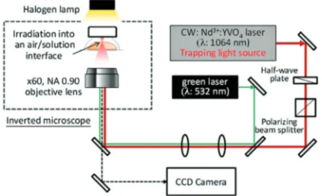
Fig. 1 shows the optical setup used in this work. A linearly-polarized CW laser beam of 1064 nm from a Nd3+:YVO4laser
(Coherent, MATRIX 1064-10-CW) was used as the trapping light source, and the output power was tuned by adjusting the angle of a half-wave plate coupled with a polarizing beam split-ter. A green laser (Laserglow technologies, LRS-0532-TFH-01,
λ = 532 nm) was also introduced into the inverted microscope through the same optical path as that of the trapping laser. These laser beams were focused on the same position by an objective lens (60× magnification, NA 0.90). After confirming that the green laser was focused at a surface layer of the solu-tion, the laser was switched off, and the trapping laser was turned on. The laser power was fixed to 1.1 W throughout the objective lens for all laser trapping experiments. The crystalli-zation behaviour was observed using a charge-coupled device (CCD) video camera (WATEC, WAT-231S2) under halogen lamp illumination.
Two kinds of crystal forms prepared by the laser were identified by Fourier transform infrared (FT-IR) spectroscopy (HORIBA, FT-720), in which the IR spectra were collected in attenuated total reflection (ATR) mode. Extinction coefficients of H2O, D2O, and respectiveL-Phe solutions were determined
by measuring their transmittance at 1064 nm passing through a glass cuvette with different optical path lengths, which was carried out using a spectrophotometer (Hitachi, U-4100).
Results and discussion
Laser trapping crystallization in supersaturated H2O solution
It is reported that the spontaneous crystallization of L-Phe aqueous solution provides two stable pseudopolymorphs of monohydrate needle-like and anhydrous plate-like forms depending upon the temperature, and that the transition temperature is about 37 °C in H2O.20Namely, each crystal is
spontaneously formed at temperatures below and above the transition point. First, we checked the pseudopolymorphism through spontaneous crystallization in our H2O samples. At
30–60 min after a spigot of the sample bottle was slightly opened for solvent evaporation, a lot of the needle-like crystals were observed and assigned to the monohydrate form accord-ing to the above paper.20
On the other hand, irradiation of the trapping laser into the air–solution interface of the solution thin film always provided a plate-like crystal at the focal spot, whose morphology is clearly different from that of spontaneous crystallization. Fig. 2 shows a series of CCD images of the crystallization behaviour under the laser irradiation. Immediately after starting the
Fig. 1 Optical setup for laser trapping crystallization ofL-Phe.
irradiation, only a small bright spot of the trapping laser due to its weak reflection at the interface was observed ( panel 1 of Fig. 2). At 245 s after starting the irradiation, the crystal became large as identified by a CCD image, when the crystal size was estimated to be a few micrometres. Further laser irradiation into the crystal central part caused the continuous crystal growth ( panels 2–4 of Fig. 2). In order to measure the thickness of the formed crystal, we examined the light reflec-tion from the upper and lower crystal faces by moving the objective lens manually in a vertical direction. However, two crystal faces were too close to be distinguished from each other, which indicates that the crystal thickness was less than the focal depth of the objective lens with a few micrometres.
When the crystal became larger than the observation area of the CCD camera (80μm × 60 μm) as determined by the high magnification objective lens, the trapping laser was turned off, and a broader area of the surrounding solution (1200 μm × 900μm) was observed using a low magnification (×4) objective lens. We found that only one crystal gradually migrated outward from the original focal position ( panels 5–6 of Fig. 2). This result strongly supports that the laser trapping provided only one plate-like crystal at the focal spot, and furthermore that no other crystallization takes place even spontaneously during the irradiation. The reproducibility of this crystallization behaviour in the H2O solution was confirmed for 12 samples,
where the laser irradiation always led to the plate-like crystal formation at the focal spot. Most recently, such plate-like crystal formation induced by laser trapping has been success-fully demonstrated even in the unsaturated H2O solution.17
Thus, we consider that the plate-like crystal formation by laser trapping in H2O was independent of the initial solution
con-centration, although the initial solution concentration gener-ally determines the laser trapping crystallization behaviour. Laser trapping crystallization in supersaturated D2O solution
For the D2O solution used in this work, no spontaneous
crys-tallization took place in the closed sample bottle at least for 30 min, and then a lot of the needle-like crystals were spon-taneously generated all over the sample bottle and the solution turned gel-like. This spontaneous crystallization behaviour was similar to that in the H2O solution as described above. On the
other hand, upon starting the irradiation of the trapping laser
into the air–solution interface of a thin film of the D2O
solu-tion, needle-like crystal formation was always observed within 30 min, which can be clearly ascribed to the laser trapping crystallization. Fig. 3a shows captured images from a CCD camera for the crystallization behaviour under laser irradiation. Initially, the CCD image only showed a small bright spot ascribed to weak reflection of the trapping laser at the surface, while the 600 s laser irradiation led to needle-like crystal formation ( panel 1 of Fig. 3a). Most importantly, the needle-like crystal formation was confirmed always at the outside of the focal spot, and never at the focal spot. This be-haviour is much different from the laser trapping crystalliza-tion of other amino acids that we have reported so far, in which their crystallization is induced always at the focal point.13–17 Further laser irradiation increased the number of the needle-like crystals with the irradiation time ( panels 2–3 of Fig. 3a). A series of these crystallization experiments was repeatedly carried out for 10 samples, and it was confirmed that the irradiation always led to needle-like crystal formation from the outside of the focal spot within 10 min.
The observation of a broader area of the surrounding solu-tion gave us critical informasolu-tion on the needle-like crystal for-mation. Fig. 3b shows the CCD image of the area within the range of a few millimetres from the focal spot. This horizon-tally long image was created by combining three CCD images captured at different distances from the focal position using the low magnification objective lens. It should be noted that numerous needle-like crystals were inhomogeneously distri-buted, and more exactly, the formation area was limited to within a 0.5–1.5 mm range from the focal spot. Conversely, the crys-tals were rarely observed in the area more than 1.5 mm away from the focal spot. This inhomogeneous distribution strongly supports that the needle-like crystal formation is not due to spontaneous crystallization, and we consider that the for-mation of a millimetre-scale, large high-concentration domain is responsible for the crystallization. Such domain formation is based on our previous experiments on a millimetre-scale dense liquid droplet of glycine formed by laser trapping.12
Fig. 2 A series of CCD images of L-Phe crystallization behaviour
induced by laser trapping in H2O solution. The arrow in the images
indi-cates the focal position.
Fig. 3 (a) A series of CCD images of L-Phe crystallization behaviour
induced by laser trapping in D2O solution. (b) A horizontally long image
made by combining three CCD images captured at different distances from the focal position.
This dense droplet is prepared by focusing a laser beam into the glass–solution interface of a supersaturated glycine–D2O
solution with the SS of 1.36, and eventually the large droplet of 5 mm in diameter with the SS of 2.7 is formed. In addition, the domain is quite stable and further irradiation causes no crystallization in spite of the considerably high con-centration. Thus, it is of great interest to note why no needle-like crystal formation is induced at the focal spot with a con-centration high enough to lead to crystal nucleation, but instead is induced away from the spot. The details of the mechanism are described later.
Pseudopolymorphism ofL-Phe and laser induced-local temperature elevation in H2O and D2O
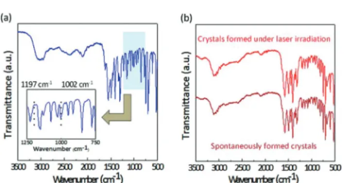
As described above, the pseudopolymorphism on the laser trapping crystallization of L-Phe clearly depends upon the solvent, that is, the plate-like and needle-like crystals were formed in H2O and D2O, respectively. Fig. 4 shows the FT-IR
spectrum of each crystal formed by laser trapping. As reported previously,17even after the focal position was completely occu-pied by the growing crystal, direct laser irradiation into the central part of the crystal led to continuous crystal growth. Namely, we made the anhydrous plate-like crystal grow to a size of a few hundred micrometres by direct laser irradiation, and the FT-IR measurement was carried out. For the mono-hydrate needle-like crystals formed within the 0.5–1.5 mm range of the focal spot, we collected a sufficient amount of crystals for the measurement. The FT-IR measurement for the crystal pseu-dopolymorph of L-Phe in H2O has been investigated.21 The
plate-like crystal showed a characteristic absorption peak at 1002 cm−1due to CH stretching, but no peak at 1197 cm−1due to hydrogen bond stretching was observed. These spectral characteristics are consistent with those of the anhydrous crystal reported previously. Therefore, we concluded that the plate-like crystal formed by laser trapping in H2O can be ascribed to its
anhydrous form, which is thermodynamically the most stable above a temperature of the transition point of 37 °C.
For the needle-like crystals prepared by laser trapping in D2O, we found that the FT-IR spectra were identical to those of
the needle-like crystals generated spontaneously from the solu-tion at room temperature. Although the actual transisolu-tion point
ofL-Phe pseudopolymorphism in D2O has not been reported
and examined yet, we confirmed that spontaneous crystalliza-tion at 35 and 40 °C provided needle-like and plate-like crys-tals, respectively. From these results, it is reasonable to consider that pseudopolymorphism in D2O solution appears
almost the same as that in H2O and that the transition point
in D2O is also similar in temperature to that in H2O. Therefore,
we consider that the laser trapping crystallization ofL-Phe in D2O provides the monohydrate crystal, which is
thermodyna-mically the most stable at room temperature, although further identification will be necessary in the future.
Here it is indispensable to estimate the local temperature elevation by light absorption of the solution because the pseu-dopolymorphism ofL-Phe is strongly affected by the tempera-ture. Fig. 5 shows the transmittance of H2O, D2O, and the
respectiveL-Phe solutions at 1064 nm passing through a glass cuvette with different optical path lengths. The transmittance decreased exponentially with increase in the path length in accordance with the Beer–Lambert law, by which we estimated the extinction coefficients of H2O and D2O to be 14.5 and
0.98 m−1, respectively. The higher extinction coefficient of H2O
is ascribed to overtone and combination absorption bands of the OH vibrational mode. The extinction coefficient of each L-Phe solution was almost identical to that of the corresponding solvent. Under experimental conditions similar to ours, the laser-induced local temperature elevation at the focal point was already estimated to be 23 and 2.6 K W−1 in H2O and D2O,
respectively.22 By assuming that the temperature elevation is simply proportional to the input laser power, the temperature elevation within the focal volume in the H2O solution can be
cal-culated to over 20 °C, which is high enough to exceed the tran-sition point of 37 °C. On the other hand, that in D2O solution is
estimated to be only 3.0 °C, which is too low to be over the tran-sition point. Thus, the local temperature elevation induced by the trapping laser irradiation should be responsible for the pseudopolymorphism ofL-Phe induced by laser trapping. Dynamics and mechanism of laser trapping crystallization through high-concentration domain formation
One of the most notable results in this work is that laser trap-ping crystallization in D2O provided the monohydrate crystals
Fig. 4 FT-IR spectra of anL-Phe plate-like crystal (a) and needle-like
crystals (b) prepared by laser irradiation.
Fig. 5 Transmittance of 1064 nm light passing through the solutions as a function of the optical path length.
always at the outside of the focal spot, and never at the focal spot. We here discuss the dynamics and mechanism of this amazing crystallization behaviour. In supersaturated solution, it is expected that the molecules easily form the relatively large and stable liquid-like clusters, which consist of solutes and solvents weakly linked by intermolecular interactions.23,24 Since the optical gradient force in our experiment is too small to trap singleL-Phe molecules, the liquid-like clusters are con-sidered to become a target for laser trapping. Their effective trapping instantly increases the local concentration within the focal volume, where aggregates of the clusters are prepared. Further laser irradiation continuously increases the local con-centration within the focal volume, the energy barrier for trig-gering liquid nucleation is overcome, and eventually the stable high-concentration domain is formed locally. The small domain is expanded from the focal volume to outside it spon-taneously due to intermolecular–cluster interactions, heat transfer, and convection flow, resulting in the formation of a large, high-concentration domain.
It can be considered that the concentration in the focal volume should be high enough to lead to crystal nucleation, but the monohydrate crystal was not formed at the focal spot. Here we should consider molecular/cluster orientation in the high-concentration domain formed through liquid nucleation. It is known that not only the concentration but also a suitable molecular orientation is necessary to adequately determine and distinguish crystal nucleation from the solution.25–27 In other words, even if the molecular concentration is high enough to trigger crystal nucleation, molecular orientation is not always preferable for triggering crystal nucleation. This knowledge is supported by our previous study on a
millimetre-scale dense liquid droplet of glycine in D2O formed by focusing
the laser beam at the surface of a glass substrate.12The droplet was formed through liquid nucleation and the SS was estimated to be 2.7 as mentioned above. The concentration is certainly high enough to lead to crystal nucleation, but it never takes place. Thus, both sufficient concentration and suitable molecular orien-tation are necessary to trigger crystal nucleation.
Coming back to this work, the high-concentration domain is also formed through liquid nucleation, so that it should have a homogeneous concentration and relatively ordered molecular/cluster orientation. As same as a dense liquid droplet of glycine, it can be considered that the orientation is unfavourable for triggering the crystal nucleation. Here we suggest that the orientation should become more disordered at the domain edge, where solutes and solvents go in and out. In particular, the orientation at the triple point of air, domain, and solution is expected to be much different from that inside the domain. Therefore, the needle-like crystal formation possibly takes place somewhere within the edge of the dense area, where the molecular/cluster orientation is preferable for triggering the needle-like crystal nucleation. The first nuclea-tion is realized far away from the focal spot due to its stochas-tic nature, and the surface of the first generated crystal induces some fluctuation in the steady state, leading to the subsequent nucleation. As a result, needle-like crystals are formed inhomogeneously around the focal spot, as shown in Fig. 3b. Fig. 6a summarizes the schematic illustrations of this prospective mechanism of laser trapping crystallization in D2O
leading to needle-like crystal formation.
Fig. 6b shows schematic illustrations for the crystallization behaviour in H2O solution, when the laser irradiation always
Fig. 6 Schematic illustrations of the prospective mechanism for laser trapping crystallization ofL-Phe in D2O (a) and H2O (b).
provides only one anhydrous crystal at the focal spot. Laser trapping of the clusters is considered to be similar to the case of the D2O solution ( panel (i) of Fig. 6b). On the other hand,
different from the D2O solution, local temperature elevation by
laser heating is expected to be high enough to exceed the tran-sition point of 37 °C. Therefore, during aggregation of the clus-ters by laser trapping, laser heating provides vigorous molecular motion and vibration of H2O and destroys the
inter-molecular interactions between H2O andL-Phe. Consequently,
H2O molecules come out from the liquid-like clusters, and the
dehydration in aggregation is completed ( panel (ii) of Fig. 6b). Further laser irradiation increases the local concentration of L-Phe without H2O and leads directly to crystal nucleation of
the anhydrous plate-like form not through liquid nucleation ( panel (iii) of Fig. 6b). Thus, dehydration on the laser trapping crystallization ofL-Phe was realized only in H2O solution since
a higher local temperature elevation is achieved by laser heating compared with D2O.
Conclusions
We successfully demonstrated the laser trapping crystallization
of L-Phe through high-concentration domain formation by
focusing a continuous-wave laser beam of 1064 nm at the surface of L-Phe–H2O and L-Phe–D2O solutions. Upon
irradiation into the H2O solution, a single plate-like anhydrous
crystal was prepared at the focal spot. This anhydrous form is thermodynamically the most stable at a temperature above a transition point of 37 °C, so we concluded that the local temp-erature elevation of over 20 °C induced by laser heating led to dehydration during laser trapping of the liquid-like clusters. On the other hand, in D2O solution, a lot of needle-like
crys-tals, which are ascribed to the monohydrate form, were formed outside of the focal spot, not at the focal spot, and were densely distributed in the area within a few millimetres of the focal spot. This indicates that an extremely large high-concentration domain is growing up to a millimetre-scale before monohydrate crystal nucleation, and that the mono-hydrate crystal formation takes place at the growing domain edge, where the orientation of molecules/clusters becomes more disordered. We believe that the present experiments on laser trapping-induced millimetre-scale dense domain for-mation and dehydration will be milestones in the study of laser trapping crystallization of molecular systems with hydro-gen-bonded networks of amino acids, protein, and so on. Our method can be applied to some compounds of membrane pro-teins that are difficult to crystallize through the milestones.
Acknowledgements
The present work is supported by the MOE-ATU Project (National Chiao Tung University) of the Ministry of Education, Taiwan, to H.M., and the National Science Council of Taiwan
to T.S. (NSC 102-2113-M-492-001-MY2) and to H.M. (NSC 100-2113-M-009-001).
Notes and references
1 A. Ashkin, Optical trapping and manipulation of neutral particles using lasers, Proc. Natl. Acad. Sci. U. S. A., 1997, 94, 4853.
2 C. Hosokawa, H. Yoshikawa and H. Masuhara, Cluster for-mation of nanoparticles in an optical trap studied by fluo-rescence correlation spectroscopy, Phys. Rev. E: Stat. Phys., Plasmas, Fluids, Relat. Interdiscip. Top., 2005, 72, 021408. 3 Y. Tanaka, H. Yoshikawa, T. Itoh and M. Ishikawa,
Laser-induced self-assembly of silver nanoparticles via plasmonic interactions, Opt. Express, 2009, 17, 18760.
4 W. Singer, T. A. Nieminen, N. R. Heckenberg and H. Rubinsztein-Dunlop, Collecting single molecules with conventional optical tweezers, Phys. Rev. E: Stat. Phys., Plasmas, Fluids, Relat. Interdiscip. Top., 2007, 75, 011916. 5 L. Pan, A. Ishikawa and N. Tamai, Detection of optical
trap-ping of CdTe quantum dots by two-photon-induced lumi-nescence, Phys. Rev. B: Condens. Matter, 2007, 75, 161305. 6 L. Jauffred, A. C. Richardson and L. B. Oddershede,
Three-dimensional optical control of individual quantum dots, Nano Lett., 2008, 8, 3376.
7 L. Jauffred and L. B. Oddershede, Two-photon quantum dot excitation during optical trapping, Nano Lett., 2010, 10, 1927. 8 Y. Tsuboi, T. Shoji and N. Kitamura, Crystallization of
lyso-zyme based on molecular assembling by photon pressure, Jpn. J. Appl. Phys., 2007, 46, L1234.
9 T. Shoji, N. Kitamura and Y. Tsuboi, Resonant excitation effect on optical trapping of myoglobin: The important role of a heme cofactor, J. Phys. Chem. C, 2013, 117, 10691. 10 S. Katsura, K. Hirano, Y. Matsuzawa, K. Yoshikawa and
A. Mizuno, Direct laser trapping of single DNA molecules in the globular state, Nucleic Acids Res., 1998, 26, 4943. 11 H. Masuhara, T. Sugiyama, T. Rungsimanon, K. Yuyama,
A. Miura and J.-R. Tu, Laser-trapping assembling dynamics of molecules and proteins at surface and interface, Pure Appl. Chem., 2011, 83, 869.
12 K. Yuyama, T. Sugiyama and H. Masuhara, Millimeter-scale dense liquid droplet formation and crystallization in glycine solution induced by photon pressure, J. Phys. Chem. Lett., 2010, 1, 1321.
13 T. Sugiyama, T. Adachi and H. Masuhara, Crystallization of glycine by photon pressure of a focused CW laser beam, Chem. Lett., 2007, 36, 1480.
14 T. Rungsimanon, K. Yuyama, T. Sugiyama and
H. Masuhara, Crystallization in unsaturated glycine/D2O
solution achieved by irradiating a focused continuous wave near infrared laser, Cryst. Growth Des., 2010, 10, 4686.
15 K. Yuyama, T. Rungsimanon, T. Sugiyama and
H. Masuhara, Selective fabrication ofα- and γ-polymorphs of glycine by intense polarized continuous wave laser beams, Cryst. Growth Des., 2012, 12, 2427.
16 K. Yuyama, K. Ishiguro, T. Sugiyama and H. Masuhara, Laser trapping dynamics of L-alanine depending on the laser polarization, Proc. SPIE-Int. Soc. Opt. Eng., 2012, 8458, 84582D.
17 K. Yuyama, T. Sugiyama and H. Masuhara, Laser trapping and crystallization dynamics ofL-phenylalanine at solution surface, J. Phys. Chem. Lett., 2013, 4, 2436.
18 Handbook of Chemistry and Physics 1st student edition, ed. R. C. Weast, CRC Press, Inc., 1988, p. C-706.
19 M. Jelinska-Kazimierczuk and J. Szydlowski, Isotope effect on the solubility of amino acids in water, J. Solution Chem., 1996, 25, 1175.
20 N. C. S. Kee, P. D. Arendt, L. M. Goh, R. B. H. Tan and R. D. Braatz, Nucleation and growth kinetics estimation for L-phenylalanine hydrate and anhydrate crystallization, CrystEngComm, 2011, 13, 1197.
21 J. Lu, Q. Lin, Z. Li and S. Rohani, Solubility of L-phenyl-alanine anhydrous and monohydrate forms: Experimental measurements and predictions, J. Chem. Eng. Data, 2012, 57, 1492.
22 S. Ito, T. Sugiyama, N. Toitani, G. Katayama and H. Miyasaka, Application of fluorescence correlation spec-troscopy to the measurement of local temperature in solu-tions under optical trapping condition, J. Phys. Chem. B, 2007, 111, 2365.
23 S. Chattopadhyay, D. Erdemir, J. M. B. Evans, J. Ilavsky, H. Amenitsch, C. U. Segre and A. S. Myerson, SAXS study of the nucleation of glycine crystals from a supersaturated solution, Cryst. Growth Des., 2005, 5, 523.
24 R. S. Berry, in Large clusters of Atoms and Molecules, ed. T. P. Martin, Kluwer Academic Publ., 1996, p. 281. 25 D. W. Oxtoby and Y. C. Shen, Density functional
approaches to the dynamics of phase transitions, J. Phys.: Condens. Matter, 1996, 8, 9657.
26 P. G. Vekilov, Dense liquid precursor for the nucleation of ordered solid phases from solution, Cryst. Growth Des., 2004, 4, 671.
27 J. Chen, B. Sarma, J. M. B. Evans and A. S. Myerson, Pharmaceutical crystallization, Cryst. Growth Des., 2011, 11, 887.