Dissociation of heme from gaseous myoglobin ions studied by infrared multiphoton
dissociation spectroscopy and Fourier-transform ion cyclotron resonance mass
spectrometry
Yi-Sheng Wang, Sahadevan Sabu, Shih-Chia Wei, C.-M. Josh Kao, Xianglei Kong, Shing-Chih Liau, Chau-Chung Han, Huan-Cheng Chang, Shih-Yu Tu, A. H. Kung, and John Z. H. Zhang
Citation: The Journal of Chemical Physics 125, 133310 (2006); doi: 10.1063/1.2221696 View online: http://dx.doi.org/10.1063/1.2221696
View Table of Contents: http://scitation.aip.org/content/aip/journal/jcp/125/13?ver=pdfcov Published by the AIP Publishing
Articles you may be interested in
Note: Photodissociation of CH3COCN at 308 nm by time-resolved Fourier-transform infrared emission spectroscopy: Is CO a primary or secondary product?
J. Chem. Phys. 138, 246102 (2013); 10.1063/1.4812378
Photodissociation of gaseous CH3COSH at 248 nm by time-resolved Fourier-transform infrared emission spectroscopy: Observation of three dissociation channels
J. Chem. Phys. 138, 014302 (2013); 10.1063/1.4768872
Infrared multiphoton dissociation tandem charge detection-mass spectrometry of single megadalton electrosprayed ions
Rev. Sci. Instrum. 82, 084104 (2011); 10.1063/1.3628667
Infrared absorption of gaseous ClCS detected with time-resolved Fourier-transform spectroscopy J. Chem. Phys. 126, 174310 (2007); 10.1063/1.2730501
Free electron laser-Fourier transform ion cyclotron resonance mass spectrometry facility for obtaining infrared multiphoton dissociation spectra of gaseous ions
Rev. Sci. Instrum. 76, 023103 (2005); 10.1063/1.1841953
Dissociation of heme from gaseous myoglobin ions studied
by infrared multiphoton dissociation spectroscopy and Fourier-transform
ion cyclotron resonance mass spectrometry
Yi-Sheng Wang, Sahadevan Sabu, Shih-Chia Wei, C.-M. Josh Kao, Xianglei Kong, Shing-Chih Liau, Chau-Chung Han, and Huan-Cheng Changa兲
Institute of Atomic and Molecular Sciences, Academia Sinica, P.O. Box 23-166, Taipei 106, Taiwan
Shih-Yu Tu and A. H. Kung
Institute of Atomic and Molecular Sciences, Academia Sinica, P.O. Box 23-166, Taipei 106, Taiwan and Department of Photonics, National Chiao Tung University, Hsinchu 300, Taiwan
John Z. H. Zhang
Department of Chemistry, New York University, New York, New York 10003 and Key Laboratory of Mesoscopic Chemistry of MOE, Institute of Theoretical and Computational Chemistry, College of Chemistry and Chemical Engineering, Nanjing University, Nanjing 210093, People’s Republic of China
共Received 7 April 2006; accepted 15 June 2006; published online 3 October 2006兲
Detachment of heme prosthetic groups from gaseous myoglobin ions has been studied by collision-induced dissociation and infrared multiphoton dissociation in combination with Fourier-transform ion cyclotron resonance mass spectrometry. Multiply charged holomyoglobin ions 共hMbn+兲 were generated by electrospray ionization and transferred to an ion cyclotron
resonance cell, where the ions of interest were isolated and fragmented by either collision with Ar atoms or irradiation with 3m photons, producing apomyoglobin ions共aMbn+兲. Both charged heme
loss共with 关Fe共III兲-heme兴+and aMb共n−1兲+as the products兲 and neutral heme loss 共with 关Fe共II兲-heme兴
and aMbn+ as the products兲 were detected concurrently for hMbn+ produced from a myoglobin solution pretreated with reducing reagents. By reference to Ea= 0.9 eV determined by blackbody
infrared radiative dissociation for charged heme loss of ferric hMbn+, an activation energy of 1.1 eV
was deduced for neutral heme loss of ferrous hMbn+with n = 9 and 10. © 2006 American Institute of Physics.关DOI:10.1063/1.2221696兴
I. INTRODUCTION
Mass spectrometry of noncovalent protein complexes is a promising approach toward understanding the nature of molecular recognition such as protein-ligand interactions and protein-protein interactions.1,2 One of the benchmark sys-tems for this study is the intact holomyoglobin ion.3–10 Ho-lomyoglobin共hMb兲 is a protein responsible for oxygen trans-port in muscular tissues. It possesses a prosthetic heme group bound noncovalently to a single polypeptide chain. Both the-oretical and experimental investigations have been carried out extensively concerning the binding characteristics of oxygen, carbon monoxide, and some other ligands to this protein.11–13 It is known that the binding affinity of these ligands depends sensitively on the conformation of the polypeptide chain that defines the binding condition for the heme moiety.14
Bound noncovalently to the polypeptide chain, the heme group is prone to detachment by either acid-or alcohol-induced denaturation of myoglobin in solution.8Prior studies have examined the binding characteristics of heme in gas-eous hMbn+ ions in the absence of a solvent. There are two
dissociation channels leading to the formation of heme-released apomyoglobin 共aMb兲 ions,
charged heme loss: hMbn+→ aMb共n−1兲+
+关Fe共III兲-heme兴+, 共1兲 neutral heme loss: hMbn+→ aMbn++ Fe共II兲-heme. 共2兲
Using an electrospray ionization共ESI兲-Fourier-transform ion cyclotron resonance共FTICR兲 mass spectrometer, Gross et al. carried out thermochemical measurements for the dissocia-tion energy of ferric hMbn+with blackbody infrared radiative
dissociation 共BIRD兲. From an analysis of the dissociation rates at different temperatures made possible by heating the ion cyclotron resonance 共ICR兲 cell and assuming that the secondary structures of the proteins are unchanged, they de-termined an Arrhenius activation energy of 0.8– 0.9 eV for charged heme loss in hMbn+ with n = 9 – 12. This measured
dissociation energy is similar to that共1.1 eV兲 of heme loss in solution.15–17The latter studies additionally showed that the interaction between heme and the proximal histidine ac-counts for⬃25% of the total dissociation energy and the rest was contributed by van der Waals attractions and hydrogen bonding of propionic acid residues of the heme group to the specific residues in the protein.16,17
With respect to Eqs.共1兲and共2兲, Chrisman et al. identi-fied these two types of heme loss with collision-induced dis-sociation 共CID兲 for both oxidized and reduced forms of hMbn+ 共n=2–10兲 in a quadrupole ion trap. Their results, in
a兲Author to whom correspondence should be addressed. Electronic mail:
0021-9606/2006/125共13兲/133310/7/$23.00 125, 133310-1 © 2006 American Institute of Physics
combination with UV-vis spectroscopic measurements, led to the conclusion that loss of charged heme primarily occurs in ferric hMbn+, whereas loss of neutral heme occurs predomi-nantly in ferrous hMbn+. There is no evidence for electron
transfer between heme and the polypeptide chain upon col-lisional activation of the charge-selected protein ions. Mark and Douglas10very recently studied Coulomb effects on the binding characteristic of 关Fe共III兲-heme兴+ and 关Fe共II兲-heme兴
in both positive and negative hMb ions in a high-pressure collision cell of a triple quadrupole tandem mass spectrom-eter. They found that the energies required to induce neutral heme loss are similar for both types of ions, while the ener-gies required to induce charged heme loss are significantly less for the positive ions, indicating that the Coulomb repul-sion between aMb共n−1兲+ and关Fe共III兲-heme兴+ lowers the
bar-rier for the heme release.10Unfortunately, information about the absolute dissociation activation energy for the neutral heme loss could not be deduced from their measurements.
In dissociation of molecular ions, sustained off-resonance irradiation共SORI兲 CID has been proven to be one of the most useful techniques because of its efficiency and low cost of implementation.18,19 A large body of theories, technological developments, and application notes of SORI-CID can be found in the literature.20–28 The technique is unique in that it repeatedly activates the ions of interest to moderate kinetic energy for multiple collisions with back-ground atoms or molecules. Mixed ions with the same mass-to-charge ratios can be excited simultaneously to the same energy, thereby allowing qualitative estimation of the relative activation energies between different dissociation channels. The method is well suited for the presently proposed study of charged versus neutral heme loss from hMbn+ because the
difference in dissociation energy between these two channels is small and needs to be measured carefully.
To determine quantitatively the relative activation ener-gies of the processes as described in Eqs. 共1兲 and 共2兲, IRMPD offers an alternative approach.28 A number of experiments29–31 have demonstrated that continuous-wave 共cw兲 CO2laser irradiation at⬃10m is a convenient means
of increasing the internal energy of trapped gaseous ions without the need of heating the ICR cell. Although under-standing of the temperature/laser intensity relationship is far from complete due to the complexity of the heating mecha-nism involved, the infrared multiphoton dissociation 共IRMPD兲 technique nevertheless can provide quite accu-rately the relative ordering of the dissociation activation en-ergies for peptide ions with similar size and structure.31 In view of this utility, we attempted in this work to deduce such information with both SORI-CID and IRMPD for hMbn+
produced from a solution containing both ferrous and ferric myoglobins. Unlike previously conducted studies,7the light source we use is a high-repetition-rate optical parametric os-cillator共OPO兲 laser for excitation of NH stretches, which are highly localized vibrational modes and should have similar absorption strengths for both oxidized and reduced forms of the proteins. Results of the quasi-cw IRMPD experiments can therefore be compared closely with BIRD measurements.
II. EXPERIMENT
The key instrument used in this experiment is FTICR mass spectrometer 共APEX IV, Bruker-Daltonics兲 equipped with a 7 T actively shielded superconducting magnet and an external ESI source.32,33Gaseous myoglobin ions were gen-erated by spraying a sample solution through an electrically grounded, gas-assisted nebulization system at an infusion rate of 100L h−1. The ESI assembly was pointed 60° off-axis toward the counter electrode, which was a negatively biased capillary defining the ion entry into vacuum. A coun-terflowing stream of heated nitrogen gas applied to the elec-trode facilitated ion desolvation. After passing through a dif-ferentially pumped region, the protonated protein ions were accumulated in a hexapole ion trap for 1 s and then pulsed into the ICR cell. Mass spectra were acquired typically by coadding 30 repeated scans.
The sample solution consisted of horse-heart myoglobin obtained from the vendor 共M1882, Sigma兲 without further purification. A stock protein solution 共3.5 mM ferric hMb兲 was first prepared with a 1:4 methanol/water mixture con-taining⬃0.0001% formic acid. ESI-FTICR mass spectra of the solution diluted to 3M 共pH 5.38兲 showed a progres-sion of multiply charged hMbn+ features peaking at n = 11
关Fig. 1共a兲兴. While some weak features corresponding to aMbn+can be found in the spectrum, most of the myoglobin
ions remained intact, consistent with the observation that myoglobin retains its native conformation at pH greater than 4 and CH3OH concentration lower than 30% in solution.8To
produce mass spectra exhibiting ferrous hMbn+ peaks, the
stock protein solution was first treated with 10 mM L-ascorbic acid for 8 h, followed by dilution to 3 M 共pH 4.92兲 for ESI. Further increase of the concentration of the reducing reagents inevitably produced myoglobin-ascorbate adduct ions and also resulted in a rapid decrease of the total ion intensities关Fig.1共b兲兴. All the protein solutions after preparation were kept in the dark below 5 ° C to prevent degradation before use.
FIG. 1. FTICR mass spectra of protonated holomyoglobin ions共hMbn+兲
produced from ESI of protein solutions共a兲 without any pretreatment and 共b兲 pretreated with L-ascorbic acid for 8 h. The peaks denoted by “ⴱ” in 共b兲 correspond to protonated apomyoglobin ions共aMbn+兲 with n=9–13.
133310-2 Wang et al. J. Chem. Phys. 125, 133310共2006兲
IRMPD spectra of hMbn+ were obtained using a
home-built pulsed OPO laser as the light source. The design and instrumentation of the infrared laser system have been de-scribed previously.34,35 In brief, the laser is composed of a periodically poled lithium niobate共PPLN兲 optical parametric oscillator in a grazing-incidence grating cavity configuration for broad wavelength tuning in the mid-infrared region. The PPLN crystal was pumped by an acousto-optic Q-switched Nd:YAG共yttrium aluminum garnet兲 laser 共210S, Lightwave Electronics兲 at a repetition rate of 4 kHz and with a pulse duration of 15 ns. The idler output wavelength of the OPO laser was tunable from 2.5 to 3.5m with a bandwidth of ⬃1 cm−1. The laser had an average output power in the
range of 80 mW共i.e., 20J/pulse兲. Prior to the IRMPD ex-periment, the ions of interest were first isolated by radio-frequency 共rf兲 cleanup sweeps and thermalized with pulsed argon gas at a peak pressure of⬃2⫻10−7Torr for 20 s. The
charge-selected protein ions were then irradiated by the tun-able OPO laser for 1 s. Upon resonant excitation, the gas-eous myoglobin ions released its heme fragment. The result-ing dissociation fractions were plotted as a function of laser wavelength to obtain the infrared action spectra.
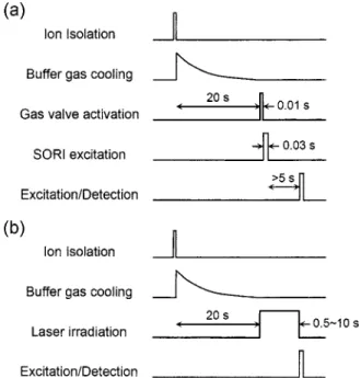
Simultaneous measurements for charged heme loss and neutral heme loss were conducted using SORI-CID under mild excitation conditions in the presence of Ar buffer gas at a pressure of⬃1⫻10−6Torr. The time sequence of the pulse
events in the CID process is shown in Fig.2共a兲, where the gas valve was activated for 10 ms prior to activation of the ions by SORI for 30 ms. In this experiment, the SORI acti-vation was operated at a rf of ⬃1 kHz lower than the ion cyclotron frequency of the protein complex. Control of the ion energy was made by varying the attenuation of the exci-tation rf amplitude 共in decibel兲 while keeping the durations of the rf excitation and the Ar gas pulsing constant.
The relative dissociation energies of charged versus neu-tral heme loss were quantified by measuring the dissociation
rates of ferric and ferrous hMbn+ions concurrently but
sepa-rately as a function of laser intensity. Figure 2共b兲 shows the time sequence of the pulse events used in the IRMPD mea-surement. Since the OPO laser was operated at a high rep-etition rate共4 kHz兲, a mechanical chopper served to control the laser irradiation period. The frequency of the laser output was fixed at 3333 cm−1 for excitation of the NH
stretches.33,36–40The laser power was changed by varying the polarization of the pump laser pulses and monitored with a power meter. Constrained by the performance of the instru-ments and the ion source conditions, only the dissociation energies of hMb9+ and hMb10+ can be measured with suffi-cient accuracy in this experiment.41
III. RESULTS AND DISCUSSIONS A. SORI-CID
Given the spectra shown in Fig. 1共b兲, we isolated hMb10+ produced from ESI of a myoglobin solution
pre-treated with reducing reagents using the rf cleanup sweep. Although the ions being isolated are of the same mass-to-charge ratio, they are actually composed of a mixture of ferric and ferrous hMbn+. Both neutral heme loss and
charged heme loss can, therefore, be monitored simulta-neously in the SORI-CID process. In this experiment, the excitation frequency used was offset from the resonance fre-quency of the precursor ion by −1 kHz, which has a negli-gible effect on the fragment ions, as confirmed by repeating the experiment with an offset frequency of +1 kHz. To pro-vide a more quantitative measure for the extent of the disso-ciation for this particular ion, we obtained the band intensity 共I兲 of each component and calculated the corresponding dis-sociation fraction as If/共Ip+ If兲, where p and f denote
precur-sor and fragment ions, respectively. Figure 3 shows the SORI-CID mass spectra obtained at different degrees of rf attenuation共in decibel兲. We indicate in the figure the rf at-tenuation instead of the absolute value of the collision energy because the exact energy involved in the SORI-induced CID process cannot be extracted from this experimental setting. However, as discussed in the Introduction, the advantage of using SORI is that both forms of the hMbn+ions are equally
excited with the same rf irradiation. Assuming that these two types of protein ions have the same collisional cross sections, which appears to be a reasonable assumption, an identical amount of collisional energy will be deposited into the pro-tein complexes that undergo either charged or neutral heme loss. High precision measurement for the relative ordering of the activation energies between these two dissociation chan-nels is then possible based on the intensities of the corre-sponding precursor and fragment ions.
In Fig. 4, we summarize the relative contributions of charged versus neutral heme loss as a function of the rf at-tenuation. The intensity ratio of the aMb10+ peak versus the
aMb9+peak is 1.2 at the highest activation energy, at which
no residual parent ions are detectable in the mass spectrum 共trace at 22 dB in Fig. 3兲. It suggests that the relative
abun-dance of gaseous ferrous hMbn+ versus ferric hMbn+ ions in the ICR cell before the SORI-CID process is 1.2:1. The dis-sociation ratio of these two channels at lower excitation
en-FIG. 2. Time sequences of pulse events in共a兲 SORI-CID and 共b兲 IRMPD experiments.
ergies should therefore reveal how their dissociation activa-tion energies differ. As shown in the lower traces in Fig.4, the production of aMb9+ is higher than that of aMb10+ by roughly 30% at the rf attenuation of 28 dB. Although the difference between these two fractions diminishes
progres-sively with the decreasing rf attenuation, this trend of the change suggests that the binding energy between aMb9+and 关Fe共III兲-heme兴+is somewhat lower than that between aMb10+
and Fe共II兲-heme. The result is in qualitative agreement with the CID measurement for hMbn+ 共n=5–9兲 using a triple quadrupole tandem mass spectrometer by Mark and Douglas.10
B. IRMPD
We started the IRMPD measurement with pure ferric hMbn+. Specifically, we acquired first the infrared photodis-sociation spectra of these ions carrying different numbers of charges. Though there have been many experimental methods42employed to characterize the structures of proteins and polypeptides in the gas phase, no infrared spectra have been reported except that of McLafferty and co-workers.36,37 These authors used a 6 T FTICR mass spectrometer and a pulsed infrared OPO laser to obtain the photodissociation spectra of multiply charged bovine ubiquitin ions in the fre-quency range of 3050– 3775 cm−1, and identified a single
broad feature at 3350 cm−1 with a full width at half
maxi-mum 共FWHM兲 of more than 100 cm−1 for free- and/or
hydrogen-bonded-NH stretching vibrations of this gaseous protein ion.33,36–40 Figure 5 shows the IRMPD spectra ob-tained in this work for ferric hMbn+ with n = 9 – 11. Two
prominent absorption bands were observed in the spectral scan range of 2850– 3650 cm−1for each ion. The band
peak-ing at 3330 cm−1again arises from N–H stretching vibrations
and the weaker feature at⬃2950 cm−1can be ascribed to CH
stretching vibrations. Compared to the spectrum observed for myoglobin in aqueous solution,43these two absorption bands are narrower共FWHM ⬃60 cm−1兲 and much better resolved
because of the absence of solvent interference.
It is noteworthy in Fig.5 that the observed band inten-sity increases nearly quadratically with the charge number from n = 9 to n = 11. In infrared action spectra as presently acquired, the observed band intensity is a convolution of the absorption strength of the vibrational mode excited and the
FIG. 3. SORI-CID mass spectra of hMb10+at different levels of rf
attenua-tion共in dB兲. The fragment ions aMb9+and aMb10+result from charged heme
loss and neutral heme loss, respectively.
FIG. 4. Fractions of charged共䊐兲 vs neutral 共䊊兲 heme loss of hMb10+, and
their ratios共䊏兲, as a function of SORI excitation energy. The dissociation fraction is defined as If/共Ip+ If兲, where Ipand If are the intensities of
pre-cursor ions and fragment ions, respectively.
FIG. 5. IRMPD spectra of hMbn+with n = 9共⫻兲, 10 共䊏兲, and 11 共䊊兲. The
peak intensities represent dissociation fractions as defined in Fig.4.
133310-4 Wang et al. J. Chem. Phys. 125, 133310共2006兲
dissociation yield of the individual ion within our detection time window. Since these three ions differ only in their charge numbers, they should have similar absorption strengths for both NH and CH stretches. The observed band intensity variation in Fig. 5 should therefore be associated with the difference in dissociation rate of these protein ions. Assuming that the absorbed photon energy dissipates very rapidly to other vibrational degrees of freedom and thermal equilibrium is reached before dissociation takes place, these observations suggest that the preexponential factor共A兲 in the first-order Arrhenius equation increases with n, given the same dissociation activation energy of Ea⬃0.9 eV at n
= 9 – 11. A plausible interpretation for such a charge number dependence is that the gaseous myoglobin ion unfolds to a greater extent when carrying more charges, a result in close agreement with BIRD measurements.5
In determining the dissociation activation energy with IRMPD using a cw CO2laser, Jockusch et al.29demonstrated
that the measured dissociation energy depends on the total laser power and yet is independent of the dimension of the laser beam used. Building on this foundation, our IRMPD-based dissociation energy measurements began with pure fer-ric hMbn+ions of n = 9 – 11. Similar to previous findings,29,31
a short induction period was required for the laser excitation to raise the temperature of the ion population to the point of dissociation, at which the weakest bonds 共i.e., the noncova-lent bonds兲 broke. No secondary fragments other than the heme group from this protein complex were observed. Given in Fig. 6共a兲 is a plot for the dissociation fraction of ferric hMb11+ as a function of laser irradiation time, showing
first-order kinetics. By acquiring the first-first-order rate constants at different laser intensities, plotting of the natural logarithm of the rate constant versus the natural logarithm of the laser intensity yields an activation energy共Ea兲 for the
photodisso-ciation process according to the equation29
Ealaser= skB d ln kd
d ln Ilaser, 共3兲
where kd is the experimentally determined dissociation rate
constant, kBis the Boltzmann constant, and Ilaseris the laser
intensity. Due to the complexity of the IRMPD mechanism involved in such a large protein ion, we treat s here as a scaling factor by reference to the BIRD measurement. This scaling is deemed justified because a quasi-cw light source is used in this experiment.
Figure 6共b兲 displays the result of the activation energy measurement for pure ferric hMbn+ions with n = 10– 12. The
measured value for hMb10+ is slightly higher than those of
hMb11+ and hMb12+. However, compared with BIRD results
共0.9±0.1 eV for hMb10+ and hMb11+ and 0.8± 0.1 eV for
hMb12+兲,5
these values suggest and averaged scaling factor of
s = 4.6± 0.5⫻103K for all three ions. It should be noted that
the scaling factor so derived is twice as large as that 共2369 K兲 reported by Paech et al.30
for four peptide ions using a cw CO2laser as the excitation source. This
discrep-ancy, clearly, is associated with the size 共mass⬎16 000 Da versus mass⬍3000 Da兲 of the biomolecules studied, the wavelength
共⬃3m vs⬃10m兲 of the infrared photons used, and the mechanism of the dissociation involved in these two types of measurements.
Based on this measured scaling factor, s = 4.6⫻103 K,
we determined the dissociation energy for the neutral heme loss with IRMPD. In this measurement, both ferric and fer-rous hMbn+ions produced from ESI of a myoglobin solution
pretreated with reducing reagents were first isolated by rf sweeps. The relative abundance of these two components was then determined with SORI-CID as depicted earlier. By exciting the NH stretches at 3333 cm−1 and monitoring the
charged and the neutral heme loss channels simultaneously, the respective activation energies were determined by fitting two sets of experimental data separately to Eq.共3兲. Figure7
shows the plots of ln共kd兲 vs ln共Ilaser兲 for both ions. For the
detachment of charged heme from the ferric components, we determined two slopes 2.39± 0.10 and 2.21± 0.20 for the n = 9 and n = 10 ions, respectively. The latter agrees well with that shown in Fig. 6共b兲 for ferric hMb10+ within our
experi-mental accuracy, which serves as an independent validation for this IRMPD method. With the use of the same scaling factor as before, we obtained an average activation energy of
Ea= 1.1± 0.1 eV for the detachment of the neutral heme
FIG. 6. IRMPD of pure ferric hMbn+.共a兲 Time trace of charged heme loss at
each of five laser intensities for the n = 11 ion only.共b兲 Plot of the natural logarithm of the first-order unimolecular dissociation rate constant, kd共s−1兲,
vs the natural logarithm of the laser intensity Ilaser共in units of W cm−2兲. The
fitted slopes are 2.42± 0.05, 2.06± 0.12, and 2.05± 0.07 at n = 10, 11, and 12, respectively. The frequency of the OPO laser excitation was fixed at 3333 cm−1.
group from the ferrous hMb9+ and hMb10+ components. In
line with the conclusion reached earlier by SORI-CID, this IRMPD measurement indicates that the difference in disso-ciation activation energy between these two channels is small, ⬃27% of the total energy 共cf. caption of Fig. 7兲 or
6 ± 1 kcal mol−1.
IV. CONCLUSION
We have demonstrated that it is possible to determine fairly accurately the relative dissociation activation energies of charged versus neutral heme loss from a mixture of ferric and ferrous hMbn+ protein ions using IRMPD assisted by SORI-CID in a FTICR mass spectrometer. The excitation was made specifically through the high-frequency vibrational modes, such as the NH stretches, which have similar absorp-tion cross secabsorp-tions among proteins with different charge numbers and oxidation states. By monitoring the dissociation kinetics of these two heme loss channels simultaneously, we conclude that the barrier for neutral heme release in ferrous hMbn+ is significantly higher than that for charged heme
re-lease in ferric hMbn+ by⬃27% at both n=9 and n=10.
Infrared photodissociation spectra at the 3m region were obtained for the first time for gaseous myoglobin ions in this work. With the availability of lasers共such as the free electron lasers兲 covering a wider range of excitation wavelength,44 employment of the approaches as presently illustrated is expected to provide additional insight into the structure and binding characteristics of this and other nonco-valent protein complexes in the gas phase. Further elucida-tion of these complex systems may come from quantum chemistry calculations, which have been shown to be accu-rate enough to predict the electronic structures of unligated or ligated ferroporphyrins.45
ACKNOWLEDGMENTS
This work is supported by grants from Academia Sinica and the National Science Council共Grant No. NSC 92-3112-B-001-012-Y兲 of Taiwan. The authors thank Professor Y. T. Lee for critical comments.
1J. A. Loo, Mass Spectrom. Rev. 16, 1共1997兲.
2G. Brenner-Weiss, F. Kirschhofer, B. Kuhl, M. Nusser, and U. Obst, J.
Chromatogr. A 1009, 147共2003兲.
3V. Katta and B. T. Chait, J. Am. Chem. Soc. 113, 8534共1991兲. 4Y.-T. Li, Y.-L. Hsieh, J. D. Henion, and B. Ganem, J. Am. Soc. Mass
Spectrom. 4, 631共1993兲.
5D. S. Gross, Y. Zhao, and E. R. Williams, J. Am. Soc. Mass Spectrom. 8,
519共1997兲.
6Y.-L. Chen, J. M. Campbell, B. A. Collings, L. Konermann, and D. J.
Douglas, Rapid Commun. Mass Spectrom. 12, 1003共1998兲.
7F. He, C. L. Hendrickson, and A. G. Marshall, J. Am. Soc. Mass
Spec-trom. 11, 120共2000兲.
8K. R. Babu and D. J. Douglas, Biochemistry 39, 14702共2000兲. 9P. A. Chrisman, K. A. Newton, G. E. Reid, J. M. Wells, and S. A.
McLuckey, Rapid Commun. Mass Spectrom. 15, 2334共2001兲.
10K. J. Mark and D. J. Douglas, Rapid Commun. Mass Spectrom. 20, 111
共2006兲.
11S. Z. Hu, K. M. Vogel, and T. G. Spiro, J. Am. Chem. Soc. 116, 11187
共1994兲.
12J. S. Olson and G. N. Phillips, Jr., JBIC, J. Biol. Inorg. Chem. 2, 544
共1997兲.
13E. Sigfridsson and U. Ryde, JBIC, J. Biol. Inorg. Chem. 4, 99共1999兲. 14E. Antonini and M. Brunori, Hemoglobin and Myoglobin in their
Reac-tions with Ligands共North-Holland, Amsterdam, 1971兲.
15C. L. Hunter, A. G. Mauk, and D. J. Douglas, Biochemistry 36, 1018
共1997兲.
16M. S. Hargrove, A. J. Wilkinson, and J. S. Olson, Biochemistry 35,
11300共1996兲.
17C. L. Hunter, E. Lloyd, L. D. Eltis, S. P. Rafferty, H. Lee, M. Smith, and
A. G. Mauk, Biochemistry 36, 1010共1997兲.
18T. Solouki, L. Pasa-Tolic, G. S. Jackson, S. G. Guan, and A. G. Marshall,
Anal. Chem. 68, 3718共1996兲.
19P. D. Schnier, J. C. Jurchen, and E. R. Williams, J. Phys. Chem. B 103,
737共1999兲.
20J. W. Gauthier, T. R. Trautman, and D. B. Jacobson, Anal. Chim. Acta
246, 211共1991兲.
21S. K. Shin and S. J. Han, J. Am. Soc. Mass Spectrom. 8, 86共1997兲. 22S. J. Pastor and C. L. Wilkins, Int. J. Mass Spectrom. Ion Process. 175,
81共1998兲.
23M. V. Gorshkov, L. Pasa-Tolic, and R. D. Smith, J. Am. Soc. Mass
Spectrom. 10, 15共1999兲.
24J. Laskin, E. Denisov, and J. H. Futrell, Int. J. Mass. Spectrom. 219, 189
共2002兲.
25X. Guo, M. C. Duursma, A. Al-Khalili, and R. M. A. Heeren, Int. J.
Mass. Spectrom. 225, 71共2003兲.
26J. Laskin and J. H. Futrell, Mass Spectrom. Rev. 22, 158共2003兲. 27A. G. Marshall,C. L. Hendrickson, and G. S. Jackson, Mass Spectrom.
Rev. 17, 1共1998兲.
28J. Laskin and J. S. Futrell, Mass Spectrom. Rev. 24, 135共2005兲. 29R. A. Jockusch, K. Paech, and E. R. Williams, J. Phys. Chem. A 104,
3188共2000兲.
30K. Paech, R. A. Jockusch, and E. R. Williams, J. Phys. Chem. A 106,
9761共2002兲.
31M. A. Freitas, C. L. Hendrickson, and A. G. Marshall, J. Am. Chem. Soc.
122, 7768共2000兲.
32O. P. Charkin, N. M. Klimenko, P. T. Nguyen, D. O. Charkin, A. M.
Mebel, S. H. Lin, Y.-S. Wang, S.-C. Wei, and H.-C. Chang, Chem. Phys. Lett. 415, 362共2005兲.
33X. L. Kong, I.-A. Tsai, S. Sabu, C.-C. Han, Y. T. Lee, H.-C. Chang, S.-Y.
Tu, A. H. Kung, and C.-C. Wu, Angew. Chem., Int. Ed. 45, 4130共2006兲.
34C. S. Yu and A. H. Kung, J. Opt. Soc. Am. B 16, 2233共1999兲. 35X. Qian, T. Zhang, C. Y. Ng, A. H. Kung, and M. Ahmed, Rev. Sci.
Instrum. 74, 2784共2003兲.
36H. B. Oh, K. Breuker, S. K. Sze, Y. Ge, B. K. Carpenter, and F. W.
McLafferty, Proc. Natl. Acad. Sci. U.S.A. 99, 15863共2002兲.
37H. B. Oh, C. Lin, H. Y. Hwang, H. Zhai, K. Breuker, V. Zabrouskov, B.
FIG. 7. Plot of the natural logarithm of the dissociation rate constants kd
共s−1兲 of charged vs neutral heme loss of hMb9+共䊏,䊐兲 and hMb10+共쎲,䊊兲 as
a function of the natural logarithm of the laser intensity Ilaser共in units of W cm−2兲. The laser excitation frequency was fixed at 3333 cm−1. The fitted
slope of charged vs neutral heme loss is 2.26± 0.12 vs 2.86± 0.12, respectively.
133310-6 Wang et al. J. Chem. Phys. 125, 133310共2006兲
K. Carpenter, and F. W. McLafferty, J. Am. Chem. Soc. 127, 4076 共2005兲.
38Y.-S. Wang, H.-C. Chang, J. C. Jiang, S. H. Lin, Y. T. Lee, and H.-C.
Chang, J. Am. Chem. Soc. 120, 8777共1998兲.
39C.-C. Wu, J. C. Jiang, I. Hahndorf, C. Chaudhuri, Y. T. Lee, and H.-C.
Chang, J. Phys. Chem. A 104, 9556共2000兲.
40C. Chaudhuri, J. C. Jiang, C.-C. Wu, X. Wang, and H.-C. Chang, J. Phys.
Chem. A 105, 8906共2001兲.
41It was found experimentally that the ratio of ferrous versus ferric hMbn+
produced from ESI of the reducing-reagent-treated solution decreases
rapidly with increasing n. At n = 11, the ratio was⬃0.3, which is too low to allow accurate measurement for the dissociation rate constant of fer-rous hMb11+by IRMPD.
42M. F. Jarrold, Acc. Chem. Res. 32, 360共1999兲.
43R. H. Austin, A. H. Xie, L. van der Meer, B. Redlich, P. A. Lindgard, H.
Frauenfelder, and D. Fu, Phys. Rev. Lett. 94, 128101共2005兲.
44J. Oomens, D. T. Moore, G. von Helden, G. Meijer, and R. C. Dunbar, J.
Am. Chem. Soc. 126, 724共2004兲.
45M.-S. Liao and S. Scheiner, J. Chem. Phys. 116, 3635共2002兲.