行政院國家科學委員會補助專題研究計畫成果報告
※ ※ ※ ※ ※ ※ ※ ※ ※ ※ ※ ※ ※ ※ ※ ※ ※ ※ ※ ※ ※ ※ ※ T T 病 毒 感 染 對 慢 性 B 型 或 C 型 肝 炎 臨 床 病 理 之 影 響 ※ ※ I n f l u e n c e o f T T v i r u s i n f e c t i o n o n ※ ※ c l i n i c o p a t h o l o g i c a l c o u r s e o f c h r o n i c h e p a t i t i s B o r C ※ ※ ※ ※ ※ ※ ※ ※ ※ ※ ※ ※ ※ ※ ※ ※ ※ ※ ※ ※ ※ ※ ※ ※ 計畫類別:■個別型計畫 □整合型計畫 計畫編號:NSC89-2315-B-002-022- 執行期間:88 年 8 月 1 日至 89 年 7 月 31 日 計畫主持人:高嘉宏 共同主持人:陳定信 執行單位:國立台灣大學醫學院 中 華 民 國 89 年 10 月 11 日中文摘要
背景及目的:TT 病毒(TTV)和 B 型肝炎病毒(HBV)或 C 型肝炎病毒(HCV)常有共 同感染現象,然而 TTV 感染對慢性 B 型或 C 型肝炎之影響仍未明瞭。本研究之 目的在於瞭解慢性 B 型或 C 型肝炎患者之 TTV 感染盛行率及 TTV 共同感染對 其臨床病理及病毒學特性之影響,此外亦探討 TTV 共同感染對慢性 C 型肝炎干 擾素治療反應之影響。 方法:共收集 100 例無症狀 B 型肝炎帶原者,220 例慢性 B 型肝炎患者和 170 例慢性 C 型肝炎患者(其中 110 例曾接受 6 個月之干擾素治療)。血清 HCV RNA 和 TTV DNA 以 PCR 法偵測,而血清 HBV DNA 和 HCV RNA 濃度則以 bDNA 法定量之。 結果:490 例慢性肝炎病毒感染患者中,21.5%的 B 肝帶原者和 30.6%的 C 肝帶 原者體內可偵測到 TTV DNA,均較健康成年人之 10%顯著為高。TTV 和 HBV 共同感染患者和 HBV 單獨感染患者在性別分布,平均年齡,輸血史,平均最高 ALT 值和肝組織學嚴重度上無明顯差異,但共同感染患者之血清 HBV DNA 濃 度似乎較低(P=0.066)。TTV 和 HCV 共同感染患者和 HCV 單獨感染患者在這些 臨床病理特點,血清 HCV RNA 濃度和對干擾素治療反應上均無明顯差異。此 外,41 例 HCV 和 TTV 共同感染患者中,29 例(70%)於干擾素治療結束時血清 TTV DNA 呈陰性,而 17 例(41%)於停藥半年後仍為陰性。進一步分析發現患者 肝功能持續正常與血清 TTV DNA 清除無關,而與血清 HCV RNA 清除有關。 結論:台灣地區之慢性 B 型和 C 型肝炎患者常合併 TTV 感染,但 TTV 共同感 染並不會影響其臨床病理病程及對干擾素之治療成效。慢性 TTV 感染之自然病 史及 TTV 共同感染是否會影響慢性 B 型或 C 型肝炎之自然病史仍有待長期之追 蹤研究。 關鍵詞:B 型肝炎,C 型肝炎,慢性肝病,肝硬化,肝癌,TT 病毒。ABSTRACT
Concomitant infection with the newly identified TT virus and the hepatitis B virus (HBV) or hepatitis C virus (HCV) is common. However, the effect of TTV infection on chronic hepatitis B or C is unknown. The prevalence of TTV infection, the effect of TTV infection on the clinical, histological and virological features of patients with chronic hepatitis B or C, and the influence of TTV infection on the HCV response to interferon alfa therapy were studied. A total of 100 asymptomatic hepatitis B surface antigen carriers, 220 patients with HBV-related chronic liver diseases, and 170 patients with HCV-related chronic liver disease and HCC were enrolled. Serum HCV RNA and serum TTV DNA were detected by polymerase chain reaction. Serum HBV DNA and serum HCV RNA level were quantified by branched DNA assays. Infection with TTV was detected in 21.5% of HBV carriers and 31% of HCV carriers. TTV infection had little effect on the clinicopathological course of chronic HBV infection. In chronic hepatitis C, clinical features, histological severity, serum HCV RNA levels, and the response to interferon alfa therapy did not differ between those with and without TTV infection. The loss of serum TTV DNA did not correlate with the biochemical response as did in the loss of serum HCV RNA. In conclusion, TTV infection is found frequently in patients with chronic hepatitis B or C in Taiwan; however, coinfection with TTV does not affect the clinicopathological course of chronic hepatitis B or C and the response to interferon alfa therapy.
Key Wor ds: chronic hepatitis B, chronic hepatitis C, chronic liver disease, hepatocellular
INTRODUCTION
Chronic liver disease and hepatocellular carcinoma (HCC) are endemic in Taiwan, and most are caused by HBV or HCV infection [Chen et al., 1987; 1990]. However, there still remains a proportion of hepatitis cases with undefined etiology, suggestive of the existence of additional causative agents [Alter 1994; Kao et al., 1996b]. Recently, a DNA virus was isolated from a patient with posttransfusion hepatitis of unknown etiology and designated TT virus (TTV) for the initials of the index patient [Nishizawa et al., 1997]. In addition, TTV genomes were detected in patients with cryptogenic posttransfusion hepatitis and the emergence of viremia coincided with the modest increases of serum alanine aminotransferase (ALT) levels [Okamoto et al., 1998]. A recent study showed that TTV genome is circular and negative stranded, and comprises 3,852 bases with a particle size of 30-50 nm, suggesting TTV is similar to the Circoviridae [Mushahwar et al., 1999]. In Japan, TTV DNA was detected in 12% of healthy blood donors, 47% of patients with fulminant non-A~G hepatitis and 46% of patients with chronic liver diseases of unknown etiology [Okamoto et al., 1998], suggesting that TTV may be the cause of some cryptogenic liver diseases. However, the clinical significance of infection with TTV alone or in combination with other hepatitis viruses has been questioned [Charlton et al., 1998; Naoumov et al., 1998; Simmonds et al., 1998; Prati et al., 1999]. Taking advantage of the extremely common chronic HBV and HCV infections in Taiwan, the prevalence of TTV infection and the possible role of TTV coinfection on the clinical, virological and histological features of patients with chronic hepatitis B or C were studied. In addition, the response of HCV and TTV to interferon-alfa treatment was evaluated.
MATERIALS AND METHODS
Patients
Serum samples were retrospectively studied from 490 patients with long-term follow-up at the gastroenterological clinics of the National Taiwan University Hospital. These included (i) 100 asymptomatic hepatitis B surface antigen (HBsAg) carriers (54 men, 46 women; mean age, 30+7 years) with normal serum ALT level for at least one year, and 220 histologically verified HBsAg-positive chronic liver disease and HCC patients (173 men, 47 women; mean age, 45+12 years); (ii) 170
histologically verified HCV-related chronic liver disease and HCC patients (102 men, 68 women; mean age, 50+10 years). Among them, 110 patients with chronic hepatitis C (71 men, 39 women; mean age, 45+12 years) had received previously 3 million units of interferon alfa (IFN) thrice weekly for 24 weeks. The presence of HCV RNA and TTV DNA in the serum were determined before initiation of IFN therapy; at the end therapy; and 24 weeks after the therapy was discontinued. The response to IFN was classified into two patterns according to the serum ALT level. Patients who had normalized serum ALT levels (< 40 U/L) at the end of therapy and during the follow-up period was considered to have a sustained biochemical response. Non-sustained response was defined as serum ALT levels that could not be normalized either at the end of therapy or during follow-up period. The diagnosis of chronic liver disease was based on clinical and pathological grounds accepted generally including chronic persistent hepatitis (CPH), chronic active hepatitis (CAH), liver cirrhosis (LC) and HCC. Those with dual infections by HBV and HCV were excluded. HBV carrier was defined by a positive reaction for HBsAg, and HCV carriers by both second-generation anti-HCV and HCV RNA positivity for at least 6 months. All the enrolled patients had no markers suggestive of autoimmune hepatitis including antinuclear antibodies, antimitochondrial antibodies and anti-smooth muscle antibodies. None had a history of alcoholism (> 50 gm/day), injection drug abuse, homosexuality, or hepatotoxic drug intake. Metabolic liver disease including hemochromatosis, Wilson's disease or α-1 anti-trypsin deficiency was excluded by clinical and laboratory data. In addition, 100 healthy adults who were at no risk for hepatitis and were with normal serum ALT level and negative for serological markers of current hepatitis viral infection were used as a control group. Serum samples taken from each subject were stored at -70 oC until use.
Serological Mar ker s
HBsAg and anti-HCV were tested with commercially available kits (Ausria II and HCV EIA II, Abbott Laboratories, North Chicago, IL).
Detection of HCV RNA and Genotyping of HCV
Serum HCV RNA was assayed by reverse transcription (RT)-PCR with primers from the most conserved 5' untranslating region of the viral genome [Kao et al., 1992], and identification of HCV genotype by type-specific primers as previously described [Kao et al., 1995]. To avoid false-positive results, the methods described by Kwok and Higuchi to prevent cross contaminations were applied [Kwok and Higuchi 1989].
Quantitation of HBV DNA and HCV RNA
Serum HBV DNA level was quantified by a branched DNA (bDNA) signal amplification assay (Hepatitis B Viral DNA, Chiron, Emeryville, CA) as described previously [Kao et al., 1998]. The HBV DNA quantification range of the bDNA assay
is 2.5 ~ 17,700 pg/mL. Serum HCV RNA level was determined by using a second generation bDNA signal amplification assay (Quantiplex-HCV, version 2.0; Chiron) with a detection limit of 0.2 MEq/mL according to the manufacturer's instructions.
Detection of TTV DNA
The presence of TTV DNA was assayed by nested PCR with primer pairs from the open reading frame (ORF)-1 of the viral genome [Kao et al., in press]. Briefly, total DNA was extracted from 100 µ L serum using QIAamp Blood kit (QIAGEN Ltd, Crawley, UK) and resuspended in 50 µ L elution buffer. For the first stage PCR, a 25 µl of reaction mixture containing 2 µl of the cDNA sample, 1x PCR buffer (10 mM tris-HCl pH 9.0, 50mM KCl, 1.5 mM MgCl2, 0.01% gelatin and 0.1% Triton X-100), 10 mM of each dNTP, 100 ng of each outer primer (outer sense: T-1s 5'-ACAGACAGAGGAGAAGGCAACATG-3'; outer antisense: T-2a 5'-CTACCTCCTGGCATTTTACC-3') and 1 unit of Taq DNA polymerase was amplified in a thermal cycler (Perkin-Elmer Cetus, Norwalk, CT) for 30 cycles. Each cycle entailed denaturation at 95 oC for 60 s, primer annealing at 55 oC for 30 s and extension at 72 oC for 60 s with a final extension step at 72°C for 7 min. After the first amplification, 1 µl of the PCR products was reamplified for another 30 cycles with 100 ng of each inner primer (inner sense: T-3s 5'-GGCAACATGTTATGGATAGACTGG-3'; inner antisense: T-4a 5'-CTGGCATTTTACCATTTCCAAAGTT-3'). The second round of PCR was done in the same manner as the first round giving a 272 bp amplification product. The amplified products were separated in 3% agarose gel electrophoresis and stained by ethidium bromide. Nucleotide sequences of selected amplified products were directly determined by using fluorescence labelled primers with a 373A Sequencer (Applied Biosystems, Foster City, CA) to verify the specificity.
Statistical Analysis
Data were analyzed by Chi-square test with Yates' correction or Student's t test where appropriate. A P value of less than 0.05 was considered significant.
RESULTS
Of 490 patients with chronic hepatitis viral infections, the overall prevalence of TTV viraemia was 21.5% and 30.6% in HBV and HCV carriers, respectively, which was significantly higher than that in healthy adults (10%, Kao et al., in press, P < 0.02 and < 0.001, respectively).
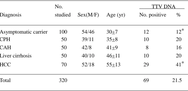
In 320 HBV carriers with various liver diseases (TABLE I), the prevalence of TTV DNA ranged from 12% to 20% among asymptomatic carriers, patients with CPH, CAH and liver cirrhosis. However, the prevalence was significantly higher in those with HCC than in asymptomatic carriers (41% vs. 12%, P < 0.001). Cases of cirrhosis with HBV and TTV coinfection had a similar percentage of transfusion history to those with HBV infection alone (20% vs. 17%). The average age of HCC patients with both HBsAg and TTV DNA was comparable to that of patients with HBsAg alone (52+13 vs. 56+12 years, P = 0.2).
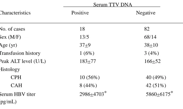
No statistically significant difference was found in gender distribution, mean age, frequency of transfusion history, mean peak serum ALT level, and histological severity when the 18 patients with HBV and TTV coinfection were compared with the 82 patients with HBV infection alone (TABLE II). However, patients with both HBV and TTV infection tended to have a lower mean serum HBV DNA level than those with HBV infection alone (P = 0.066).
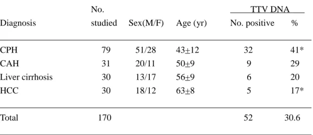
In 170 patients with HCV-related chronic liver diseases, the prevalence of TTV DNA decreased with the severity of chronic liver disease (TABLE III). It was highest in patients with CPH (41%) and was lowest in patients with HCC (17%, P = 0.034). Cases of liver cirrhosis with HCV and TTV coinfection had a higher frequency of past transfusion than those with HCV infection alone (67% vs. 21%, P = 0.09). The mean age of HCC patients possessing both HCV RNA and TTV DNA was significantly lower than that of patients possessing HCV RNA alone (56+5 vs. 64+8 years, P = 0.04).
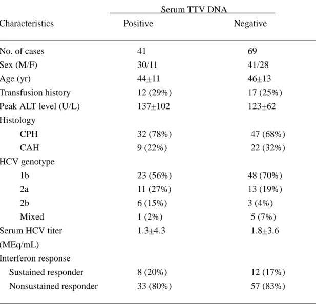
Among 110 patients with chronic hepatitis C, HCV genotypes 1b, 2a, 2b and mixed infection were found in 71, 24, 9, and 6, respectively. When these patients were stratified by the presence or absence of TTV DNA, there was no significant difference in clinicopathological features including gender distribution, mean age, percentage of transfusion history, mean peak serum ALT level, histological severity, and distribution of HCV genotypes (TABLE IV). Although patients with HCV and TTV coinfection had a lower mean serum HCV RNA level than those with HCV infection alone, the difference was not statistically significant (P = 0.5). In addition, the biochemical response to interferon-alfa therapy did not differ between patients with and without TTV infection (TABLE III). The sustained response rate was 20% and 17% in
patients with TTV DNA and in those without TTV DNA, respectively.
Of the 41 patients with HCV and TTV coinfection before initiation of interferon therapy, 29 (70%) lost serum TTV DNA at the end of therapy as determined by PCR assay, and 17 (41%) remained serum TTV DNA negative after stopping therapy for 6 months. However, no correlation was found between the sustained biochemical response and the loss of serum TTV DNA. Six months after stopping therapy, serum TTV DNA was not detectable in 4 of 8 patients (50%) who had sustained biochemical responses compared to 13 of 33 patients (39%) who did not respond (P=0.9). On the contrary, a correlation was seen between sustained biochemical response and sustained loss of serum HCV RNA (Fig. 1).
DISCUSSION
Although a transfusion-transmissible flavi-like RNA virus, GB virus-C (GBV-C) or hepatitis G virus (HGV) [Simons et al., 1995; Linnen et al., 1996], has been claimed to be associated with fulminant and chronic hepatitis in initial reports [Yoshiba et al., 1995], most of the subsequent studies indicated that GBV-C does not cause liver disease as do classical hepatitis viruses [Kao et al., 1996a; 1997; Wang et al., 1996; Alter et al., 1997]. And thus, the search of a new hepatitis virus goes on. A novel virus in association with hepatitis flares was discovered from a patient with posttransfusion hepatitis of unknown etiology [Nishizawa et al., 1997]. The virus has been characterized recently and is similar to the Circoviridae [Mushahwar et al., 1999]. TTV may replicate in liver cells, because its DNA is detected in the liver in titers 10 to 100 times higher than in the corresponding serum from some patients with chronic non-A~E hepatitis [Okamoto et al., 1998]. These findings render TTV an attractive candidate virus in causing liver disease. In contrast, subsequent studies have challenged the initial observations, and an overwhelming body of evidence has indicated that TTV infection is not associated with liver damage [Charlton et al., 1998; Naoumov et al., 1998; Simmonds et al., 1998; Prati et al., 1999].
Although reliable serologic assays are not available, previous studies based on PCR procedures to detect TTV DNA in serum samples from different populations have shown that the virus is transfusion-transmissible, globally distributed and can induce persistent viremia in humans [Okamoto et al., 1998; Simmonds et al., 1998; Prati et al., 1999]. In general, TTV is common in populations at risk of infection with blood-borne viruses, such as hemophiliacs or patients on maintenance hemodialysis, and abusers of intravenous drugs [Hohne et al., 1998; Okamoto et al., 1998; Poovorawan et al., 1998; Takayama et al., 1999]. However, transmission modes other than parenteral routes have been suggested due to the high prevalence of TTV viraemia in healthy population [Zuckerman 1999].
Many hepatitis viruses share the same modes of transmission, thus multiple viral infection may occur in a given patient [Pontisso et al., 1993]. Coinfection of TTV has been frequently observed in patients with chronic hepatitis B and C [Naoumov et al., 1998]. Taking advantage of the extremely common chronic HBV and HCV infections in Taiwan, we investigated the presence of simultaneous TTV infection in HBV and HCV carriers. In the present study, we consistently found a significantly higher overall prevalence of TTV infection in patients with chronic hepatitis B (22%) or C (37%) than in healthy adults (10%) [Kao et al., in press], implying that HBV, HCV and TTV may share common modes of transmission. By contrast, the prevalence in asymptomatic HBsAg carriers (12%) was similar to that in the general population
(TABLE I). These findings are not unanticipated because most HBsAg carriers in Taiwan contracted HBV infection during their perinatal periods or early childhood [Chen 1987], and superinfection of TTV may occur thereafter.
Although active infection of TTV is observed frequently in patients with chronic hepatitis B and C, the interaction between TTV and HBV or HCV in such patients remains unclear. The data showed a relatively constant TTV DNA prevalence (16~20%) among patients with HBV-related CPH, CAH, and cirrhosis (TABLE I), and a significantly increased prevalence in patients with HCC compared to asymptomatic HBV carriers (41% vs. 12%, P < 0.001), implying an association between TTV and HCC. However, such a result does not necessarily represent a causative role of TTV in the development of HCC among HBV carriers. The comparable mean age between HCC patients with both HBsAg and TTV DNA and those with HBsAg alone (52+13 vs. 56+12 years, P = 0.2) supported this interpretation. Accordingly, the higher prevalence of TTV infection in HCC patients may simply reflect greater chances of parenteral and non-parenteral exposure to TTV during their illness than was experienced by asymptomatic HBV carriers.
Among patients with HBV-related CPH and CAH, the clinical and histological features were comparable between those with and without TTV coinfection (TABLE II). Previous studies have demonstrated that HCV superinfection could exert a suppressive or inhibitory effect on the replication of preexisting HBV [Pontisso et al., 1993]. Whether this phenomenon also holds true for TTV infection is unknown. In this study, there was a tendency of lower serum HBV DNA levels in patients with both HBV and TTV infection than those with HBV infection alone (2986+4703 pg/mL vs. 5860+6175 pg/mL, P = 0.066). Taken together, these observations suggested that TTV may have no influence on the clinicopathological course of chronic HBV infection.
Contrary to the patients with HBV infection, in those with chronic HCV infection, the prevalence of serum TTV DNA decreased from 41% steadily to 17% among patients with CPH, CAH, LC and HCC (TABLE III). The lower TT viremia in HCV-induced end-stage liver disease is not due to the liver status per se, as this did
not happen in those caused by HBV. This steady decrease of viremia from chronic hepatitis to HCC was not seen in GBV-C either (data not shown). And thus, there might be specific interactions between TTV and HCV. Perhaps HCV may suppress the replication of TTV in a given patient as in the situation of HCV superinfection on top of HBV carriage [Pontisso et al., 1993].
In chronic hepatitis C, TTV infection did not influence the severity of liver disease (TABLE IV). Serum ALT levels and histological severity in patients with TTV coinfection did not differ from those in patients with HCV infection alone. This
result is consistent with the view that liver lesions are related to HCV rather than to TTV infection, leading to the suspicion that TTV has little or no pathogenecity. This is further confirmed by the absence of correlation between serum TTV DNA positivity and normalization of serum ALT level during and after interferon alfa therapy (Fig. 1). In contrast, serum HCV RNA positivity was associated closely with the biochemical response, indicating the hepatitis activity in these patients was related primarily to HCV infection (Fig. 1).
The influence of TTV coinfection on the HCV response to interferon-alfa therapy is unknown. Our results showed that the sustained response rate to interferon alfa therapy was similar between chronic hepatitis C patients with and without TTV infection, suggesting TTV does not interfere the HCV response to interferon alfa. In addition, the sustained virological response rate of TTV to interferon alfa remains virtually unexplored. Our data showed that 29 of 41 chronic hepatitis C patients (70%) with TTV coinfection lost their serum TTV DNA at the end of interferon alfa therapy, and 17 (41%) had a sustained virological remission after stopping therapy for 6 months. These facts indicated that TTV is actually an interferon-sensitive virus, and if proven to be pathogenic, further studies to better define the optimal dose and duration of interferon usage are then needed.
In summary, these results showed that in Taiwan TTV infection is frequent in patients with chronic hepatitis B or C; however, coinfection of TTV has no effect on the clinicopathological status of chronic hepatitis B or C and the therapeutic response to interferon alfa. The natural course of chronic TTV infection and whether its coinfection affects the natural history of chronic type B or type C hepatitis await long-term longitudinal studies.
ACKNOWLEDGEMENTS
This study was supported by grants from the Department of Health, and the National Science Council, Executive Yuan, TAIWAN.
REFERENCES
Alter HJ (1994): Transfusion transmitted hepatitis C and non-A, non-B, non-C. Vox Sanguinis 67:19-24.
Alter HJ, Nakatsuji Y, Melpolder J, Wages J, Wesley R, Shih JW, Kim JP (1997): The incidence of transfusion-associated hepatitis G virus infection and its relation to liver disease. . New England Journal of Medicine 336:747-754.
Charlton M, Adjei P, Poterucha J, Zein N, Moore B, Therneau T, Krom R, Wiesner R (1998): TT-virus infection in North American blood donors, patients with fulminant hepatic failure, and cryptogenic cirrhosis. Hepatology 28:839-842. Chen DS (1987): Hepatitis B virus infection, its sequelae, and prevention in Taiwan.
In: Okuda K, Ishak KG, eds. Neoplasms of the Liver. Tokyo: Springer-Verlag, p.p. 71-80.
Chen DS, Kuo G, Sung JL, Lai MY, Sheu JC, Chen PJ, Yang PM, Hsu HM, Chang MH, Chen CJ, Hahn LC, Choo QL, Wang TH, Houghton M (1990): Hepatitis C virus infection in an area hyperendemic for hepatitis B and chronic liver disease: the Taiwan experience Journal of Infectious Diseases 162:817-822.
Hohne M, Berg T, Muller AR, Schreier E (1998): Detection of sequences of TT virus, a novel DNA virus, in German patients. Journal of General Virology 79 :2761-2764.
Kao JH, Chen PJ, Yang PM, Lai MY, Sheu JC, Wang TH, Chen DS (1992): Intrafamilial transmission of hepatitis C virus: the important role of infections between spouses. Journal of Infectious Diseases 166:900-903.
Kao JH, Chen PJ, Lai MY, Yang PM, Sheu JC, Wang TH, Chen DS (1995): Genotypes of hepatitis C virus in Taiwan and the progression of liver disease. Journal of Clinical Gastroenterology 21:233-237.
Kao JH, Chen PJ, Chen DS (1996a): GBV-C in the aetiology of fulminant hepatitis. Lancet 347:120.
Kao JH, Lai MY, Hwang YT, Yang PM, Chen PJ, Sheu JC, Wang TH, Hsu HC, Chen DS (1996b): Chronic hepatitis C without detectable anti-hepatitis C antibodies by second generation assay: a clinicopathologic study. Digestive Disease and Science 41;161-165.
Kao JH, Chen PJ, Lai MY, Chen W, Liu DP, Wang JT, Shen MC, Chen DS (1997): GB virus-C/hepatitis G virus infection in an area endemic for viral hepatitis, chronic liver disease, and liver cancer. Gastroenterology 112:1265-1270.
Kao JH, Chen PJ, Lai MY, Chen W, Chen DS (1998): Effects of GB virus-C/hepatitis G virus on hepatitis B and C viremia in multiple hepatitis virus infections. Archives of Virology 1998;143:797-802.
Kao JH, Chen W, Hsiang SC, Chen PJ, Lai MY, Chen DS: Prevalence and implication of TT virus infection: minimal role in patients with non-A~G hepatitis in Taiwan. Journal of Medical Virology (in press).
Kwok S, Higuchi R (1989): Avoiding false positive with PCR. Nature 339:237-238. Linnen J, Wages J, Zhang-Keck ZY, Fry KE, Krawczynski KZ, Alter H, Koonin E,
Gallagher M, Alter M, Hadziyannis S, Karayiannis P, Fung K, Nakatsuji Y, Shih JW, Young L, Piatak M, Hoover C, Fernandez J, Chen S, Zou JC, Morrris T, Hyams KC, Ismay S, Lifson JD, Hess G, Foung SKH, Thomas H, Bradley D, Margolis H, Kim JP (1996): Molecular cloning and disease association of hepatitis G virus: a transfusion-transmissible agent. Science 271:505-508.
Mushahwar IK, Erker JC, Muerhoff AS, Leary TP, Simons JN, Birkenmeyer LG, Chalmers ML, Pilot-Matias TJ, Dexai SM (1999): Molecular and biophysical characterization of TT virus: Evidence for a new virus family infecting humans. Proceedings of the National Academy of Sciences of the United States of America 96:3177-3182.
Naoumov NV, Petrova EP, Thomas MG, Williams R (1998): Presence of a newly described human DNA virus (TTV) in patients with liver disease. Lancet 352:195-197.
Nishizawa T, Okamoto H, Konishi K, Yoshizawa H, Miyakawa Y, Mayumi M (1997): A novel DNA virus (TTV) associated with elevated transaminase levels in posttransfusion hepatitis of unknown etiology. Biochemical and Biophysical Research Communications 241:92-97.
Okamoto H, Nishizawa T, Kato N, Ukita M, Ikeda H, Iizuka H, Miyakawa Y, Mayumi, M (1998): Molecular cloning and characterization of a novel DNA virus (TTV) associated with posttransfusion hepatitis of unknown etiology. Hepatology Research 10:1-16.
Pontisso P, Ruvoletto MG, Fattovich G, Chemello L, Gallorini A, Ruol A, Alberti A (1993): Clinical and virological profiles in patients with multiple hepatitis virus infections. Gastroenterology 105:1529-1533.
Poovorawan Y, Theamboonlers A, Jantaradsamee P, Kaew-in N, Hirsch P, Tangkitvanich P (1998): Hepatitis TT virus infection in high-risk groups. Infection 26:355-358.
Prati D, Lin YH, De Mattei C, Liu JK, Farma E, Ramaswamy L, Zanella A, Lee H, Rebulla P, Allain JP, Sirchia G, Chen B (1999): A prospective study on TT virus infection in transfusion-dependent patients with beta-thalassemia. Blood 93:1502-1505.
Simmonds P, Davidson F, Lycett C, Prescott LE, MacDonald DM, Ellender J, Yap PL, Ludlam CA, Haydon GH, Gillon J, Jarvis L M (1998): Detection of a novel
DNA virus (TTV) in blood donors and blood products. Lancet 352:191-195. Simons JN, Leary TP, Dawson GJ, Pilot-Matias TJ, Muerhoff AS, Schlauder GG,
Desai SM, Mushahwar IK (1995): Isolation of novel virus-like sequences associated with human hepatitis. Nature Medicine 1:564-569.
Takayama S, Yamazaki S, Matsuo S, Sugii S (1999): Multiple Infection of TT Virus (TTV) with Different Genotypes in Japanese Hemophiliacs. Biochemical and Biophysical Research Communications 256:208-211.
Wang JT, Tsai FC, Lee CZ, Chen PJ, Sheu JC, Wang TH, Chen DS (1996): A prospective study of transfusion-transmitted GB virus C infection: similar frequency but different clinical presentation compared with hepatitis C virus. Blood 1996;88:1881-1886.
Yoshiba M, Okamoto H, Mishiro S (1995): Detection of the GBV-C hepatitis virus genome in serum from patients with fulminant hepatitis of unknown aetiology. Lancet 346:1131-1132.
Zuckerman AJ. The acronym TTV. Transfusion transmitted virus. Lancet 1999;353:932.
TABLE I. Prevalence of Serum TT Virus (TTV) DNA in 320 Hepatitis B Surface Antigen Carriers with Different Liver Diseases
_____________________________________________________________________ No. TTV DNA Diagnosis studied Sex(M/F) Age (yr) No. positive % _____________________________________________________________________ Asymptomatic carrier 100 54/46 30+7 12 12* CPH 50 39/11 35+8 10 20 CAH 50 42/8 41+9 8 16 Liver cirrhosis 50 40/10 46+11 10 20 HCC 70 52/18 55+13 29 41* _____________________________________________________________________ Total 320 69 21.5 _____________________________________________________________________ * P < 0.001
TABLE II. Demographic and Clinical Data of 100 Cases of Chronic Hepatitis B with and without TT Virus (TTV) DNA
_____________________________________________________________________ Serum TTV DNA
Characteristics Positive Negative
_____________________________________________________________________
No. of cases 18 82
Sex (M/F) 13/5 68/14
Age (yr) 37+9 38+10
Transfusion history 1 (6%) 3 (4%) Peak ALT level (U/L) 183+77 166+52 Histology CPH 10 (56%) 40 (49%) CAH 8 (44%) 42 (51%) Serum HBV titer 2986+4703* 5860+6175* (pg/mL) _____________________________________________________________________ * P = 0.066
TABLE III. Prevalence of Serum TT Virus (TTV) DNA in 170 Hepatitis C Virus Carriers with Different Liver Diseases
_____________________________________________________________________ No. TTV DNA Diagnosis studied Sex(M/F) Age (yr) No. positive % _____________________________________________________________________ CPH 79 51/28 43+12 32 41* CAH 31 20/11 50+9 9 29 Liver cirrhosis 30 13/17 56+9 6 20 HCC 30 18/12 63+8 5 17* _____________________________________________________________________ Total 170 52 30.6 _____________________________________________________________________ * P = 0.034
TABLE IV. Demographic and Clinical Data of 110 Cases with Chronic Hepatitis C with and without TT Virus (TTV) DNA
_____________________________________________________________________ Serum TTV DNA
Characteristics Positive Negative
_____________________________________________________________________
No. of cases 41 69
Sex (M/F) 30/11 41/28
Age (yr) 44+11 46+13
Transfusion history 12 (29%) 17 (25%) Peak ALT level (U/L) 137+102 123+62 Histology CPH 32 (78%) 47 (68%) CAH 9 (22%) 22 (32%) HCV genotype 1b 23 (56%) 48 (70%) 2a 11 (27%) 13 (19%) 2b 6 (15%) 3 (4%) Mixed 1 (2%) 5 (7%) Serum HCV titer 1.3+4.3 1.8+3.6 (MEq/mL) Interferon response Sustained responder 8 (20%) 12 (17%) Nonsustained responder 33 (80%) 57 (83%) _____________________________________________________________________
FIGURE LEGEND
Figure 1. Changes of serum alanine aminotransferase (ALT) levels, HCV RNA and TTV DNA in 3 representative patients having chronic hepatitis C coinfected with TTV and receiving treatment with 3 million units of interferon alfa (IFN) thrice weekly for 24 weeks. A correlation was seen between serum ALT levels and serum HCV RNA response; however, such correlation was not found for serum TTV DNA.