行政院國家科學委員會專題研究計畫 成果報告
肝栓塞 TACE 手術後之血動力研究
計畫類別: 個別型計畫 計畫編號: NSC91-2213-E-002-125-YF 執行期間: 91 年 08 月 01 日至 93 年 07 月 31 日 執行單位: 國立臺灣大學工程科學及海洋工程學系暨研究所 計畫主持人: 許文翰 報告類型: 精簡報告 報告附件: 國際合作計畫研究心得報告 處理方式: 本計畫可公開查詢中 華 民 國 93 年 12 月 9 日
肝栓塞 TACE 手術後之血動力研究
Liver Hemodynamic studies on after patient’s hepatic transarterial
chemoembolisation (TACE)
執行期間:91 年 8 月 1 日至 92 年 12 月 31 日
計劃編號:91-2213-E-002-125-YF
計劃主持人:許文翰 國立台灣大學工程科學及海洋工程系所 教授
Abstract
For the treatment of microcirculation disorders, the three-dimensional representation of the hepatic circulation is crucial for the physian. Such a representation is provided in this paper for an exploration of the blood flow, blood volume and exchange between sinusoids and hepatocytes in the hepatic acinus model.
1. Introduction
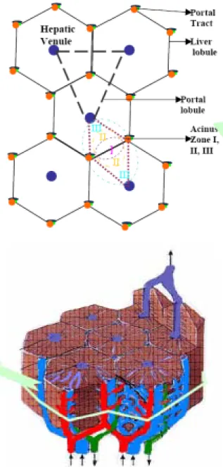
In this paper, we analyse the 3-D mixing mechanism of blood from hepatic arteriole and portal venule in the hepatic vascular bed [1]. Also, emphasis given to the flow patterns from the space of sinusoid upto the hepatic venule drainage. In the subsequent paragraphs, focus is given to the hepatic circulation especially on hepatic arterioles and hepatic / portal venules. Selle et al., [1] presented the analysis and visualization of hepatic vasculature and provided the shading techniques emphasizing the curvature of vessles. In the case of liver failure, one promising alternative is the development of an extracorporeal bioartificial liver (BAL). This is essentially a bioreactor containing cultured hepatocytes that function as an extracorporeal liver on a temporary basis [2] as liver cells has the capacity to regenerate itself [3]. Hence for a quantitative diagnosis, surgical planning and post operative treatments, a precise knowledge of microcirculation is important. A detailed hepatic acinus structure along with their zones is shown in Figure1.
Figure 1. Simple liver Acinus
2. Modeling
The parallelization of the liver into lobules, together with the complex hepatic vascular tree, provides the microscopic picture about the flow distribution. At the most gross level, there is an unusual presence of two vascular
inputs, the portal and arterial flows, rather than the single arterial input. At the microscopic level, the acinar arrangement of the vascular system creates a unique series of microenvionments, which are acknowledged to be of paramount importance in controlling the functional characteristics of the parenchymal cells. In the current study we considered a simple liver acinus, which represents a small parenchymal mass consisting of pre/terminal hepatic arteriole, pre/terminal portal venule along with fenestrated endothelial cells and hepatic venule. The present study focuses on the acinus structure of the liver. Usually, the acinus mass is divided into three zones. The representation of Acinus structure is show in Figure 2
Figure 2. Acinus structure
3. Methods
We developed a three-dimensional computational fluid dynamics model of an acinus structure using commercially available software, CFD-ACE (CFD Research Corp., Huntsville, AL). The model solves the full Navier-Stokes equations for unsteady flow of a Newtonian fluid using a finite volume approach. The idealized geometry consists of rigid micrometer cylinders simulating a portal tract, space of sinusoid and hepatic venule. No slip conditions are imposed on the walls of the cylinder. Fully developed flow distribution is assumed for the hepatic arteriole and portal venule.
4. Results and Discussion
The hepatic arteriole flow has eight times greater than the flow rate of portal venule interms of mass flow, velocity and pressure. But the transmittent activity is initiated by the hepatic arteriole due to the sphincter mechanism present at the end of each arteriole especially in liver. This mechanism is analysed in detail for the unsteady case. Also when this sphincter is closed there is normal portal venule flow. The functional working difference of zone 1, 2 and 3 are analysed interms of mass flow rate, velocity and pressure distribution.
Acknowledgement
The authors acknowledge support under NSC Grant NSC 91-2213-E-002-125-YF. References
[1] Selle D., Preim, B., Schenk A., Peitgen, H. O.: Analysis of vasculature for liver surgical planning, IEEE Transactions on Medical Imaging 21(11): 2002. [2] Ledezma G. A., Folch A., Bhatia S. N.,
Balis U. J., Yarmush M. L., Toner M: Numerical Model of Fluid Flow and Oxygen Transport in a Radial-Flow Microchannel Containing Hepatocytes. Journal of Biomechanical Engineering 121: 58-64 , 1999.
[3] Michalopoulos G.K., DeFrances M.C.: Liver regeneration. Science, 276: 60, 1997.