國
立
交
通
大
學
材料科學與工程學系
博 士 論 文
幾丁聚醣複合物奈米結構與可控制藥物釋放行
為之研究
Study on the nanostructural evolution and
controlled drug release behavior of chitosan
nanocomposites
研究生:劉昆和
指導教授:陳三元 博士
幾丁聚醣複合物奈米結構與可控制藥物釋放行為之研究
Study on the nanostructural evolution and controlled drug
release behavior of chitosan nanocomposites
研 究 生:劉昆和 Student:Kun-Ho Liu
指導教授:陳三元 Advisor:San-Yuan Chen
國 立 交 通 大 學
材 料 科 學 與 工 程 研 究 所
博 士 論 文
A ThesisSubmitted to Department of Materials Science and Engineering College of Engineering
in Partial Fulfillment of the Requirements for the Degree of
Doctor of Philosophy
in Materials Science and Engineering
January 2009
Hsinchu, Taiwan, Republic of China
I
中文摘要
幾丁聚醣近來在醫藥應用方面受引起很大的注意,因為具有很好
的生物相容性使得幾丁聚醣廣泛應用在眼科的局部治療、植入物以及
注射藥物領域。而且,由於幾丁聚醣能夠被人體體內酵素(特別是溶
解酵素)所代謝,因此也被認為是生物可分解性材料。另外,幾丁聚
醣表面帶正電荷的性質使其擁有很好的生物吸附性,在應用上能夠長
時間的停留在體內以提升其利用率。幾丁聚醣也擁有能夠幫助傷口癒
合以及抗菌等優點。最重要的是,幾丁聚醣大量存在於自然界中讓製
作成本變得非常便宜。
幾丁聚醣具有陽離子特性使它能夠形成具有酸鹼敏感與電場敏
感特性的聚電解質水膠。本論文的第一部分著重在討論具有電敏感性
的幾丁聚醣/奈米黏土複合水膠的合成(第四章)。表面帶負電的黏土顆
粒能夠當作幾丁聚醣的交鍊劑來增加複合物的交鍊程度,並且提升其
機械強度、膨潤收縮特性和疲勞特性。實驗結果中可以發現,比起純
幾丁聚醣來說,複合物在電場不斷開關的循環刺激下能夠擁有較佳的
抗疲勞特性。除此之外,我們也利用另一種無機黏土(蒙脫土)來增加
幾丁聚醣的抗疲勞特性以及長時效穩定藥物釋放行為(第五章)。脫層
後的蒙脫土也能夠當作幾丁聚醣的交鍊劑,結果發現在電場的刺激
下,不同的交鍊程度會強烈影響維他命
B12 的釋放行為。當蒙脫土
II
的含量增加,藥物擴散指數
n 值和複合物的對於電場的反應性將會減
低。另外,具有較高蒙脫土含量的複合物在電場不斷開關的循環刺激
下電反應性會減弱,但是能夠擁有較佳的抗疲勞特性。除此之外,在
第六章中,利用乳化法和溶膠水膠法的製程可以成功製備出顆粒尺寸
介於
50-130 奈米的幾丁聚醣-四乙氧基矽複合奈米顆粒。四乙氧基矽
經過水解縮合反應,能夠和幾丁聚醣形成網狀結構,進而增加藥物的
包覆效率,同時也提供較佳的機械強度,使其在電場關閉時不讓藥物
流出載體。
幾丁聚醣因為具有氫氧根和氨根的基團,因此可以很容易被用來
做為改質的處理,使其在生藥應用上具有很大的利用價值。雙性幾丁
聚醣同時擁有親水性和疏水性的特性使其能夠在水溶液中自組裝形
成類似微胞狀的團聚物,這樣的特性主要是由於疏水基團分子或疏水
基團分子間的作用力所造成的。在第七章中,我們利用親水性甲基羧
基基團與疏水性己醯基團的嫁接物,製備出一種新穎的幾丁聚醣衍生
物,能夠在水溶液中自組裝形成空心的奈米膠囊。同時也藉由實驗方
法鑑定出此雙性幾丁聚醣衍生物的臨界團聚濃度和表面電性。而此自
組裝特性和結構穩定性主要來自於分子間作用力和熱力學的作用。而
更有趣的發現指出,改變不同的疏水基團鏈長(變化從 2 個碳到 12 個
碳)或嫁接量,可以操控幾丁聚醣奈米團聚物的自組裝行為(第八章)。
III
從結果中可以發現,嫁接量和碳鏈數的乘積值(X
DH× X
Cn),能夠用來
當作一個指標來決定奈米團聚物的結構: 當乘積超過 1.5 時,奈米團
聚物會從實心的結構轉變成空心的結構變化。在藥物傳遞應用中,抗
癌藥物小紅莓在此奈米結構下被證明可以大大提升吸附性以及包覆
率。
關鍵詞
: 幾丁聚醣、奈米空心膠囊、電刺激、智慧型釋放、黏土有機
混合物
IV
Abstract
Chitosan (CS) is currently receiving a great of interest for medical and pharmaceutical application. Indeed, it is known for being biocompatible allowing it use in various medical applications such as topical ocular applications, implantation or injection. Moreover, chitosan is metabolised by certain human enzymes, especially lysozyme, and is considered as biodegradable. Due to its positive charges at physiological pH, chitosan is also bioadhesive, which increases retention at the site of application. Chitosan also promotes wound-healing and has bacteriostatic effects. Finally, chitosan is very aboundant, and its production of low cost and ecologically interesting.
CS with cationic characteristics is capable to form polyelectrolyte hydrogel that owing pH-sensitivity and electric-sensitivity. The first part in this thesis is focusing on the synthesis of electrostimulus-responsive hybrid composites composed of chitosan (CS) and clay (chapter 4). The addition of negatively charged clay as an ionic cross-linker strongly affect the cross-linking density as well as the mechanical property, swelling–deswelling behavior and fatigue property of the hybrids. Compared with pure CS, a significant improvement in the anti-fatigue property against cyclic electric stimulations of the hybrid was found. In addition, an inorganic phase, MMT, was incorporated in the CS matrix to enhance the anti-fatigue property and corresponding long-term stable release kinetics (chapter 5). The exfoliated silica nanosheets are able to act as cross-linkers to form a network structure between the CS and MMT, and this difference in the cross-linking density strongly affects the release of vitamin B12 under electrostimulation. Further
V
increasing the MMT content reduced both the diffusion exponent n and the responsiveness of the nanohydrogel to electrostimulation. A consecutively repeated ‘‘on” and ‘‘off” operation shows that the electroresponsiveness of the nanohydrogel with higher MMT concentrations was reduced, but its anti-fatigue behavior was considerably improved. In addition, nanoparticles (NPs) with particle size of 50-130 nm composed of chitosan (CS) and tetraethyl orthosilicate (TEOS) were prepared through emulsion and sol-gel process (chapter 6). TEOS that hydrolyzed and condensed to form network structure with the CS improved the drug loading capacity and also provided the mechanical enhancement of the nano-sphere to restrict drug release when the electric field was switched off.
In addition, CS appears to be more useful in biomedical applications because of its both hydroxyl and amino groups that can be easily modified. Amphiphilic CS consisting of hydrophilic and hydrophobic segments can form micelle-like self-assemblies due to non-covalent association arising from intra- and/or intermolecular interactions among hydrophobic segments in aqueous media. In chapter 7, a new type of amphiphilic chitosan, which was synthesized through the use of both hydrophilic carboxymethyl and hydrophobic hexanoyl substitutions, was employed to self-assemble into a hollow nanocapsule in an aqueous environment. Critical aggregation concentration (cac) and zeta potential were experimentally identified for the amphiphilic chitosan (CHC). The self-assemble mechanism, together with the corresponding nanostructural stability, of this unique CHC nanocapsule was also proposed in terms of intermolecular interaction and thermodynamic reason. Further, a more interesting finding where through the use of acyl chain of varying chain lengths, from C2 to C12, for intramolecular substitution, the
VI
self assembly behavior and the resulting nanostructure of the chitosan nano-aggregate can be well manipulated (chapter 8). It was found that the critical value of (XDH × XCn), i.e., a product of “degree of acyl substitution” and “carbon number of acyl chain”, can be employed as an indicator for structural variation of the nano-aggregates: when (XDH × XCn) exceeded 1.5, the architecture of the nano-aggregates underwent a structural transformation from solid nano-particle to hollow nano-capsules. An improved affinity and capacity of doxorubicin drug encapsulation can be technically designed according to the nature of the resulting nanocapsules for controlled delivery.
Keywords: chitosan,, nanocapsule, electric-stimuli, controlled release, clay/organic composites.
VII
Acknowledgments
首先我要感謝我的父母,因為他們無怨無悔的支持以及不求回報的付出,才 讓我能夠順利完成博士學位。也感謝我的家人在我求學生活中面臨低潮的時候, 願意鼓勵我激勵我。此外,還要感謝我的指導教授陳三元老師以及劉典謨老師, 在研究過程中,提供我最大的幫助以及最重要的建議與指導,讓我能夠更深入了 解科學的涵意與研究的精神。最後感謝所有實驗室的同學以及學弟妹們,因為有 你們,讓我的研究生活變得更多采多姿,也讓我的生命更加豐富,謝謝你們!VIII
Contents
中文摘要………..Ⅰ Abstract………..Ⅳ Acknowledgments………Ⅶ Contents………Ⅷ Figure Captions………ⅩⅠ Table Captions………ⅩⅤ Chapter 1. Introduction………...1Chapter 2. Literature Review and Theory………...5
2.1 Environmental sensitive of chitosan hydrogel………..5
2.2 Inorganic clay-organic (chitosan) hybrid composites………10
2.3 Self-assembles of modified chitosan derivatives………...12
2.4 Chitosan nanoaggregates for drug delivery system……….….14
Chapter 3. Experimental methods………..17
3.1 Flowchart of Experiment Process………..………..17
3.2 Materials……….….18
3.3 Characteristics Analysis………19
Chapter 4. Effect of Clay Content on Electro-stimuli Deformation and Volume Recovery Behavior of Clay-chitosan Hybrid Composite………...25
4.1 Introduction………...………….25
4.2 Preparation of CS-clay films………..…28
4.3 Chemical interaction of the CS and clay in the hybrid………...29
4.4 Degree of cross-linking in the hybrid………...31
IX
4.6 Cyclic deformation and recovery of hybrid films………..…..39
Chapter 5. Drug release behavior of chitosan–montmorillonite nanocomposite hydrogels following electrostimulation…….44
5.1 Introduction………..44
5.2 Preparation of CS-MMT nanohydrogels………..46
5.3 Structural Characterization……….48
5.4 Drug release behavior under an applied electric field………51
5.5 Repeating electrical-stimulation on release behavior………55
Chapter 6. Electrical-Sensitive Nanoparticle Composed of Chitosan and TEOS for Controlled Drug Release………...61
6.1 Introduction………..61
6.2 Fabraction of drug-loaded and non-loaded CS/TEOS nanoparticles.64 6.3 Results and Discussion……….…66
Chapter 7. Self-Assembled hollow nanocapsule from amphiphatic carboxymethyl-hexanoyl chitosan as drug carrier………75
7.1 Introduction………..75
7.2 Synthesis of CHC………77
7.3 Preparation of drug-loaded and non-loaded CHC hollow nanocapsules………..78
7.4 Formation of CHC Nano-aggregates……….80
7.5 Nanostructural development of the CHC Nano-aggregates………....87
7.6 DOX Loading and Release Behavior of CHC Nanocapsules...……91
Chapter 8. Hydrophobic Effect on the Structural Evolution of Acylated-Carboxymethyl Chitosan and its Self-Assemble Forming of Doxorubicin-loading Nanocapsule…………....94
X
8.2 Synthesis of carboxymethyl chitosan (CC)………96
8.3 Synthesis of acylated carboxymethyl-chitosan (ACC)………..…96
8.4 Preparation of drug-loaded and non-loaded ACC nano-aggregates ...………97
8.5 Structural Characteristics of ACC………..101
8.6 Self-assemble behavior of ACC………103
8.7 Molecular configuration of water in ACC……….………..106
8.8 Self-Assembly Map………...111
8.9 Doxorubicin loading………115
Chapter 9. Conclusions...117
9.1 electric-sensitive of clay/chitosan hybrids……….117
9.2 electric-sensitive of TEOS/CS nanoparticles………117
9.3 Acylated-carboxymethyl chitosan nanocapsule………118
Reference...119
Curriculum Vitae...127
XI
Figures Captions
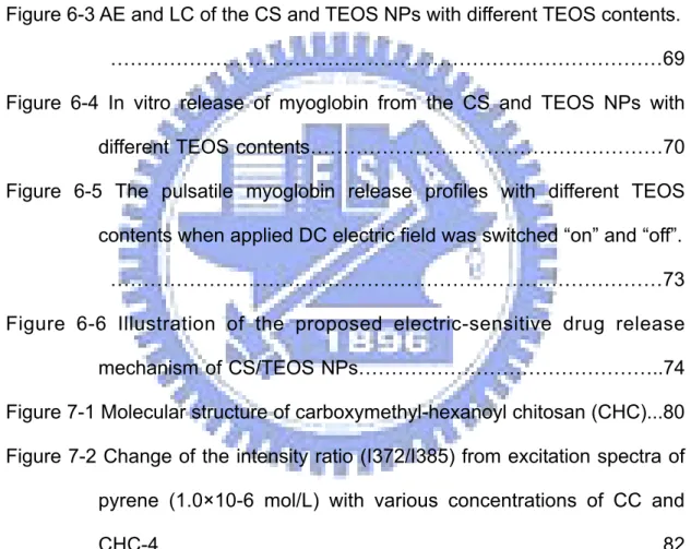
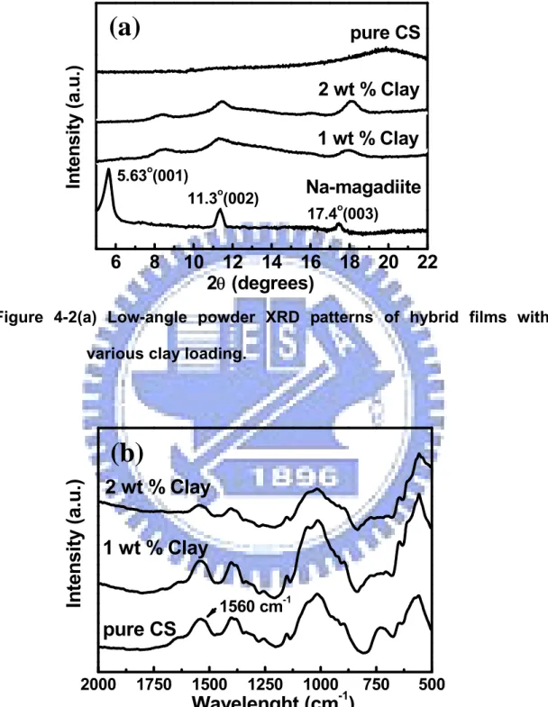
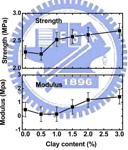
Figure 4-1. Zeta-potential profile of nanoclay (magadiite) in various pH values ………29 Figure 4-2(a) Low-angle powder XRD patterns of hybrid films with various clay loading……….31 Figure 4-2(b) FTIR spectra of hybrid films with various clay loading………….31 Figure 4-3 Mechanical properties of hybrid film. Initial cross-sectional area (10 mm2) was used for calculating the modulus and strength………...32 Figure 4-4 Clay content dependencies of the number of cross-linked chain (N*) ………34 Figure 4-5(a) Swelling kinetics of hybrid film with different Cclay at pH 7.4….35 Figure 4-5(b) Plots of ln(W0/Wt) against time of hybrid film with different Cclay at pH 7.4………..36 Figure 4-5(c) Equilibrium swelling ratio of hybrid film with different Cclay at pH 7.4……….…37 Figure 4-6(a) Deswelling behavior in 1.5h of hybrid film with different Cclay
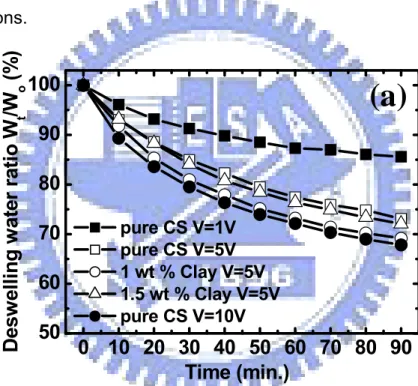
under applied voltage of 1, 5, and 10V………38 Figure 4-6(b) Deswelling water ratio in 1.5h of hybrid film with different Cclay under applied voltage of 1, 5, and 10V………39 Figure 4-7(a) Relative ratio of swelling after cyclic on-off switching of
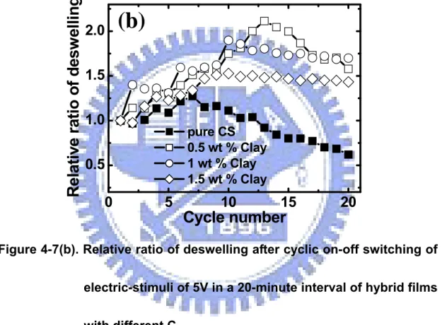
electric-stimuli of 5V in a 20-minute interval of hybrid films with different Cclay……….40 Figure 4-7(b). Relative ratio of deswelling after cyclic on-off switching of electric-stimuli of 5V in a 20-minute interval of hybrid films with different Cclay……….41
XII
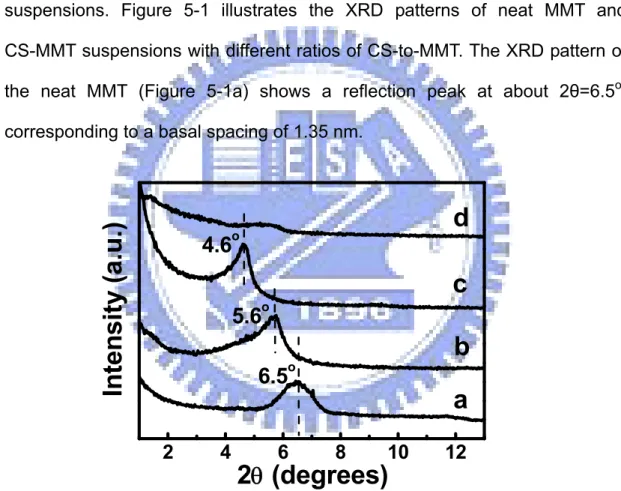
Figure 4-8. Weight changes of pure chitosan and hybrid film with 1 wt % clay addition under cyclic on-off switching of electric-stimuli of 5V…….43 Figure 5-1 Low-angle powder XRD patterns of (a) neat MMT and the CS/MMT nanocomposites with the CS and MMT ratios of (b) 0.5:1 (c) 2:1 and (d) ball milling for 24 h………48 Figure 5-2 Time dependence of optical absorbance A (relative to its initial value,
A0) of the (a) exfoliated MMT (b) original MMT and (c) monolayer or bilayer intercalated-MMT suspensions………50 Figure 5-3 Side-angle powder XRD patterns of pure CS, CS-MMT1 and
CS-MMT4………51 Figure 5-4 (a) Time-dependent cumulative release curves of vitamin B12 from the nanohydrogels upon an applied electric voltage, showing the different release profiles with different CMMT. (b) Dependences of the loaded MMT contents on the diffusion exponent n and voltage-induced swelling ratios of the nanohydrogels. (The filled boxes indicated the diffusion exponent n and the empty circles indicated the V-induced swelling ratio)………53 Figure 5-5 Real (…) and the ideal (—) pulsatile drug release profiles of pure CS hydrogels as an electric field was switched “on” and “off”…….56 Figure 5-6 (a) Standard release rates of drug from the nanohydrogels (CS,
CS-MMT1, CS-MMT2, and CS-MMT4) after cyclic on-off switching operations. (b) Dependences of the loaded MMT contents on the drug standard release rate in the first on-off cycle (filled boxes) and anti-fatigue coefficient of the nanohydrogels (empty circles)……...58 Figure 5-7 Standard release rates of drug from pure CS and CS-MMT2 under cyclic on-off switching of electric-stimuli……….59
XIII
Figure 6-1. The illustrations of the formation of CS and TEOS NPs. (a) the formation of TEOS network structure, (b) the original CS polymer entwined into the network, (c) the formation of NPs by ionic gelation of TPP………..67 Figure 6-2 (a) SEM image of pure CS NPs. The (b) lower- magnification and
higher-magnification TEM image of the CS and TEOS NPs (23%). ………68 Figure 6-3 AE and LC of the CS and TEOS NPs with different TEOS contents.
………69 Figure 6-4 In vitro release of myoglobin from the CS and TEOS NPs with
different TEOS contents………70 Figure 6-5 The pulsatile myoglobin release profiles with different TEOS
contents when applied DC electric field was switched “on” and “off”. ………73 Figure 6-6 Illustration of the proposed electric-sensitive drug release
mechanism of CS/TEOS NPs………..74 Figure 7-1 Molecular structure of carboxymethyl-hexanoyl chitosan (CHC)...80 Figure 7-2 Change of the intensity ratio (I372/I385) from excitation spectra of pyrene (1.0×10⁻⁶ mol/L) with various concentrations of CC and CHC-4………..82 Figure 7-3 Zeta potential of carboxymethyl-hexanoyl chitosan (CHC) with
different DH……….84 Figure 7-4 TEM images of self-aggregates prepared, respectively, from (a) CC
and (b) CHC-4 by ultrasonication in water………..86 Figure 7-5 Fraction of mean size 1 and 2 of CC and CHC nano-aggregates..87 Figure 7-6 Schematic illustration of formation process of CHC hollow
XIV
nanocapsules………..88 Figure 7-7 SEM images of CHC-4 nanocapsules……….91 Figure 7-8 (a) DOX encapsulation efficiency and (b) DOX release profiles of
CHC hollow nanocapsules………....92 Scheme 8-1 Acylation Reaction of carboxymethyl chitosan……….101 Figure 8-1 FTIR spectra of CC, C2-0.5, and C12-0.5……….102 Figure 8-2 Dependence between the DH of ACC as Cn=2, 6 and 12 and (a) the critical aggregation concentration, CAC and (b) ln(CAC), respectively………105 Figure 8-3 DSC curves of CC and ACC derivates (Cn-0.5) measured at water content=200%. Dash lines represent the curve fitting by Lorentzian curve-fitting procedure……….107 Figure 8-4 Dependence between the content of bound water, Wnf, max, and the hydrophobic side chain with different Cn of ACC in DH=0.25 and 0.5……….109 Figure 8-5 Plot of CAC (○) and Wnf, max (■) of ACC derivates with different values of (XDH × XCn)………112 Figure 8-6 TEM images of (a) C2-0.5, (b) C6-0.5, and C6-0.5 after dehydration;
and (d) SEM image of the ACC nano-aggregates after electron-induced rupturing………..113 Figure 8-7 Schematic illustration of formation process of ACC
nano-aggregates………..114 Figure 8-8 DOX encapsulation efficiency of ACC nano-aggregates with various values of (XDH × XCn)………..116
XV
Table Captions
Table 6-1. Kinetics contants (K), and release exponents (n) following linear regression of release of myoglobin from CS and TEOS NPs….72 Table 7-1 Sample Name and Corresponding Estimation of Substitution Degree by 1H-NMR………80 Table 7-2 Characterization of CC and CHC self-assembled hollow
nanocapsules………84 Table 8-1 The preparation conditions and the characteristics of ACC
1
Chapter 1
Introduction
Research on site-specific and temporal control of drug delivery systems is receiving a major impetus towards the development of new and/or improved drug therapies. “Intelligent” drug carriers which released the right amount of drug at the right time and/or at the right place may enable us to precisely control the delivery of drugs. These “smart” drug delivery vehicles would also enable the tailoring of medical treatment to individual patients. However, the precise control of drug levels in the body can only be achieved if the drug carrier responds in a reproducible and predictable fashion to an internal or external chemical, physical or biological stimulus. In addition, the carrier should be non-toxic, non-irritant biodegradable, biocompatible, easy to administer and should not need to be removed when drug-depleted.
Hydrogels have been used extensively in the development of the smart drug delivery systems, since hydrogels containing such ‘sensor’ properties can undergo reversible volume phase transitions or gel–sol phase transitions upon only minute changes in the environmental condition. Many physical and chemical stimuli have been applied to induce various responses of the smart hydrogel systems. Therefore, environment-sensitive hydrogels are ideal candidates for developing self-regulated drug delivery systems. However, the most significant weakness of all these external stimuli-sensitive hydrogels is that their response time is too slow. Thus, fast-acting hydrogels are necessary, and the easiest way of achieving that goal is to make thinner and smaller hydrogels, which indicates that nanotechnology is becoming more important
2
for such high sensitive drug- delivery structure.
Recently, nanotechnology has become a popular term representing the main efforts of the current science and technology. One of the important areas of nanotechnology is “nanomedicine,” which, according to the National Institute of Health (NIH) Nanomedicine Roadmap Initiative, refers to highly specific medical intervention at the molecular scale for diagnosis, prevention and treatment of diseases [1]. The importance of nanotechnology in drug delivery is in the concept and ability to manipulate molecules and supramolecular structures for producing devices with programmed functions. The approach which may come closest to the original intention may probably be found in the application of dispersed nanoparticles loaded with an active ingredient [2]. Within the solid matrix of the particles, the active ingredient can be dissolved, entrapped or encapsulated.
In addition, a significant drawback of most environmentally stimulus-sensitive hydrogels is that the responsivity and reversibility often decrease with both time and number of on–off operation cycles as the gel fatigues considerably. It is necessary to obtain a most suitable composition and viscoelasticity at a certain cross-linking density for optimal responsiveness that the deformation and relaxation properties of hydrogel with cyclic on–off operations. Herein, considerable attention was paid to inorganic–organic hybrid materials because their solid-state properties could be tailored in relation to the nature and relative content of their constitutive components. The strong chemical bonds (covalent or ionic) or interactions such as van der Waals forces, hydrogen bonding, or electrostatic forces, often exist between the organic and inorganic components that usually leads to some novel nanocomposites with improved performance properties.
3
Chitosan (CS), poly- β (1,4)-2-amino-2-deoxy-D-glucose, is the deacetylated product of chitin, poly(N-acetyl-D-glucosamine), a natural polymer found in the exoskeletons of crustaceans and insects and in the cell wall of fungi and microorganisms. The chemically active groups in the chitosan structure are the free amine groups, located in the C2 position of the glucose residue in the polysaccharide chain, and the hydroxyl groups, with both being susceptible to modification [3]. CS, a cationic biopolymer with very low toxicity, has been evaluated extensively as an excipient for drug delivery systems. Hence, in this study, a new class of “electrically-charged” hybrid composite based on chitosan (CS) and clay will be first investigated and developed by taking the advantage of the electrochemical properties, i.e., surface charge, and nanostructural properties, i.e., layer of the clay particles, as illustrated in Chapter 4. Moreover, the effects of various cross-linking degrees caused by different clay content of the hybrid composite on the electrically-stimulated swelling-deswelling behavior, mechanical deformation were elucidated. In Chapter 5, the release kinetics and mechanism of the vitamin B12 in terms of MMT contents were investigated under a given electric-field stimulus. Furthermore, the anti-fatigue behavior with respect to the repeated field stimuli of the resulting nanohydrogel in terms of MMT addition was also elucidated. In chapter 6, nanoparticles (NPs) with particle size of 50-130 nm composed of CS and TEOS were obtained through emulsion and sol-gel process. Therefore, the mechanism of the drug release from the CS/TEOS NPs effectively controlled by electric-stimuli operations was demonstrated in this work.
In Chapter 7, a new type of chitosan hollow structure, i.e., carboxymethyl-hexanoyl chitosan (CHC), which was modified first by hydrophilic carboxymethylation, followed by hydrophobic modification with
4
hexanoyl groups to add amphiphilic character, was employed to study its self-aggregation behavior to form nanocapsule in aqueous solution and nanostructural evolution. The stability of nancapsules and formation mechanism of the CHC macromolecules were explored through the use of critical aggregation concentration (CAC), zeta-potential, electron microscopy, and dynamic light scatter (DLS). By taking the advantage of self-aggregation nature, the CHC was employed to encapsulate doxorubicin (DOX), an anticancer agent of broad spectrum with reasonable therapeutic index and intriguing biological and physicochemical actions, to further understand its loading efficiency and release behavior. Then, in Chapter 8, the structural evolution of the chitosan-based nano-aggregates was systematically investigated in terms of different degrees of acyl substitution (DH) and number of carbon of the acyl ligand (Cn). In addition, the doxorubicin (DOX)-encapsulated capacity of the nano-aggregates of varying degrees of hydrophobicity, in terms of DH and Cn, was also studied.
5
Chapter 2
Literature Review and Theory
2.1 Environmental sensitive of chitosan hydrogelControlled drug delivery systems, which are intended to deliver drugs at predetermined rates for predefined periods of time, have been used to overcome the shortcoming of conventional drug formulations. In fact, it would be most desirable if the drugs could be administered in a manner that precisely matches physiological needs at proper times (temporal modulation) and/or at the proper site (site-specific targeting). In addition, it would be highly beneficial if the active agents were delivered by a system that sensed the signal, and then acted to release the right amount of drug in response. Hydrogels have been used extensively in the development of the smart drug delivery systems. A hydrogel is a network of hydrophilic polymers that can swell in water and hold a large amount of water while maintaining the structure. The drugs can be protected in hydrogel from hostile environments, e.g., the presence of enzymes and low pH in the stomach, and controlled-released by changing the gel structure in response to environmental stimuli. The types of environment-sensitive hydrogels are also called “Intelligent” or “smart” hydrogels. Many physical and chemical stimuli have been applied to induce various responses of the smart hydrogel systems, where the physical stimuli include temperature, electric fields, solvent composition, light, pressure, sound and magnetic fields; while the chemical or biochemical stimuli include pH, ions and specific molecular recognition events [4]. Smart hydrogels have been used in diverse applications, such as in marking artificial muscles [5], chemical
6
valves, immobilization of enzymes and cells [6], and concentrating dilute solutions in bioseparation.
All the pH-sensitive polymers contains pendant acidic (e.g. carboxylic and sulfonic acids) or basic (e.g. ammonium salts) groups that either accept or release protons in response to change in environmental pH. The polymers with a large number of ionizable groups are known as polyelectrolytes. Chitosan is an N-deacetylated product of chitin mainly composed of D-glucosamine (GlcN) residue. Chitosan, a cationic polyelectrolyte, is capable of forming hydrogels useful for drug delivery. Hypothetically, chitosan is positively charged in solution due to its protonated amine groups. The residual amino groups of chitosan will be ionized in acidic buffers, which contributes to the electrostatic repulsion between adjacent ionized residual –NH2 groups of chitosan leading to chain expansion and eventually increases the water uptake of the gel. Since the swelling of polyelectrolyte hydrogels is mainly due to the electrostatic repulsion among charges present on the polymer chain, the extent of swelling is influenced by any condition that reduce electrostatic repulsion, such as pH, ionic strength, and type of counterions [7]. Albertsson et al. [8] had reported that physically crosslinked chitosan hydrogels with pH-sensitive were synthesized by grafting D,L-lactic acid (LA) and/or glycolic acid (GA). The hydrophobic side chains caused stronger effect on the water state and swelling behavior of chitosan hydrogels. In addition, semi-interpenetrating networks (semi-IPN) have also attracted a lot of attentions. The IPNs, by their original definition, are composed two or more chemically distinct components held together ideally and solely by their permanent mutual entanglements. Agnely groups [9] had synthesized a chitosan-poly(ethylene oxide) (PEO) semi-IPN of its swelling properties at pH 1.2 and pH7.2. Comparison with a chitosan
7
reference network, it was concluded that the semi-IPN had a promising potential because of its higher pH-dependent swelling properties. Kim et al. [10] had prepared the IPN hydrogels composed of poly (vinyl alcohol) (PVA) and chitosan by UV irradiation and found that the swelling ratio increased with increasing the molar ratio of hydrophilic groups of chitosan in IPNS. In addition, γ-irradiation copolymerization of chitosan and polyvinyl pyrrolidone (PVP) exhibited pH-sensitive behavior in aqueous solution were synthesized by Mun et al. [11]. It was shown that increasing of PVP concentration in feed composition also as radiation dose accompanied of increase of yield of gel fraction and decrease of swelling degree.
Most polymers increase their water-solubility as the temperature increases. Polymers with LCST (lower critical solution temperature), however, decrease their water-solubility as the temperature increases. Hydrogels made of LCST polymers shrink as the temperature increases above the LCST. This type of swelling behavior is known as inverse (or negative) temperature-dependence. The inverse temperature-dependent hydrogels are made of polymer chains that either possess moderately hydrophobic groups or contain a mixture of hydrophilic and hydrophobic segments. At lower temperatures, hydrogen bonding between hydrophilic segments of the polymer chain and water molecules are dominates, leading to enhanced dissolution in water. As the temperature increases, however, hydrophobic interactions among hydrophobic segments become strengthened, while hydrogen bonding becomes weaker. The net result is shrinking of the hydrogels due to inter-polymer chain association through hydrophobic interactions. In additionm, as the polymer chain contains more hydrophobic constituent, LCST becomes lower [12]. The LCST can be changed by adjusting the ratio of hydrophilic and
8
hydrophobic segment of the polymer. In addition, hydrogels that are responsive to both temperature and pH can be made by simply incorporation ionizable and hydrophobic (inverse thermo-sensitive) functional groups the same hydrogels. Wang et al. [13] reported the preparation and characterization of PNIPAM/CS hydrogels with semi-interpenetrating and full-interpenetrating polymeric networks. Leung et al. [14] prepared PNIPAM/CS core-shell microgel via a new method; both thermo-sensitivity and pH-sensitivity were characterized. Lin et al. [15] reported the synthesis of multi-responsive core-shell copolymer latex with the crosslinked copolymer of NIPAM and CS as the core and the copolymer of methacrylic acid and methyl methacylate as the shell. Chu et al. [16] reported that chitosan-based hydrogel having both temperature and pH sensitivity were prepared by blending chitosan with temperature sensitive PNIPAM and polyethylene glycol (PEG). Khurma et al. [17] prepared the chitosan-based semi-IPN hydrogels containing different amounts of PEG and found that the equilibrium water content and the amount of freezing water in the swollen hydrogels increased with the increase in PEG concentration in the gels. In addition, Gao et al. [18] developed a novel copolymer p(CS-Ma-DMAEMA) with chitosan, maleic anhydride (Ma) and 2-(dimethylamino)ethyl methacrylate (DMAEMA) by grafting and copolymerization.
Hydrogels sensitive to electric current are usually made of polyelectrolytes, as are pH-sensitive hydrogels. Electro-sensitive hydrogels undergo shrinking or swelling in the presence of an applied electric field. An electric field as an external stimulus has advantages such as the availability of equipment which allows precise control with regards to the magnitude of current, duration of electric pulses, intervals between pulses, etc. Under the
9
influence of an electric field, electro-responsive hydrogels generally deswell or bend, depending on the shape of the gel and its position relative to the electrodes. Bending occurs when the main axis of the gel lies paralled to (but dose not touch) the electrodes whereas deswelling occurs when the hydrogel lies perpendicular to the electrodes [19]. Upon the application of an electric field above a threshold value, polyelectrolyte gels generally deswell as water is synthesized from the gel. Anionic gels shrink at the anode [20] while cationic gels shrink at the cathode [21]. Since volume changes of responsive hydrogels are usually diffusion-controlled [22], the deswelling equilibrium is reached slowly, i.e., gel response to an electric stimulus is slow. The extent of the gel deswelling increases with the magnitude of the electric field, but is not linearly proportional to it. Gong et al. [23] and Budtova et al. [24] have shown that the extent of deswelling depends on the amount of charge transported through the gel, rather than on the voltage applied. When the electric field is removed, the gel absorbs fluid and swells. Thus, upon sequential switching “on” and “off” of the electric field, the gel deswells and swells, following the electric field protocol. The magnitude of the gel response, as well as the degree of reversibility often decreases with time and with increasing number of on-off cycles as the gel fatigues. Three main mechanisms of electro-induced gel deswelling, namely: (1) the establishment of a stress gradient in the gel, (2) changes in local pH around the electrodes and (3) electroosmosis of water coupled to electrophoresis, have been suggested. The mechanical response of polyelectrolyte hydrogels to an applied electric field can be used to control drug release from these gels. The effects of electrical stimulation on drug release depends to a large extent on the mechanisms by which the gel responds to the stimulus, the mechanisms via which drug is released from the
10
gel and any interactions between the drug and the gel network [21]. The main mechanisms of drug release are forced convection of drug out of the gel along with syneresed water, diffusion, electrophoresis of charged drugs, and liberation of drugs upon erosion of electro-erodible gels. Block et al. [21] first use chitosan as matrices for electrically-modulated drug delivery. It was found that gel syneresis was pronounced, particularly at higher milliamperage (mA) and for chitosan gels with lower degrees of acetylation. In addition, the release of the model drugs from the gel matrix was in the order benzoic acid (anionic)>hydrocortisone (neutral)>lidocaine (cationic), which is consistent with the electrokinetically competing forces that are involved in these gels. In addition, the IPN system could be a promising candidate to meet many requirements because it can induce quite strong mechanical properties. Therefore, Chitosan/Polyallylamine IPN [25], Poly(vinyl alcohol)/Chitosan IPN [26], Chitosan/Poly(diallyldimethylammonium chloride) semi-IPN [27], and Chitosan/Hyaluronic acid complex [28] were synthesized and characterized for their electrical sensitivity.
2.2 inorganic clay-organic (chitosan) hybrid composites
Over the last decade, the utility of inorganic nanoparticles as additives to enhance polymer performance has been established and now provides numerous commercial opportunities, ranging from advanced aerospace systems to commodity plastics. Low-volume additions (1-5 wt.-%) of highly anisotropic nanoparticles, such as layered silicates or carbon nanotubes, provide property enhancements with respect to the neat polymer that are comparable to those achieved by conventional loadings (1-40 wt.-%) of traditional fillers. In addition, unique value-added properties not normally
11
possible with traditional fillers are also observed, such as reduced permeability, tailored biodegradability, optical clarity, self-passivation, electrical conductivity, electrostatic discharge, remote-actuated shape recovery, and flammability, oxidation, and ablation resistance. Although much attention gas been paid to polymer/clay nanocomposites, relatively little attention has been paid to biopolymer/clay nanocomposites. These are the cases of polylactide/clay nanocomposites [29], cotton/clay nanocomposites [30], poly(butylenes succinate)/clay nanocomposites [31] and plant oils/clay nanocomposites [32].
Chitosan has been extensively investigated for several decades for molecular separation, food packaging film, artificial skin, bone substitutes, water engineering and so on owing to its good biocompatibility, biodegradability, as well as multiple functional groups. However, its properties, such as thermal stability, hardness and gas barrier properties are frequently not good enough to meet those wide ranges of applications. Up to now, there is only a limited number of reports about the enhancement of properties of chitosan using polymer-layer silicate nanocomposite (PLSN) technology [33]. Asira had a preliminary study about chitosan-clay nanocomposites and reported a markedly improved tensile property but inferior thermal property of composites to that of pure chitosan [34]. Ruiz-Hitzky and his coworkers synthesized functional chitosan/mintmorillonite nanocomposites, which can effectively act as active phase for an electrochemical sensor in the detection of different anions [33]. Wang et al. [35] successfully synthesized chitosan/montmorillonite nanocomposites and reported that nanodispersed clay improved the thermal stability and enhanced the hardness and elastic modulus of the matrix systematically with increased clay loading, up to a loading of 10 wt.%. Because of the polycationic nature of chitosan in acidic
12
media, this biopolymer also appears as an excellent candidate for intercalation in Na+-montmorillonite by mwans of cationic exchange processes [36]. On the other hand, an acidic pH value is necessary to provide –NH3+ groups in the chitosan structure. In such conditions, the adsorption process is mainly controlled by a cationic exchange mechanism due to the Coulombic interactions between the positive –NH3+ groups of the chitosan and the negative sites in the clay structure.
2.3 Self-assembles of modified chitosan derivatives
Chitosan usually has high molecular weight and strong network of intermolecular or intramolecular hydrogen bonds. Its poor solubility in water and common organic solvents has so far limited its widespread utilization. As a result, there have been many publications about the methods to enhance the solubility of chitosan, one of which was derivatization. For example, its solubility can be dramatically enhanced by introducing the carboxymethyl (CM) groups to the chitosan. The structure of CM-chitosan is similar to amino acids with amino group and carboxyl group in the molecule, and the difference from chitosan is the carboxymethyl group linked to the nitrogen or oxygen atom. Its good water-solubility, non-cytotoxicity and good bioactivity as functional biomaterial made CM-chitosan an important derivative [37]. Several biological properties of CM-chitosan [38] as well as its synthesis [39] have already been reported. Further, the aggregation behavior of CM-chitosan in neutral aqueous solution was investigated by Zhu et al. [40]. It was proposed that the driving force for the aggregation of CM-chitosan in the dilute solution is a combination of the effects of intermolecular H-bonding of CM-chitosan, electrostatic repulsions between COO- groups on the CM-chitosan chains and hydrophobic
13
interaction among the hydrophobic moieties in CM-chitosan such as acetyl groups and glucosidic rings. Thus, CM-chitosan nanoparticle as carrier for the anticancer drug, doxorubicin (DOX), was evaluated by Du et al. [41]. It was found that the DOX release rate can be hindered by CM-chitosan nanoparticle with high molecular weight (MW) and degree of substitution (DS).
Polymeric amphiphiles consisting of hydrophilic and hydrophobic segments can form micelle or micelle-like self-assemblies with a hydrophobic core and a hydrophilic shell due to the intra- and/or intermolecular interactions of hydrophobic segments in aqueous media [42]. Hydrophobically modified chitosan derivatives such as alkylated chitosan [43] and deoxycholic-modified chitosan [44] have been focusing on recently due to their amphiphilic nature. This novel kind of polymeric amphiphiles can form monodisperse self-aggregated nanoparticles in aqueous media, and their morphology can be controlled by the chemical structures of hydrophobically modified chitosan such as the molecular weight of chitosan, the types and the DS of hydrophobic groups [45]. Zhang et al. [46] reported the synthesis of Cholesterol-modified chitosan conjugate with succinyl linkages (CHCS). CHCS formed monodisperse nanoparticles in aqueous media and showed a potential as a sustained-release carrier of epirubicin in vitro. Delair et al. [47] reported the synthesis of biocompatible nanoparticles from the pH-induced self-complexation of the amphoteric polysaccharide N-sulfated chitosan. These particles were assembled by electrostatic interactions between the protonated amino residues and the sulfate functions and stabilized by an excess of surface sulfate groups. Chung et al. [48] prepared the polymeric nanoparticles from amphiphilic chitosan derivatives fluorescenin isothiocyanate (FITC)-conjugated glycol chitosans that provided a novel and
14
simple method for size control of self-assembled nanoparticles. Park et al. [49] had prepared hydrophobically modified chitosan using linolenic acid as hydrophobic group. The hydrogel nanoparticles formed with hydrophobized chitosan was used as water soluble proteins such as BSA carriers. Fang et al. [50] prepared the amphiphilic graft copolymer using chitosan as hydrophilic segment and poly (L-lactic acid) (PLLA) as hydrophobic segment through a protection-graft-deprotection route. The results indicated both hollow and solid spherical micelles were present in aqueous solutions.
However, due to the rigidity of the molecular chains in water, it is difficult for hydrophobically modified chitosan derivatives to form perfect spherical-shaped self-aggregated nanoparticles [51]. Hence, chitosan was hydrophilically modified by O-carboxymethylation to increase the flexibility of chitosan molecular chains in water, then followed by the hydrophobic modification with cholesterol to yield novel polymeric amphiphiles, cholesterol-modified O-carboxymethyl chitosan conjugates, which were used to prepare self-aggregated nanoparticles in water by probe sonication [52]. The results showed that the negatively charged carboxymethyl groups are advantageous for the formation of well-shaped and stable self-aggregated nanoparticles.
2.4 Chitosan nanoaggregates for drug delivery system
A general and widely accepted classification of nanoparticles has been given that the nanoparticles are solid colloidal particles with a diameter between 10 and 1000 nm. Among these colloidal particles, those formed by a shell-like wall with a liquid content may be considered as nanocapsules, while their solid counterparts are often referred to as nanospheres. Among the
15
nanosized polymer materials, hollow polymeric nanospheres (nanocapsules) obtained particular interest because of their great potential in biomedical utilization, for instance, to encapsulate large quantities of therapeutic and diagnostic agents in their hollow cavities and release them at later stage. Such encapsulation can greatly increase drug bioavailability, protect agent from destructive factors upon parenteral administration, and modify their pharmacokinetics and biodistribution in body. Various methods, such as the self-assembly of block copolymers in selective solvent [53], layer-by-layer deposition of polyelectrolytes on temporary core [54] (which will be removed permanently at later stage), and microemulsion as well as microemulsion polymerization [55], has been developed to fabricate hollow polymeric spheres. Jiang et al. [56] demonstrated a simple and direct method for fabricating hollow polymeric nanospheres with biocompatible and biodegrafable macromolecules. In the approach, hollow polymeric nanospheres were formed in a completely aqueous system without the aid of surfactants, organic solvents, precursors of block and graft copolymers, template cores, or emulsion phase, and decreasing their potential toxicity. This hollow CS/poly(acrylic acid) (PAA) nanospheres as the drug carrier and their drug release pattern in vitro and in vivo with DOX as a model drug was further investigated [57]. The nanospheres showed a continuous release of the entrapped DOX up to 10 days in vitro and showed comparable in-vitro cytotoxicity against HepG2 cells compared to the free DOX.
Upon entrapment of therapeutic agents or drugs, oral administration is the most convenient and comfortable means of administering protein drugs and eliminates pain caused by an injection, the stress associated with multiple daily injections, and possible infections [58]. However, peptide drugs are
16
poorly absorbed after oral administration because of their susceptibility to enzymatic degradation and their low permeability across the intestinal epithelium. Mucoadhesive polymers represent one class of biomaterials with an interesting potential for the design of trans-mucosal nanoparticulate carriers. These polymers offer the possibility to facilitate the interaction of the nanocarrier with the intestinal mucosa and, hence, its access to the underlying epithelium. Indeed, this mechanistic principle has been adopted to explain the efficacy of particles made of acrylic polymers [59], polyanhydrides [60], and chitosan [61] as carriers for the trans-mucosal delivery of peptides. Alonso et al. [] had shown that high molecular weight (MW>100kDa) chitosan nanocapsules are efficient vehicles for improving the oral absorption of salmon calcitonin. Sung et al. [62] reported a simple ionic-gelation method to prepare nanoparticles composed of chitosan and poly(γ-glutamic acid) (γ-PGA) for oral insulin delivery. The in vivo results indicated that the insulin-loaded nanoparticles could effectively reduce the blood glucose level in a diabetic rat model. However, outside the range of pH 2.5-6.6, the nanoparticles became unstable and disintegrated. To overcome this problem, chitosan was conjugated with trimethyl groups for the synthesis of N-trimethyl chitosan (TMC) [63]. Nanoparticle self-assembled by the synthesized TMC and (γ-PGA) for oral delivery of insulin was successfully prepared and showed that TMC/γ-PGA nanoparticles were able to open the tight junctions between Caco-2 cells, and their effect on the tight junction’s integrity appeared to be reversible.
Chapter 3
Experiment Methods
3.1 Flowchart of Experiment Processchitosan
Inorganic/organic hybrid
Modification
Film
nanoparticle
Self-assembly
Electrical-stimuli release
nanocapsule
Ratio of I/O
Degree of hydrophobicity
Swell and Deswell behavior
Drug release behavior
Drug enc
a
p
s
ulation
Surface potential
W
ater st
ate
Particle size distribution
Fatigue property
Morphol
ogy
18
3.2 Materials
The chitosan used in this study was supplied by Aldrich-Sigma and used without purification. The same type of chitosan had been used by Darder et al. who pointed out that the chitosan has an average molecular weight of 342500 g mole-1 and a deacetylation degree (DD) of ca. 75% [33].
As inorganic clay, Na-magadiite was purchased from Chang-Chun Petrochemical Co. (Hsinchu, Taiwan). The mean particle size and size distribution of the clay were measured by UPA (Ultrafine Particle Size Analyzer), Honeywell. The mean particle size of the clay (Na-magadiite) is estimated about 3 μm and a narrow particle size distribution of 2-4 μm can be observed.
Na+-Montmorillonite, supplied by Nanocor Co., is a Na+ form of layered smectite clay with a cationic exchange capacity (CEC) of 120 mequiv (100 g)–1. The MMT platelet shows a surface dimension of about 200-500nm in length and several ten nm in width.
Sodium phosphate for the preparation of buffers and acetic acid were purchased from Aldrich Chemicals. 2-propanol, sodium hydroxide, chloroacetic acid, acetic anhydride, hexanol anhydride, decanoic anhydride, and dodecanoic anhydride purchased from Sigma Co, USA, were reagent grade.
19
3.3 Characteristics Analysis
The crystallographical structures of CS-clay hydrogel were determined using XRD diffractometer (XRD, M18XHF, Mac Science, Tokyo, Japan) equipped with a Cu Kα radiation source (λ=0.154 nm). The diffraction data were collected from 2θ= 1~30o at a scanning rate of 2o per minutes.
FTIR spectra were recorded with KBr pellets on a Bomem DA8.3 spectrometer (Canada) for tablet analysis in the spectral region (4000-400 cm-1) with 64 scans recorded at a resolution of 4 cm-1.
Malvern Zreasizer HS3000 photon correlation spectrometer with an applied voltage of 100V and a 5-cm quartz cell was used to determine the zeta potential values of clay (Na-magadiite) in the phosphate buffer solution (PBS) with different pH value (pH= 4, 6, 7, and 10). The concentration of clay in aqueous suspension was fixed at 0.1 wt%.
The tensile mechanical properties of the hybrid composites were measured using a complete system of MTS Tytron 250 and TestStar IIs system in the following conditions: crosshead speed, 10 mm min-1; test temperature, 25°C. The initial cross section (10 mm2) was used to calculate the tensile strengths and the tensile modulus. All results reflect the average of three measurements. The difference between each measurement is <5%.
The stability of the CS-MMT suspensions were characterized using the optical absorbance of a UV-VIS spectrometer (SP-8001, Metertech Inc.), at a wavelength of 550 nm. Square glass cuvettes with a path length of 1 cm were used as sample holder. All the data shown in the work is an average value of three measurements and the measurement error is well below 5%.
The mean size and size distribution of the nanocapsules were measured by dynamic light scattering (DLS) Nanoparticle Size Analyzer (LB-550,
20
HORIBA, Japan). All measurements were done with a wavelength of 633.0 nm at 25oC with an angle detection of 90o. Each sample was repeatedly measured three times.
For elemental analyses, samples were extensively dried (80oC, 24h) prior to submission of samples. Elemental analyses were performed with a Heraeus Vario III-NCH elemental analyzer (Germany).
Morphological evaluation was performed by Transmission Electron Microscopy (TEM) (JEOL2100, Japan) and Scanning Electron Microscopy (SEM) (S6500, JEOL, Japan). Sample solutions were dropped onto the carbon-coated 300 mesh copper grids and dried at 50oC, then examined without being stained for TEM analysis. For SEM observation, sample was suspended into anhydrous ethanol, then dip-coated on the silicon substrate. After evaporation at 50oC for 24 h, dried samples were coated with gold (~ 20 nm thickness) for analysis.
Proton nuclear magnetic resonance spectroscopy (1H-NMR) spectra were acquired to confirm the sites and degrees of substitution recorded by NMR spectrometer (Varian unityinova 500) at 270 MHz. The samples were dissolved at a concentration of 10 mg/ml in D2O and the spectra were performed at 353 K.
Differential scanning calorimeter (DSC, Perkin-Elmer instrument) was employed to identify the content and structural configuration of water molecules. The ACC solutions of 1.3% (w/v) were prepared by dissolving the obtained derivates in DI water. These suspensions were then cast onto petri dishes and dried at room temperature for 24 h, to form final dried samples. Those dried samples were later subjecting to swelling in DI water with various time duration of 1, 2, 3, 4, and 5 min of swelling, respectively, corresponding to
21
various amount of water content. Samples were quenched from room temperature to 213K and conditioned at 213 K for 10 min prior to the DSC test. DSC curves were then obtained by heating the sample to 300 K at a scanning rate of 10 K min-1. The maximum content of non-freezable bound water (Wnf,max) can be determined by detecting the endothermic peak assigned to the first-order phase transformation of water in the samples with various contents of water. The endothermic peak of freezable bound water is not detected until a critical amount of water is added to the sample. The critical amount of water is correlated with the number of tight water binding sites.
Measurement of swelling behavior of CS-clay films
The film was first cut into round-shaped plate (its radius about 1 cm). The average thickness and the average weight of the obtained dried films were measured about 0.20 mm and 0.12 g, respectively. In order to measure the swelling ratio, each sample was weighed before and after immersion in phosphate buffer solution. After the excessive surface water had been removed with filter paper, the weight of swollen samples was measured at various time intervals. The procedure was repeated five times, until no further weight gain was detected. The swelling ratio was determined according to the following equation:
Swelling Ratio (%) = [(Ws-Wd)/Wd] × 100
where Ws and Wd represent the weight of swollen and dried samples, respectively. All results reflect the average of five measurements. The difference between each measurement is <5%.
22
Electric-stimuli response behavior of CS-clay films
The film, pre-equilibrated and swollen in PBS, was cut into round-shaped plate (its radius about 1 cm) and weighed. Two platinum electrodes (its radius about 1.5 cm) were kept in contact with opposite surface of the film. The released water from hybrid films was continually removed using filter paper and the weight change of swollen hybrid films was checked periodically under electric field. The deswelling water ratio was evaluated as Wt/Wt0 where Wt0 and Wt were the initial weight of fully swollen films and the weight of films at deswelling time t, respectively. All results reflect the average of five measurements. The difference between each measurement is <5%.
Swelling under an applied voltage
The dry nanohydrogel, pre-equilibrated and swollen in 20 ml PBS (phosphate-buffered solution, pH7.4) for equilibrium, was cut into round-shaped plate (1 cm radius) and weighed. Two ring-shaped platinum electrodes (outer radius is 1.5 cm and inner radius is 0.5 cm) were kept in contact with opposite surface of the swollen nanohydrogel in the PBS. The electric voltage of 5V was applied from a dc power source for one hour. After the excessive surface water had been removed with filter paper, the weight of swollen samples was measured. The procedure was repeated three times, until no further weight gain was detected. All the data shown is an average of three measurements, where the measurement error is well below 5%.
23
Drug Release under an applied voltage
2% vitamin B12 (relative to the total weight of the final suspensions) was added into those final suspension (prepared for forming nanohydrogel in the second stage.) prior to drying. The drug-loading nanohydrogel, pre-equilibrated and swollen in 20 ml PBS for five minutes, was cut into round-shaped plate (radius of 1 cm) and weighed. Two ring-shaped platinum electrodes (outer radius is 1.5 cm and inner radius is 0.5 cm) were kept in contact with opposite surface of the swollen nanohydrogel in the PBS. The electric voltage of 5V was applied from a dc power source [64]. At appropriate time intervals, 3 ml solution was extracted from the container and analyzed using a UV spectrophotometer (Agilent 8453) at a specific wavelength λ=361 nm.
For on-off switching operation, the drug-loading nanohydrogel swollen in 20 ml PBS was then kept in contact with two platinum electrodes for electric-stimulation. A repeated operation between switching on and switching off of the electric-stimuli of 5V were carried out for ten cycles and the time durations of switching between “on” and “off” are both five minutes. The amount of drug release was measured spectroscopically at various time intervals. Since there has a risk of measurement error of drug release rate between different batches of drug loads in the nanohydrogels upon repeated on-off operation, where the difference in drug concentration between each operation for different samples will cause a variation, mostly reduction, of the release rate for later stage of the on-off electric-stimuli release, we then defined a standard release rate (Rsd) by normalization of the release rate upon each cycle of test.
(
)
[
M
M
M
]
t
R
sd=
i−1−
i i−1 i≧1Here, Mi is the residual drug amount in the nanohydrogel for the ith electric-stimulation and t is the time of applied voltage of 5V (five min). The obtained values are an average of three measurements and the difference between each measurement is <5%.
Measurement of self-aggregation behavior
The pyrene solution (1.0 × 10⁻⁴ M) in methanol was added into the test tubes, and evaporated under a stream of nitrogen gas to remove the solvents. Then, solutions of CHC self-aggregates in distilled water were, respectively, added into the above test tubes, brought the final concentration of pyrene to 1.0 × 10⁻⁶ M, which was nearly equal to the solubility of pyrene in water at 22oC [65]. The mixtures were sonicated for 30 min in an ultrasonic bath and shaken in a shaking air bath for 1 h at room temperature. Pyrene emission spectra were obtained using a fluorescence spectrophotometer (Hitachi FL-4500, Japan). The probe was excited at 343 nm, and the emission spectra were recorded in the range of 350-500 nm at an integration time of 1.0 s. The excitation and emission slit opening were 10 and 2.5 nm, respectively.
25
Chapter 4
Effect of Clay Content on Electro-stimuli Deformation
and Volume Recovery Be-havior of Clay-chitosan
Hybrid Composite
4.1 Introduction
“Intelligent” or “smart” hydrogels which can control drug release by changing the gel structure in response to environmental stimuli have been used in diverse applications, such as artificial muscles [66], bio-separation [67], and drug delivery system [68]. The environmental stimuli such as pH, pressure, temperature, light, magnetic fields and electric fields cause smart hydrogels to undergo macroscopic deformation and produce contractile force. In those stimuli, electric field is one of the most frequently employed stimulus methods to trigger desirable mechanical deformation of those “smart” hydrogels for specific engineering purposes. A typical example is “artificial” muscle which can be applied directly as a medical device with simple mechanical movement or employed as an electrically-controllable matrix or “switch” for drug delivery in vitro and in vivo [69]. An electric field as an external stimulus provides advantages with the enhanced feasibility of equipment currently available in the market that allows precise control of a number of parameters included the magnitude of current, duration of pluses, intervals between pulses, etc, which can be directly translated as a precise of the deformation behavior of the hydrogels.
26
is that the responsivity and reversibility often decrease with both time and number of on-off operation cycles as the gel fatigues considerably. Generally, the deformation under a stimulus is increased with a larger molecular mobility in a gel of smaller cross-linking density. It is well known that the extent of gel deswelling increases with the magnitude of the electric voltage, but is not linearly proportional to it. Gong et al. [23] reported that the extent of deswelling depended on the amount of charge transported through the gel, rather than on the voltage applied. Furthermore, after removal of the stimulation, volume recovery occurs because the gel absorbs fluid and swells, which is also increased with the low cross-linking density. Sutani et al. [70] reported that the differences, including cross-linking density, the mobility and flexibility of the network and the viscosity properties of the gel, might affect the deformation and relaxation properties of gel with cyclic on-off operations. At the same time, it was also proved that there is a most suitable composition and viscoelasticity at a certain cross-linking density for the optimal electro-responsiveness. Shiga et al. found that an acrylacid-acrylamide gel swelled or deswelled under electric-stimulation, depending on the concentration of ions in the gel [71]. In order to further understand the dynamics of ion species in ionic gels, Doi et al. [72] proposed a semi-quantitative theory to explain the swelling and shrinking (deswelling) behaviors of electric-response gels. However, the actual mechanism to result in the fatigue of gel under stimulation is still not clear. Moreover, the fatigue problem of those hydrogels has to be circumvented for a reliable performance in medicine.
During the last decade, it was well-known that a considerable attention has been paid to inorganic-organic hybrid materials because it is possible to tailor their solid-state properties in relation to the nature and relative content of
27
constitutive components. Low-volume additions (1-5 wt.-%) of highly anisotropic nanoparticles, such as layered silicates, provide property enhancement with respect to the neat polymer that are comparable to those achieved by conventional loadings (15-40 wt %) of traditional fillers. Besides, unique value-added properties not normally possible with traditional fillers are also observed, such as enhanced strength, electrical conductivity, electrostatic discharge, remote-actuated shape recovery, and ablation resistance [73, 74]. Wang et al. [35] successfully synthesized chitosan/montmorillonite nanocomposites and reported that the nano-dispersed clay improved the thermal stability and enhanced the hardness and elastic modulus of the matrix systematically with the increase of clay loading, up to loading of 10 wt%. However, higher clay loading in the matrix perhaps enhances the possibility of inhomogeneous distribution of clay. In this case, this increase in both hardness and elastic modulus of the chitosan/clay nanocomposite imparts sufficient rigidity to the nanocomposite. This will then deteriorate desirable flexibility of the composite upon controlled contractile-expansion deformation under environmental stimuli. Therefore, it is more technically interesting to prepare such a nanocomposite system which is flexible and mechanically strong enough to be operated reliably under cyclic environmental stimuli, such as electrical stimulation.
By taking the advantage of the electrochemical properties, i.e., surface charge, and nanostructural properties, i.e., layer of the clay particles, a new class of “electrically-charged” hybrid composite based on chitosan (CS) and clay was studied. The inorganic material, clay, used in this investigation is poly-silicate magadiite (Na2Si14O29 ⋅ nH2O), which is composed of one or multiple negatively charged sheets of SiO4 tetrahedra with abundant
28
silanol-terminated surface compensated by either Na+ or H+ in the interlayer spacing. The cationic biopolymer chitosan composed mainly of β-(1, 4)-linked 2-deoxy-2-amino-d-glucopyranose units is a deacetylated product of chitin. In the present study, with the incorporation into CS matrix, it is expected to enhance the thermal stability and mechanical properties of the resulting hybrid composite compared to neat CS polymer. Moreover, electrically-stimulated swelling-deswelling behavior, mechanical deformation, and clay content of the hybrid composite will be elucidated.
4.2 Preparation of CS-clay films
To prepare the CS-clay films, 1g CS was first dissolved in 40ml 1% acetic acid solution and then followed by centrifuging to remove the insoluble material. This ensures that the chitosan used to prepare the hybrid films can be completely dissolved without detectable insoluble fractions. Then, a small amount of clay (0.005g, 0.01g, 0.015g, 0.02g, and 0.03g) was added into 10ml distilled water to form suspension. Next, the mixed suspension added into the prepared CS solution with clay content of 0.5 wt %, 1 wt %, 1.5 wt %, 2 wt %, and 3 wt %, followed by stirring at 60oC until a uniformly-distributed CS-clay suspension was obtained. This solution was then cast onto petri-dishes (the radius is ca. 1.5 cm), and dried at 30oC for 24 h. The dried films were then immersed into an aqueous solution of 1M NaOH to remove residual acetic acid. The obtained products were washed with distilled water and dried for a week at 40oC in vacuum.
4.3 Chemical interaction of the CS and clay in the hybrid
Magadiite has a layered structure with negatively charged silicate layers compensated by interlayer sodium ions. Zeta-potential profile in Fig. 4-1 shows the change of the electrical charge of the clay in buffer solution with different pH values. The isoelectric point of the clay is determined to be about 5.3, which indicates that above pH 5.3 the net charge of the clay is negative. The poly-cationic nature of CS makes it an excellent candidate for interaction with the negatively-charged Na-magadiite clay by means of electrostatic attraction [75]. Thus, the clay in CS matrix can be acted as an effective multi-functional cross-linker [76]. For this purpose, aqueous medium with a low pH value is employed to generate ionized –NH3+ groups in the CS structure. An electrostatic attraction is expected to take place as a result of the Coulombic interactions between the positively-charged –NH3+ groups of the CS and the negatively-charged sites in the clay structure, which mainly controls the adsorption process.
29
Figure 4-1. Zeta-potential profile of nanoclay (magadiite) in various pH values.
3
4
5
6
7
8
9
10 11
-50
-40
-30
-20
-10
0
10
20
30
40
50
IEP=5.3
Z
eta
p
o
ten
tia
l (m
V
)
pH values
30
Figure 4-2(a) shows the low-angle XRD patterns of the Na-magadiite, neat CS and CS-clay hybrid films. The d001 spacing is obtained using the first rational orders corresponding to the (001) reflection. From the XRD pattern of Na-magadiite, three reflection peaks at about 2θ=5.63o, 11.3o, and 17.4, corresponding to the reflection plane of (001), (002), and (003), can be observed. Herein, the peak at 5.63o is corresponding to a basal spacing of 1.55 nm. After incorporating small amount of the clay (1 and 2 wt %), the basal plane of clay at 2θ=5.63o disappeared, substituted by a new weakened broad peak at around 2θ=8.1o. The movement of the basal reflection of clay from 2θ=5.63o to 8.1o (the basal spacing =1.1 nm) is believed to be a result of replacement of Na+ by H+. When the clay placed in contact with acids, even weak and diluted acids, an exchange reaction of the interlayer sodium ions by protons will take place, it is believed that during the preparation of chitosan-based film, due to the presence of acetic acid, sodium ions are exchanged by protons, yielding the layered silicic acid called H-magadiite [77]. On the other hand, it is found that the intensity of the peak at 2θ~20o which was identified as semi-crystalline CS decreased with the incorporation of the clay. From above results, it is believed that the dispersion of layer-type H-magadiite in the CS matrix may slightly deteriorate the crystallinity of CS. A further examination using IR spectroscopy reveals a strong absorption peak at λ=1560 cm-1, which corresponds to the vibration of the protonated amine group (δNH3) in CS and is broadened with the increase of clay addition (2 wt %), as shown in Figure 4-2(b). This correlation strongly suggests that the –NH3+ groups in the CS were interacted electro-statically with the negatively-charged sites of clay surface. This electrostatic interaction between the CS and the clay particles ensures the formation of bonding in between, which further generates