Journal of Infection (I992) z4, 23-29
A m p l i f i c a t i o n o f v i r a l R N A f o r t h e d e t e c t i o n o f d e n g u e t y p e s i a n d z v i r u s
C h i a C. Pao,*~ D i n g l S h y a n Yao,* C h i e h - Y u Lin* a n d C h w a n - C h u e n KingT
* Department of Biochemistry, Chang Gung Medical College and t Institute of Public Health, National Taiwan University School of Medicine, Taipei,
Taiwan, Republic of China Accepted for publication I7 July I99I
S u m m a r y
In vitro DNA amplification by means of the polymerase chain reaction (PCR) was used to amplify dengue types I and 2 viral genomes in cultured cells and in the serum of persons infected with dengue virus. Results of the present investigation suggest that the PCR method is type-specific in detecting dengue virus and has a detection sensitivity of less than IOO plaque-forming units (pfu) for both serotypes of the virus. The PCR method may be useful for detecting and typing dengue virus in clinical and epidemiological specimens.
Introduction
D e n g u e viruses are members of the family Flaviviridae (flaviviruses) which consists of over 6o viruses and they are known to infect human beings. T h e diseases caused by dengue virus infection range from non-descript febrile illness and dengue fever to dengue haemorrhagic fever ( D H F ) and the more serious dengue shock syndrome (DSS) in endemic areas where more than one serotype may exist. 1' 2 D e n g u e fever is probably the most important tropical a r t h r o p o d - b o r n e viral disease of human beings. Large epidemics may involve up to several million people resulting in a major public health problem. Because of the increased incidence of both dengue fever and D H F / D S S , a simple and reliable m e t h o d for rapid and accurate diagnosis of dengue virus infection is needed.
Serological and immunological methods for the diagnosis of dengue virus infection include haemagglutination inhibition (HI) and plaque-reduction neutralization ( P R N ) tests as well as I g M - c a p t u r e E L I SA. Direct detection of dengue viruses depends on their isolation in susceptible cell lines or mosquitoes followed by their identification with specific a n t i b o d i e s ) -5 Recently, D N A - based hybridisation techniques have also been used for detecting and typing dengue viruses with some success. ~,7
D N A amplification by means of a polymerase chain reaction (PCR) is a recent development which has expanded the research and diagnostic applications of molecular biology. P C R is a simple and elegant method that offers rapidity and incredible sensitivity through in vitro amplification of target nucleotide sequences, s'9 We report here the results of using P C R to detect :~Address correspondence to: Professor Chia C. Pao, Chang G u n s Medical College, Department of Biochemistry, 259 Wen Hwa Road, KweiShan, TaoYuan, Taiwan, Republic of China.
24 C. C. P A 0 E T A L .
dengue type I ( D E N - I ) and type 2 (DEN-2) viruses which have recently caused localised infections in Taiwan.
M a t e r i a l s a n d m e t h o d s Viruses
C6/36 Aedes albopictus cells (Hawaii clone) were kindly provided by D r M a y Chu of the Division of Vector-Borne Infectious Diseases, Centres for Disease Control, Fort Collins, CO, U.S.A. T h e cells were grown as a monolayer with Dulbecco's minimal essential m e d i u m supplemented with 5 % fetal calf serum, ioo units/ml penicillin, I o o # g / m l , streptomycin, and 25/~g/ml amphotericin B (GIBCO, Grand island, NY, U.S.A.). D E N - I and D E N - 2 viruses, isolated from patients, were passaged once in monkeys and then inoculated into Toxorhynchites arnboinemis. T h e infected T. amboinemis were ground, filtered and then passaged once in L L C - M K 2 cells, three times in Vero E6 cells and finally inoculated into C6/36 H cells at a multiplicity of infection of approximately o'oI. D e n g u e viruses were recovered from the C6/36 H cells on the eighth day after inoculation. T h e yields were aPproximately 7"5 × IO6 plaque-forming units (pfu) per ml for D E N - I and I'5 ~< IO 7 p f u / m l for D E N - 2 . Viral R N A was partly purified by direct phenol and chloroform extraction followed by lithium chloride precipitation before being used for PCR. 1°
S e r U m s a m p l e s
A sample of serum was obtained, after informed consent, from each of five patients I-5 days after the onset of dengue fever. T h e samples were stored at - 8 o °C before being used for serological confirmation of dengue virus infection by direct fluorescent antibody staining and for the detection of dengue viruses by PCR. Viral nucleic acid was extracted from the serum and purified by means of a mixture containing 5o #1 serum, 25 #1 ribonuclease inhibitor, vanadyl-ribonuclease complex (2oo mM, purchased from BRL), 5/zl carrier transfer R N A (2"5 #g/#l, purchased from Sigma Chemicals Inc) and I7o/zl sterile distilled water with 250 #1 water-saturated phenol. Purified nucleic acid was then recovered by ethanol precipitation before being subjected to PCR analysis.
P r i m e r s a n d p r o b e s
Oligonucleotides were synthesised from previously described viral se- quences so as to serve as primers for PCR. 11'12 T h e sequences of these oligonucleotides have been compared with other previously described flavivirus sequences so as to ensure non-homology. T h e sequences of the primers and lengths of flanking D N A are shown in Table I. Oligonucleotides complementary to internal segments of the amplified sequences were also synthesised and used as probes in Southern blot hybridisation analysis. These oligonucleotides were end-labelled with [gamma-32P]ATP and T 4 poly- nucleotide kinase to approximately I × Io 6 cpm per pmole before their use as probes. 1~ T h e sequences of the probes for D E N - I and D E N - 2 are C C T G C C G T C C T G C G C A A A C T G T G C A T T G A A G C T A A A A T A T and
Detection of dengue virus by PCR
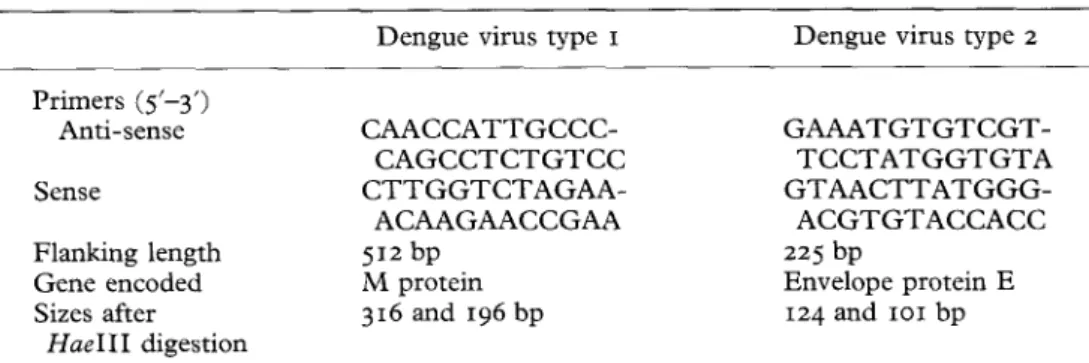
25 Table IPrimers for amplifying dengue virus by the polymerase chain reaction
Dengue virus type I Dengue virus type 2 Primers (5'-3') Anti-sense Sense Flanking length Gene encoded Sizes after H a e I I I digestion C A A C C A T T G C C C - C A G C C T C T G T C C C T T G G T C T A G A A - A C A A G A A C C G A A 512 bp M protein 316 and I96 bp G A A A T G T G T C G T - T C C T A T G G T G T A G T A A C T T A T G G G - A C G T G T A C C A C C 225 bp Envelope protein E I24 and IoI bp
Lengths of amplified dengue virus nucleotide sequences after restriction endonuclease HaeIII treatment were calculated from known nucleotide sequences of the viral genome and the locations of the restriction endonuclease sites.
bp = Base pairs.
G G A A T G G G A C T G G A G A C A C G A A C T G A A A C A T G G A T G T C A T , respectively.
Reverse transcription o f viral RNA
For the first-strand c D N A synthesis by reverse transcription, a mixture was prepared containing R N A purified from either virus or serum samples, IOO pmoles antisense primer, 5o mM Tris hydrochloride buffer p H 8"3, 75 mM potassium chloride and 3 mM magnesium chloride. This mixture was heated for io min in a 95 °C water bath and cooled rapidly on ice. c D N A synthesis was achieved after the mixture had been adjusted to contain I5 nmoles each of the four deoxyribonucleoside triphosphates, 2o units of ribonuclease inhibitor (BRL, Gaithersburg, Maryland, U.S.A.), and 200 units of cloned Moloney murine leukaemia virus ( M M L V ) reverse transcriptase (BRL, Gaithersburg) in a final volume of 3o #1, which was then incubated at 42 °C for 3 ° min. T h e c D N A was stored at - 2 o °C before being used for amplification.
Nucleic acid amplification by the p o l y m e r a s e chain reaction (PCR)
PCR amplification of c D N A was performed with thermostable
Taq
D N A polymerase in a P e r k i n - E l m e r Cetus T h e r m a l Cycler (Perkin-Elmer Cetus, Norwalk, C T , U.S.A.) T h e I o o # l amplification reaction mixture contains Io mM Tris hydrochloride buffer pH 8"3; 5o mM potassium chloride; I'5 mM magnesium chloride; o'oI % gelatin; 20 pM each of the two oligonucleotide primers; 2"5 nmoles each of the four deoxyribonucleoside triphosphates; r unit ofTaq
D N A polymerase (Perkin-Elmer Cetus, Norwalk) and c D N A prepared from the reverse transcription of putative dengue viral genome. T h e temperature of the reaction mixture was first raised to 94 °C for i min to denature the D N A , then cooled to 63 °C for 2 min to allow annealling of primers to putative target D N A sequences. This procedure was followed by raising the temperature to 72 °C for 3 rain in order to extend the D N A chain. T h e whole process was repeated 31 times with a final period of incubation at2 6 C. C. PAO E T A L .
72 °C for IO min. A I5/zl volume of the amplified reaction mixture was separated by electrophoresis in a 2"5 % agarose gel. T h e D N A in the agarose gel was visualised by means of U V light after ethidium bromide staining and then transferred on to nitrocellulose m e m b r a n e filters for Southern blot hybridisation analysis.
S o u t h e r n blot h y b r i d i s a t i o n
Southern blot hybridisations were performed according to published pro- cedureslm 14 with the following modifications: after hybridisation, each filter w a s washed sequentially in 2 X S S C ( I x S S C is o . i 5 M sodium chloride and o'oI5 M sodium citrate) and o-1% sodium dodecyl sulphate twice for Io min at room temperature; and t h e n in o.I x SSC and o-1% sodium dodecyl sulphate three times for I5 min at 55 °C. T h e sequences of the oligonucleotide probes used in Southern blotting are listed in Table I. Restriction endonucleases were purchased from Boehringer M a n n h e i m (Mannheim, Germany).
P r e c a u t i o n s against c o n t a m i n a t i o n and f a l s e - p o s i t i v e results in P C R Because of the high degree of amplification of the PCR procedures, special precautions were taken to minimise sample-to-sample contamination and P C R - p r o d u c t carry-over in order to avoid false-positive results. These measures included the separate dispensing of all reagents, physical separation of pre- and post-PCR reactions, and meticulous laboratory techniques. Furthermore, all reagents were irradiated with UV light so as to inactivate any double-stranded D N A which may have been present before sample D N A was added and the PCR started. 15 As a negative control, 50/zg h u m a n D N A or i #g Escherichia coli D N A was included in each assay and always yielded negative results. Multiple reagent controls were also included in each P C R assay and gave negative results. Repeated D N A amplification assays performed on the same specimens at different times produced the same results.
Results
Plate I shows the amplification of dengue virus R N A after viral R N A genome was first converted to c D N A by reverse transcription with M M L V reverse transcriptase. T h e D E N - I and D E N - 2 positivity were indicated by the presence of 512 base pairs (lane 2) and 225 base pairs (lane 3) D N A , respectively. Based on the n u m b e r of plaque-forming units in the virus preparation, the sensitivity of detection by PCR was estimated to be < moo pfu for either type of dengue virus. T h e primers used in this study do not cross amplify h u m a n D N A or Japanese encephalitis virus which belongs to the same fiavivirus family and is often prevalent in the same geographical area (Plate I, lanes 4 and 5).
T h e authenticity of the amplified D N A was established by two independent methods. First, the amplified putative dengue virus D N A was subjected to restriction endonuclease analysis. When the amplified D E N - I and D E N - 2 D N A were treated with restriction endonuclease HaeIII, the original 512 (Plate 2, lane 2) and 225 (Plate 2, lane 5) base pair D N A disappeared and were
Journal of Infection
1
2
3
4
5
6
P l a t e I
Plate i. Agarose gel analysis o f d e n g u e virus-specific P C R amplification. Primers u s e d a n d the lengths of nucleotide sequences amplified are listed in T a b l e I. H a e I I I - d i g e s t e d P h i X - I 7 4 D N A are u s e d as D N A size s t a n d a r d s in lanes I a n d 6. T h e sizes are (from top to bottom) 1353; lO78; 872; 6o3; 31o; 281, 271; 234; 194; 118 and 72 base pairs. L a n e s 2 a n d 3 relate to amplified p r o d u c t s of d e n g u e virus types i a n d 2 R N A , respectively. L a n e s 4 and 5 relate to h u m a n D N A a n d Japanese encephalitis virus R N A , respectively. D N A b a n d s on t h e
b o t t o m o f the gel are p r i m e r - d i m e r s .
Journal of Infection
1
2
3
4
5
6
7
8
P l a t e 2
Plate 2. Agarose gel electrophoresis o f amplified d e n g u e virus nucleotide sequences before a n d after restriction endonuclease digestion. Conditions for restriction endonucleases H a e I I I
digestion are those r e c o m m e n d e d by the e n z y m e m a n u f a c t u r e r s . Calculated lengths after
H a e I I I digestion are listed in T a b l e I. L a n e s I a n d 8 are restriction endonuclease H a e I I I -
digested p h a g e P h i - X I74 D N A u s e d as D N A size standard. T h e sizes are (from top to bottom) 1353, IO78~ 872~ 603, 31o, 281, 27I , 234, 194, I I 8 a n d 72 base pairs. L a n e s 2 a n d 3 are amplified d e n g u e virus type i before a n d after restriction endonuclease H a e I I I digestion, respectively. L a n e s 5 a n d 6 are amplified d e n g u e virus type 2 before a n d after restriction
endonuclease H a e I I I digestion, respectively. L a n e s 4 and 7 are blanks.
Journal of Infection
Plate 3co I
Plate 3- Autoradiographs of Southern blot hybridisation tests of amplified dengue virus type
I (a) and dengue virus type 2 (b) D N A with internal oligonucleotide probes. Lanes I in both (a) and (b) are undigested and lanes 2 are hybridisation of HaeIII-digested amplified dengue virus with internal oligonucleotide probes, respectively. Lanes 3 in b o t h panels are blanks. Probe sequences are indicated in Table I. T h e exposure time was 2 h for (a) and 2o min for
(b) at r o o m temperature without an intensifying screen.
Detection of dengue virus by PCR
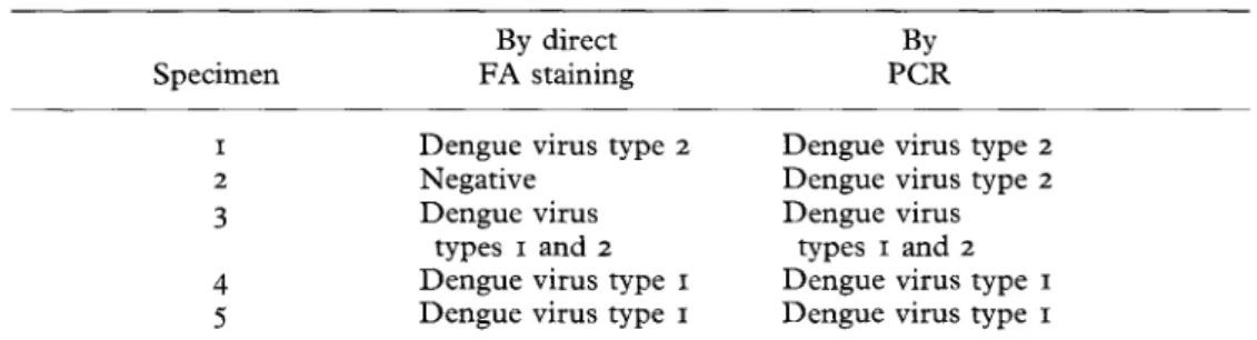
27 Table IIDetection and typing of dengue viruses by direct fluorescent antibody
(FA) staining and by the polymerase chain reaction (PCR)
By direct By
Specimen F A staining P C R
1 Dengue virus type 2 Dengue virus type 2
2 Negative Dengue virus type 2
3 Dengue virus Dengue virus
types I and 2 types I and 2 4 Dengue virus type I Dengue virus type I 5 Dengue virus type I Dengue virus type I
replaced with D N A of 316 and I96 (Plate 2, lane 3) and of 124 and IOi base pairs (Plate z, lane 6), respectively. T h e sizes of D N A fragments resulting from the actual restriction endonuclease digestion m a t c h e d exactly with those calculated from k n o w n viral genome nucleotide sequences and locations of restriction sites.
Secondly, amplified dengue virus D N A was analysed by S o u t h e r n blot hybridisation with internal oligonucleotide probes complementary to nucleo- tide sequences located between the two primers. Results indicated that amplified D N A from each type of dengue virus hybridised only with its own h o m o l o g o u s probe [lanes x in both (a) and (b) of Plate 3].
F u r t h e r m o r e , S o u t h e r n blot hybridisation showed that the 316 base pair and I24 base pair D N A fragments, resulting from
HaeIII
digestion of amplified D E N - I and D E N - 2 , hybridise with their respective oligonucleotide probes [lanes 2 in (a) and (b) of Plate 3]. T h e s e results also confirmed the locations ofHaeIII
restriction sites with respect to the locations of viral sequences that are c o m p l e m e n t a r y to the oligonucleotide probes.T h e detection and typing of dengue viruses by F A staining in five serum samples, obtained from patients d u r i n g the acute phase of infection, was c o m p a r e d with their detection by the P C R m e t h o d . T h e results are shown in Table II.
D i s c u s s i o n
D e n g u e presents a major public health p r o b l e m and is probably the most i m p o r t a n t a r t h r o p o d - b o r n e viral disease in terms of h u m a n morbidity and m o r t a l i t y ? T h e increasing incidence and transmission of dengue viruses indicate the need for a rapid and reliable m e t h o d for detecting and identifying the serotypes of dengue viruses. Serological and immunological assays, such as H I , P R N T or E L I S A , are widely used in the laboratory diagnosis of dengue virus infections and in sero-epidemiological surveys. H I tests and E L I S A , however, are often complicated by the presence of highly cross-reactive anti- flavivirus antibodies and often are not dengue type-specific. 16' 17 P R N T and conventional virus isolation m e t h o d s offer direct identification of dengue viruses b u t are t i m e - c o n s u m i n g and suffer from low rates of d e t e c t i o n ? 8 Virus culture is also expensive and often requires expertise and e q u i p m e n t that are
28 c. C. PAO E T AL.
n o t always available where t h e y are m o s t needed. It has also been suggested t h a t successful isolation of d e n g u e viruses f r o m the serum of infected patients is sometimes difficult because o f the presence o f high titred cross-reactive antibodies. 18 D N A probe h y b r i d i s a t i o n is a relatively sensitive m e t h o d for the detection o f viral genomes a n d has been applied to the detection, t y p i n g and m e a s u r e m e n t of d e n g u e virus R N A . 6'7 E v e n so, it is laborious a n d cross- h y b r i d i s a t i o n has been r e p o r t e d between d e n g u e t y p e 2 a n d type 4 in c D N A slot blot hybridisation. 7
T h e specificity of our P C R amplification of the d e n g u e viral g e n o m e was indicated by the restriction endonuclease digestion analysis and by S o u t h e r n blotting h y b r i d i s a t i o n with oligonucleotides representing the central p o r t i o n o f the amplified viral genome. T h u s , the specificity o f our analysis relies n o t only on the amplification o f f r a g m e n t s o f p r e d i c t e d sizes b u t also on the ability of the amplified p r o d u c t s to generate restriction endonuclease D N A f r a g m e n t s o f correct sizes a n d to h y b r i d i s e with specific oligonucleotide probes. Results suggest that our P C R protocols n o t only have high specificity for d e n g u e virus b u t also are able to d e t e r m i n e the types o f d e n g u e viruses. T h e ability o f P C R for t y p i n g m a y be very useful since antigenic and genetic heterogeneities have been s h o w n to exist a m o n g different isolates or strains of d e n g u e viruses. 19 Results o f this s t u d y also indicate that the sensitivity o f the P C R m e t h o d compares very favourably with either conventional virus isolation or serological analysis.
T h e results p r e s e n t e d here suggest t h a t the P C R D N A amplification m e t h o d is rapid, specific a n d sensitive a n d m a y prove useful for detecting d e n g u e viruses in clinical specimens. It m a y also find a place in the surveillance o f d e n g u e viral activity in vectors a n d in epidemiological field specimens in which the concentrations o f virus m a y be low.
(This study was supported by Medical Research Grant CMRP-286 from Chang Gung Medical College and Memorial Hospital, and by Research Grant NSC79-o412-BI82 - o2 from the National Science Council, Republic of China, awarded to C.C.P. The authors acknowledge the technical assistance of Mr Chuan-Liang Kao, Dr Wei-Jung Chen, Ms Chia-Chi Ku and Ms Li-Jung Chien. The authors also acknowledge the encouragement and support of Dr Delon Wu and Dr Chau-Hsiung Chang.)
R e f e r e n c e s
I. Halstead SB. Pathogenesis of dengue: challenges to molecular biology. Science I988 ; 239: 476-480.
2. Halstead SB. Selected primary health care: strategies for control of disease in the developing world. XI. Dengue. Rev Infect Dis I987; 6: 251-264.
3. Tesh RB. A method for the isolation and identification of dengue viruses, using mosquito cell cultures. Am J Trop Med Hyg I979; 28: IO53-Io59.
4- Kuberski TT, Rosen L. A simple technique for the detection of dengue antigen in mosquitoes by immunofluorescence. Am J Trop Med Hyg I977; 26: 533-537-
5. Henchal EA, McCown JM, Seguin MC, Gentry MK, Brandt WE. Rapid identification of dengue virus isolates by using monoclonal antibodies in an indirect immunofluorescence assay. Am J Trop Med Hyg I983; 32: I64-I69.
6. Kerschner JA, Vorndam AV, Monath TP, Trent DW. Genetic and epidemiological studies of dengue type 2 viruses by hybridization using synthetic deoxyoligonucleotides as probes.
Detection of dengue virus by PCR
z9 7. Henchal EA, Narupiti S, Feighny R, Padmanabhan R, Vakharia V. Detection of denguevirus RNA using nucleic acid hybridization. J Virol Methods x987; I5: I87-2oo.
8. Saiki SK, Gelfand DH, Stoffel S e t al. Primer-directed enzymatic amplification of D N A
with a thermostable DNA polymerase. Science I988; 239: 487-49I.
9. Eisenstein BI. The polymerase chain reaction : a new method of using molecular genetics
for medical diagnosis. N Engl J Med I99O; 3zz: I78-r83.
Io. Chomczynski P, Sacchi N. Single-step method of RNA isolation by acid guanidium
thiocyanate-phenol-chloroform extraction. Anal Biochem I987; ,62 : I56-I59.
I I. Mason PW, McAda PC, Mason T L , Fournier MJ. Sequence of the dengue-I virus genome in the region encoding the three structural proteins and the major nonstructural protein N S I . Virology I987; I 6 I : z6z-z67.
I2. Yaegashi T, Vakhari VN, Page K, Sasaguri Y, Feighny R, Padmanabhan R. Partial
sequence analysis of cloned dengue virus 2 genome. Gene I986; 46: 257-z67.
r 3. Maniatis T, Fritsch EF, Sambrook J (Eds) Molecular cloning : a laboratory manual. Cold
Spring Harbor, N.Y. : Cold Spring Harbor Laboratory, I98Z.
I4. Pao CC, Lin SS, Lin CY, Maa JS, Lai CH, Hsieh T T . Identification of human
papillomavirus D N A sequences in peripheral blood mononuclear cells. Am J Clin Pathol
I99I ; 95 : 540-546.
I5. Sarkar G, Sommer SS. Shedding light on PCR contamination. Nature I99O; 343: z7.
I6. Henchal EA, Pumak JR. The dengue viruses. Clin Microbiol Rev I99o; 3: 376-396.
I7. Innis BL, Nisalak A, Nimmannitya S e t al. An enzyme-linked immunosorbent assay to
characterize dengue infections where dengue and Japanese encephalitis co-circulate. Am J
Trop Med Hyg I989; 4o: 418-427.
I8. Waterman SH, Kuno G, Gubler D J, Sather GE. Low rates of antigen detection and virus
isolation from the peripheral blood leukocytes of dengue fever patients. Am J Trop Med
Hyg I985; 34: 380-384.
I9. Trent DW, Grant JA, Rosen L, Monath TP. Genetic variation among dengue z viruses of