Quantitative diagnosis and analysis of mutations affecting
drug resistance to rifampicin and isoniazid of clinical
Mycobacterium tuberculosis isolates in Taiwan
Chao-Wen Hsueh&Shih-Shen Lin&Chun-Hsiu Lin&Shang-Tao Chien&Wen-Sheng Liu&Fu-Gong Lin& Hsiu-Ting Tsai&Chien-Fu Huang&Chi-Chiang Yang
Abstract A SYBR Green I-based real-time quantitative PCR assay was utilized using senX3-regX3 and IS6110 as targets to identify Mycobacterium tuberculosis in 135 clinical samples from southern Taiwan. The bacterial loads of true positive samples estimated in this assay ranged from 33 to 7.7×10⁵CFU/ml, whereas false negative samples ranged from 10 to 25 CFU/ml. The specificity and sensitivity were 100% and 88.5%, respectively. Moreover, a multiplex allele-specific PCR was applied to detect mutation at three major hot spots (affecting codons 516,
526 and 531) of the rpoB gene affecting resistance to rifampicin, as well as codon 315 of the katG gene (resistance to isoniazid). The mutation rates of rpoB and katG were 69.6% (32/46) and 45.7% (21/46), respectively. The dual-gene mutation (rpoB and katG) rate was 41.3% (19/46). Among the hot spots on the rpoB gene, the mutation rate for rpoB 531(17/46, 37%) was higher than that of rpoB 516 (10/46, 21.7%) and rpoB 526 (8/46, 17.4%). Furthermore, for the four codons assayed (rpoB 516, 526, 531, and katG), 11 mutations were identified as
Chao-Wen Hsueh, Shih-Shen Lin, Chien-Fu Huang and Chi-Chiang Yang contributed equally.
C.-W. Hsueh
Division of Internal Medicine,
Kaohsiung Armed Forces General Hospital, Kaohsiung,
Taiwan, Republic of China S.-S. Lin
School of Dentistry, Chung Shang Medical University, Taichung,
Taiwan, Republic of China C.-H. Lin
Department of Pathology,
Kaohsiung Armed Forces General Hospital, Kaohsiung,
Taiwan, Republic of China S.-T. Chien
Asia-Pacific Biotech Developing, Kaohsiung,
Taiwan, Republic of China W.-S. Liu
Institute of Preventive Medicine, National Defense Medical Center, Taiwan, Republic of China
F.-G. Lin
Department of Pulmonary Medicine, Kaohsiung Armed Forces General Hospital, Kaohsiung,
Taiwan, Republic of China H.-T. Tsai
School of Nursing, Chung Shan Medical University, Taichung,
Taiwan, Republic of China C.-F. Huang
Department of Biological Science and Technology, I-Shou University,
Kaohsiung,
Taiwan, Republic of China
C.-C. Yang (*)
School of Medical Laboratory and Biotechnology, or Department of Clinical Laboratory,
Chung Shan Medical University Hospital, 110, Section 1, Chien-Kuo North Road, Taichung, Taiwan 40201, Republic of China
single mutation (23.9%), 18 as double mutations (39.1%), 3 as triple mutations, and none as quadruple mutations. Our results reveal the prevalence of tuberculosis drug-resistant mutations occurring on rpoB and katG genes in southern Taiwan.
Keywords Isoniazid . PCR . Rifampicin . Taiwan . Tuberculosis
Introduction
Tuberculosis (TB) is a potentially deadly infectious disease caused by mycobacteria, mainly Mycobacterium tuberculo-sis, and causes more than 1 million deaths annually around the world. The situation is worsening, with increasing circulation of drug-resistant strains of M. tuberculosis, which has made TB more unmanageable. Thus, the emergence of drug-resistant, especially multi-drug resistant, TB poses a serious threat to the control of this disease. Determination of drug-resistant status is beneficial to patients during therapy, especially in those patients sus-pected of having acquired resistant strains (Soini and Musser2001; Kremer and Besra2002).
Rifampicin (RIF) together with isoniazid (INH) are the most usual cocktail of drugs used to treat TB. Many studies have reported that, in up to 95–98% of strains resistant to RIF, resistance is caused by mutations in the rpoB gene locus, which encodes a β-subunit of RNA polymerase (Telenti et al. 1993; Ramaswamy and Musser 1998). Moreover, mutations in three codons (516, 526 or 531) of the rpoB gene explained the majority (70–95%) of RIF-resistant strains (Cavusoglu et al.2002). INH resistance is more frequently associated with mutations in the katG gene, in particular with a mutation in codon 315 (60–70%; Dobner et al.1997; Rattan et al.1998).
Nevertheless, the major problem in developing a PCR strategy to identify M. tuberculosis is the choice of target sequences, which should ideally be present in all M. tuberculosis strains and absent from all other bacteria. In this study, a SYBR Green I-based real-time PCR technique was used for quantitative detection of M. tuberculosis in 135 clinical specimens from southern Taiwan. The PCR amplifies two distinct regions of the M. tuberculosis genome: one fragment of the IS6110 multicopy element (Matsiota-Bernard et al. 1998) and the intergenic region (IR) of senX3-regX (Escalante et al. 1998). Moreover, a multiplex allele specific-polymerase chain reaction (MAS-PCR) method was introduced to analyze the mutation hot spots of TB drug-resistant genes, i.e., rpoB 516, 526, and 531, and katG 315. The present study reveals the mutation rates at the hot spots of the rpoB and katG genes in southern Taiwan.
Materials and methods
Samples
A total of 135 samples was collected from the Department of Pathology at Kaohsiung Armed Forces General Hospital of southern Taiwan in 2008, as approved by the Institutional Review Board for Medical Ethics and Human Investigation. Among the samples, 46 isolates collected from the M. tuberculosis complex with positive results with the Myco-bacterial growth indicator tube (MGIT; Becton Dickinson;
http://www.bd.com) were analyzed biochemically consecu-tively in the laboratory, and further identified as multidrug-resistant tuberculosis (MDR-TB) by conventional drug susceptibility testing. A total of 89 specimens were negative with both acid-fast bacilli and TB bacterial culture, including Pseudomonas aeruginosa, Streptococcus pneumoniae, Streptococcus pyogenes, Klebsiella pneumoniae, Bordetella pertussis, Staphylococcus aureus, and non-tuberculosis Mycobacterium (NTM).
Bacterial genomic DNA isolation
For DNA extraction, sputum samples or cultured bacte-rial pellets were digested in 200–500 µl N-acetyl-L -cystein/NaOH solution, incubated at 70°C for complete lysis, centrifuged at 400 g for 10 min, and the lysates were purified with the GenElute™ Bacterial Genomic DNA Kit (Sigma, St. Louis, MO). Finally, the DNA was stored in a−20°C freezer.
Multiplex PCR for confirmation of two gene candidates
Genomic DNA extracted from two M. tuberculosis isolates and six other bacterial strains (Pseudomonas aeruginosa, Streptococcus pneumoniae, Streptococcus pyogenes, Klebsiella pneumoniae, Bordetella pertussis, and Staphylococcus aureus) were analyzed as positive and negative controls, respectively, and used as templates for multiplex PCR. The multiplex PCR was performed for co-amplification of senX3-regX3 or IS6110 with bacterial 16 S rRNA in the same PCR reactions using gene-specific primer pairs: 16Sr-F: 5′-CCACACTGGAACTGAGACAC-3′ and 16Sr-R: 5′-GGTATCTAATCCTGTTTGCTCC-3′ for 16 S rRNA; TAQM3 and TAQM4 for the IS6110 multicopy element; and TAQregT2 and TAQreg2L for senX3-regX3 region of the M. tuberculosis genome. The PCR mixture contained 100 mM dNTPs, 150 pmol primers, MgCl2
(1.5 mM), buffer (1X), and 1.25 U Taq polymerase (Yeastern Biotech, Taipei, Taiwan). The PCR conditions were initiation for 5 min at 94°C; 35 cycles of 95°C for 15 s, 55°C for 30 s, and 72°C for 30 s; and final elongation at 72°C for 7 min.
Plasmid preparation
Genomic DNA (1 μl; approximately 0.1 µg/µl) extracted from the mixed samples of M. tuberculosis isolates was amplified by PCR in a GeneAmp 2400 thermal cycler (Applied Biosystems, Foster City, CA) using primers TAQM3 (5′-AGGCGAACCCTGCCCAG-3′) and TAQM4 (5′-GATCGCTGATCCGGCCA-3′) to yield a 122-bp frag-ment of the IS6110 multicopy elefrag-ment (GenBank accession no. X52471), or primers TAQregT2 (5′-GTAGCG ATGAG GAGGAGTGG-3′) and TAQreg2L (5′-ACTCGGCGA GAGCTGCC-3′) to yield a 146-bp fragment of the senX3-regX3 region of the M. tuberculosis genome (EMBL accession no. G2190478). The PCR mixture contained 100 mM dNTPs, 150 pmol primers, MgCl2 (1.5 mM),
buffer (1X), and 1.25 U Taq polymerase (Yeastern Biotech). After 10 min of incubation at 95°C, 30 cycles of amplification were performed with a thermal profile of 94, 65 and 72°C (30 s each). A final extension step of 10 min at 72°C was performed. PCR products were cloned directly into the pGEM-T easy plasmid using the pGEM-T Easy Vector System kit (Promega, Madison, WI) according to the manufacturer’s instructions. Plasmids pIS6110 and psenX3-regX3 IR were purified with the Qiaprep Spin Miniprep kit (Qiagen, Hilden, Germany), double strand sequenced, and employed to establish the standard curves of gene expres-sion of these two candidate genes and quantify the mycobacterial DNA load.
Establishing standard curves for real-time PCR
To generate reference curves for M. tuberculosis quantifi-cation, constructs bearing partial sequences of IS6110 and senX3-regX3 gene were first quantified using UV spectro-photometry. Standard reference curves were established by the serial dilution method, using 10-fold dilutions for each preparation performed in the same run as the clinical samples to be tested in each real-time PCR assay. In general, M. tuberculosis genome equivalent to 101 to 106 copies would be detected in each test, and the consistency of the reference curves for each dilution could be confirmed by the linear relationship (r2≥0.99) and Ct values found relative to the amplified bacterial genome obtained.
SYBR Green I-based real-time PCR assay
The SYBR Green I-based real-time PCR assay was modified from the method of Broccolo et al. (2003). Briefly, the quantitative assay involves SYBR Green I-based PCR methods, and two M. tuberculosis specific amplicons—IS6110 and senX3-regX3 IR—were amplified in each sample. All reactions were optimized to obtain the best amplification kinetics under the same cycling
con-ditions (2–3 min at 95°C, and 40 cycles of 15 s at 95°C and 1 min at 60°C per cycle) and composition of the reaction mixture. All reactions were performed in a final volume of 50 µl, which contains 100 mM (each) dATP, dCTP, and dGTP; 200 mM dUTP; 6 mM MgCl2; iTaq DNA
polymerase 50 units/ml (Bio-Rad Laboratories, Hercules, CA), SYBR Green I dye, 1 µM ROX internal reference dye, forward and reverse primers (TAQM3, TAQM4 for IS6110 multicopy element; TAQregT2, TAQreg2L for senX3-regX3) in variable 100–500 nM, and 1–5 µl of DNA template. The principle of real-time PCR has been described elsewhere (Su et al. 2008). Each sample was tested in triplicate to confirm average expression levels.
Bacterial load estimation
Three samples of M. tuberculosis isolates equivalent to 1× 10⁶CFU/ml M. tuberculosis detected by BACTEC™ MGIT™ 960 Mycobacterial Detection System (BD Bioscien-ces, Sparks, MD) were used as positive controls for estimation of bacterial load. In general, serial dilutions equivalent to 101 to 106copies of M. tuberculosis genome were made, and gene expression of the two candidate genes was measured as previously described at least three times. Therefore, the averaged fluorescent signals corresponding to each dilution were obtained and used as reference values for bacterial load estimation of the test samples. Thus, the final bacterial load was determined by the averaged values acquired from two genes.
MAS-PCR assay
A two-step MAS-PCR assay was performed for analysis of purified DNA. The assay consisted of four independent allele-specific targeted PCRs, which included three rpoB codons (codons 516, 526 and 531) and one katG codon (codon 315). Primers and thermal cycler programs used in this assay are listed in Table1. Accordingly, in the absence of mutation at positions rpoB 531, 526, 516 or katG 315, wild-type allele-specific fragments of 167 bp, 181 bp, 214 bp, and 87 bp respectively, would be amplified by the RIR primer and an inner forward primer of the rpoB gene, and the kat315w and katGA primer set for the katG gene. If mutations occurred, there would be no allele-specific PCR products. For the rpoB gene, two outer primers—ROF and RIR—would amplify the entire rpoB region, yielding a predicted fragment of 249 bp in all strains, and constant amplification of this 249 bp fragment could be used as quality control for MAS-PCR. The two-step PCR conditions for the rpoB gene included preliminary amplification of the larger portion of rpoB with outer primers ROF and ROR. This 281-bp fragment serves as a template for three subsequent allele-specific PCRs for three rpoB codons, performed as follows: an initial denaturation at 96°C for 3 min; 30 cycles of 95°C for 50 s, 62°C for 40 s,
and 72°C for 30 s; and a final elongation at 72°C for 3 min for the first-step PCR. From the first PCR reaction mixture, 1 µl would be taken out for the second step of the PCR amplification, and the second step of the MAS-PCR consisted of three specific simultaneous PCR reactions using the same conditions: initial denaturation at 95°C for 5 min; 35 cycles of 95°C for 15 s, 66°C for 30 s, and 72°C for 30 s; and a final elongation at 72°C for 7 min. For katG 315 MAS-PCR, the conditions consisted of two PCR programs, and the first program was as follows: initial denatured at 95°C for 1 min, 10 cycles of 95°C for 15 s, 63°C for 45 s, and 72°C for 35 s; then 25 cycles of 95°C for 20 s, 59°C for 45 s, and 72°C for 40 s; and a final elongation at 72°C for 5 min. A 231-bp katG 315 fragment was thus amplified. Then, 1 µl of the first PCR reaction would be used for the secondary run. The secondary program was initially denatured at 95°C for 1 min, followed by 35 cycles of 95°C for 15 s, 56°C for 40 s, and 72°C for 30 s, and finally elongated at 72°C for 3 min, generating a wild-type specific katG 315 product of 87 bp.
Statistical analysis
Fisher’s exact test was introduced to analyze the signifi-cance of different mutations. Statistical signifisignifi-cance was defined as a P-value less than 0.05.
Results
Design of a rapid analytical tool for Mycobacterium tuberculosis
The goal of this study was to design a rapid, high-throughput and quantitative PCR-based platform that can
detect M. tuberculosis load directly in clinical sputum specimens. It was reported that a region corresponding to the IS6110 multicopy insertion element can be found in almost all M. tuberculosis complex (Dalovisio et al.1996; Abbadi et al.2009a; Shamaei et al.2009), and senX3-regX3 IR also belongs to the M. tuberculosis complex (Escalante et al. 1998; Suresh et al. 2006a). Therefore, these two candidate genes were chosen as the targets for real-time PCR amplification. The first stage in constructing this detection platform was to ensure that these two candidate genes can be amplified specifically from M. tuberculosis, but not the other NTM pathogens. Thus, a MAS-PCR was introduced to detect these two genes. Two TB samples as positive controls, and six bacterial strains (Pseudomonas aeruginosa, Streptococcus pneumoniae, Streptococcus pyo-genes, Klebsiella pneumoniae, Bordetella pertussis, and Staphylococcus aureus) as negative controls were tested. The PCR product lengths of the two amplicons were 146 bp and 121 bp, respectively. A 485 bp product specific to bacterial 16S rRNA was also amplified as an internal control for equal genomic DNA loading in each multiplex PCR reaction. Figure1shows the specific PCR products of senX3-regX3 and IS6110, which were amplified success-fully in the two TB samples, but were negative in the negative controls. As a result, these two genes were used for the establishment of the SYBR Green I-based real-time PCR assay.
Quantitative PCR for Mycobacterium tuberculosis load
To establish the highly sensitive and specific SYBR Green I-based real-time PCR method, an experiment for deter-mining the baseline of gene expression between different sample subgroups (TB and non-TB) of each gene was Table 1 Polymerase chain reaction (PCR) primers, product length and programs
rpoBGene locus
Description Primers Product length PCR program
Forward ROF 5′-GTCGCCGCGATCAAGGA-3′ 249 bp 95°C 50′′, 62°C 40′′, 72°C 30′′; ×30
Reverse RIR 5′-TGACCCGCGCGTACAC-3′
Forward ROF 5′-GTCGCCGCGATCAAGGA-3′ 281 bp 95°C 50′′, 62°C 40′′,72°C 30′′; ×30
Reverse ROR 5′-GGTACGGCGTTTCGATGAAC-3′
rpoB 516 Inner allele-specific
primer
5′-GCTGAGCCAATTCATGGA-3′ 214 bp 95°C 15′′, 66°C 30′′, 72°C 30′′; ×35
rpoB 526 Inner allele-specific
primer
5′-GTCGGGGTTGACCCA-3′ 181 bp 95°C 15′′, 66°C 30′′,72°C 30′′; ×35
rpoB 531 Inner allele-specific
primer
5′-ACAAGCGCC GACTGTC-3′ 167 bp 95°C 15′′, 66°C 30′′, 72°C 30′′; ×35
katG Forward TB86 5′-GAAACAGCGGCGCTGGATCGT-3′ 231 bp 95°C 15′′, 63°C 45′′, 72°C 35′′; ×10
Reverse katGA 5′-CGTACAG GATCTCGAGGAAACTGT-3′ 95°C 20′′, 59°C 45′′, 72°C 40′′; ×25
katG 315 Inner allele-specific
primer
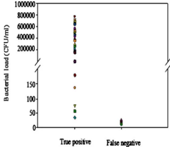
performed. The averaged means of baseline Ct values of senX3-regX3 and IS6110 were 29.0 ± 1.7 and 25.9 ± 1.2 CFU/ml, respectively, for the negative control. The cut-off values of Ct of senX3-regX3 and IS6110 were set at 30.7 and 27.1 CFU/ml, respectively, to clearly discriminate positive and negative control groups. The final bacterial load was determined by the averaged values acquired from two genes. The positive case was defined as dual positive expression of these two genes. Moreover, the M. tubercu-losis load was quantified in triplicate to evaluate the accuracy of both IS6110 and senX3-regX3 assays. Accord-ing to the criteria described above, 6 samples exhibited a false negative result among 46 positive samples, whereas none were false positive among 89 negative samples. The specificity and sensitivity were demonstrated to be 100% and 88.5%, respectively. Furthermore, the bacterial loads of true positive samples estimated in this assay ranged from 33 to 7.7×10⁵CFU/ml, whereas the bacterial loads of false negative samples (which were below 28.9 CFU/ml) ranged from 10 to 25 CFU/ml (Fig.2).
Genotyping drug resistance to two major antibiotics—RIF and INH—in Taiwan
To genotype the drug resistance to RIF and INH among TB patients in Taiwan, 46 positive samples were analyzed by MAS-PCR for the three major hot spots of rpoB gene mutation—codons 516, 526 and 531—as well as codon 315
of the katG gene. The mutation rates of rpoB and katG were 69.6% (32/46) and 45.7% (21/46), respectively. The dual-gene mutation (rpoB and katG) rate was 41.3% (19/46). Among the hot spots of the rpoB gene, the mutation rate for rpoB 531 (17/46, 37%) was higher than that of rpoB 516 (10/46, 21.7%) and rpoB 526 (8/46, 17.4%). Three-codon mutations of the rpoB gene were not identified. In summary, in the four codons (rpoB 516, 526, 531, and katG) assayed in this study, 11 mutations were identified as single mutation (23.9%); 18 as double mutations (39.1%); 3 as triple mutations but there were no quadruple mutations (Table2). Fig. 1 Specific PCR products of senX3-regX3 and IS6110 were
amplified successfully in the TB+control samples (TB1 and TB2), but
not in other common pathogens easily isolated from the human respiratory tract by multiplex PCR. The PCR product lengths of the two amplicons were 146 bp and 121 bp, respectively. A 485 bp product specific for bacterial 16S rRNA was amplified ubiquitously to
indicate equal genomic DNA loading in each multiplex PCR reaction. The lowest band in each sample lane represents primers. Lanes: M DNA size marker (100 bp ladder), PA Pseudomonas aeruginosa, SP Streptococcus pneumoniae, SPy Streptococcus pyogenes, KP Klebsi-ella pneumoniae, BP BordetKlebsi-ella pertussis, SA Staphylococcus aureus, NC negative control
Fig. 2 Bacterial loads of positive samples estimated by SYBR Green I-based real-time PCR assay
Discussion
Conventional methods used to detect M. tuberculosis test involve microscopic observation and examination of bacteria culture results on solid or liquid media. In recent years, increasing attention has been paid to the application of new molecular techniques for diagnosis, as well as clinical monitoring and research study of M. tuberculosis infections. Several rapid diagnostic tests utilizing different M. tubercu-losis genomic targets, including the IS6110 insertion sequen-ces and senX3-regX3 IR, have been reported (Dalovisio et al.
1996; Su et al. 2008; Abbadi et al. 2009a). Since some studies showed that a particular amplification of IS6110 repeat element does not lead to a complete specific identification for M. tuberculosis (Shamaei et al.2009), we chose not only the fragment of IS6110 repeat element but also a DNA fragment belonging to the senX3-regX3 region, which enabled more precise quantitative detection of M. tuberculosis specimens (Escalante et al.1998; Suresh et al.2006b). This PCR detection system has been shown to yield good levels of sensitivity (90–100%) on acid-fast bacilli positive samples (Dalovisio et al.1996). Our SYBR Green I-based real-time PCR results suggest that it is a good detection method with high sensitivity and specificity, and that it is also suitable for late-stage analyses as double confirmation with methods currently used in clinics.
Resistance to two key anti-TB drugs—RIF and INH— is likely to influence treatment results and hospital health control. It is necessary to determine rapidly the resistance of acquired strains from patients. The resistance in nearly all identified strains is attributed mainly to mis-sense mutations, small deletions or insertions of certain frag-ments within hot spot regions of the rpoB and katG genes (Telenti and Iseman 2000; Ramaswamy et al. 2004; El Sahly et al. 2006). In 2002, the incidence and mortality rate of tuberculosis was 74.6 and 5.68, respectively, per 100,000 population in Taiwan (Jou et al.2006). Because of this high incidence and the increasing prevalence of MDR-TB cases, the laboratory-based Taiwan Surveillance of Drug Resistance in TB (TSDRTB) program was established in 2003. The TSDRTB program demonstrated that drug resistance rates were 11.3% in 2004 and 10.1%
in 2005 for INH; 7.5% in 2004 and 6.2% in 2005 for RIF; 20.4% in 2004 and 18.1% in 2005 for any first-line drug; and 5.3% in 2004 and 4.0% in 2005 for MDR-TB (Jou et al. 2006). The resistance rates are higher than those reported by the third TB global drug resistance surveil-lance. Global surveillance reported that the median prevalence of drug resistance was 6.6% for INH, 2.2% for RIF, 10.4% for any drug, and 1.7% for MDR-TB (WHO2004).
The MAS-PCR assay described here is easy to imple-ment and can be performed as a routine examination for anti-TB treatment. Our MAS-PCR assay can be used as a high-throughput diagnostic tool to detect the drug-resistant mutations of TB. The mutation frequencies at codons 516, 526 and 531 within rpoB and codon 315 within katG in resistant M. tuberculosis strains varied and correlated with geographical location or ethnic population (Cooksey et al.
1997; Matsiota-Bernard et al. 1998). In Egypt, rpoB mutations were found in 76% of RIF-resistant isolates, and katG mutations were found in 47.1% of INH-resistant isolates (Abbadi et al. 2009a,b). In Iran, drug resistance to RIF was identified in 21.7% isolates, and drug resistance to INH was identified in 27.7% isolates (Shamaei et al.2009). Furthermore, studies conducted in India have demonstrated that the most common mutations in the rpoB gene were in codons 531 (59%), 526 (22%), and 516 (11.5%) (Suresh et al. 2006a,b). Recently, Liaw et al. (2005) reported that, of the 41 RIF-resistant strains isolated in northern Taiwan, 87.8% showed mutations in rpoB—identified as codons 531 (68.3%), 526 (9.8%), 522 (4.9%), and 513 (4.9%). In the present study, rpoB mutations were found in 69.6% of isolates, katG mutations were found in 45.7% of isolates. Among the 32 rpoB gene-mutated isolates, the most common mutations in the rpoB gene were in codons 531 (17/32, 53.1%), 516 (10/32, 31.3%), and 526 (8/32, 25%). Our results are similar to those of Liaw et al. (2005) regarding the prevalence of mutations in rpoB; however, we found mutations in codon 516 to be more prevalent than those in codons 513 and 522 in our study. Moreover, multidrug resistance to both RIF and INH was found in 41.3% (19/46) of the isolates. This percentage is more higher than that found in a recent study in northern Taiwan; Su et al. (2008) reported that 159 isolates out of 611 samples (26.0%) were resistant to both INH and RIF as MDR-TB, and 94.6% of RIF-resistant isolates were also resistant to INH. This present study reveals the prevalence of M. tuberculosis drug-resistant mutations occurring in rpoB and katG genes in southern Taiwan. The isolates analyzed in this study are truly representative of isolates from southern Taiwan. Compared with the results of the TSDRTB program in 2003–2005 and Liaw et al. (2005), our findings suggested that the drug-resistance trends of M. tuberculosis remain serious in Taiwan. Conducting a Table 2 Number of codon mutations found in 46 tuberculosis (TB)
samples
Mutation No. Percentage (%)
Single mutation 11 23.9
Double mutation 18 39.1
Triple mutation 3 6.5
Quadruple mutation 0 0
nation-wide anti-TB drug resistance survey in Taiwan to control and prevent drug-resistant TB is thus recommended.
Acknowledgments We thank Asia-Pacific Biotech Developing Inc.
for laboratory support. This study was also financially supported by clinical research grants from the Kaohsiung Armed Forces General Hospital. We declare no conflict of interest.
References
Abbadi S, El HG, Gomaa N, Cooksey R (2009a) Strain differentiation of Mycobacterium tuberculosis complex isolated from sputum of
pulmonary tuberculosis patients. Int J Infect Dis 13:236–242
Abbadi SH, Sameaa GA, Morlock G, Cooksey RC (2009b) Molecular identification of mutations associated with anti-tuberculosis drug resistance among strains of Mycobacterium tuberculosis. Int J
Infect Dis 13:673–678
Broccolo F, Scarpellini P, Locatelli G, Zingale A, Brambilla AM, Cichero P, Sechi LA, Lazzarin A, Lusso P, Malnati MS (2003) Rapid diagnosis of mycobacterial infections and quantitation of Mycobacterium tuberculosis load by two real-time calibrated PCR assays. J Clin Microbiol 41:4565–4572
Cavusoglu C, Hilmioglu S, Guneri S, Bilgic A (2002) Characteriza-tion of rpoB mutaCharacteriza-tions in rifampin-resistant clinical isolates of Mycobacterium tuberculosis from turkey by DNA sequencing
and line probe assay. J Clin Microbiol 40:4435–4438
Cooksey RC, Morlock GP, Glickman S, Crawford JT (1997) Evaluation of a line probe assay kit for characterization of rpoB mutations in rifampin-resistant Mycobacterium tuberculosis
iso-lates from New York city. J Clin Microbiol 35:1281–1283
Dalovisio JR, Montenegro-James S, Kemmerly SA, Genre CF, Chambers R, Greer D, Pankey GA, Failla DM, Haydel KG, Hutchinson L, Lindley MF, Nunez BM, Praba A, Eisenach KD, Cooper ES (1996) Comparison of the amplified Mycobacterium tuberculosis (MTB) direct test, amplicor MTB PCR, and IS6110-PCR for detection of MTB in respiratory specimens. Clin Infect Dis 23:1099–1106 Dobner P, Rusch-Gerdes S, Bretzel G, Feldmann K, Rifai M, Loscher
T, Rinder H (1997) Usefulness of Mycobacterium tuberculosis genomic mutations in the genes katg and inha for the prediction of isoniazid resistance. Int J Tuberc Lung Dis 1:365–369 El Sahly HM, Teeter LD, Pawlak RR, Musser JM, Graviss EA (2006)
Drug-resistant tuberculosis: a disease of target populations in
Houston, Texas. J Infect 53:5–11
Escalante P, Ramaswamy S, Sanabria H, Soini H, Pan X, Valiente-Castillo O, Musser JM (1998) Genotypic characterization of
drug-resistant Mycobacterium tuberculosis isolates from Peru.
Tuberc Lung Dis 79:111–118
Jou R, Chuang PC, Wu YS, Yan JJ, Luh KT (2006) Drug-resistant
Mycobacterium tuberculosis, Taiwan. Emerg Infect Dis 12:871–872
Kremer LS, Besra GS (2002) Current status and future development of antitubercular chemotherapy. Expert Opin Investig Drugs 11:1033–1049
Liaw YS, Tsai-Wu JJ, Hsueh PR, Wang SK, Yang PC, Luh KT (2005) Characterization of a 411-bp fragment of the rpoB gene in clinical isolates of Mycobacterium tuberculosis in a university
hospital in northern Taiwan. J Formos Med Assoc 104:792–797
Matsiota-Bernard P, Vrioni G, Marinis E (1998) Characterization of rpoB mutations in rifampin-resistant clinical Mycobacterium
tuberculosis isolates from Greece. J Clin Microbiol 36:20–23
Ramaswamy S, Musser JM (1998) Molecular genetic basis of antimicrobial agent resistance in Mycobacterium tuberculosis:
1998 update. Tuberc Lung Dis 79:3–29
Ramaswamy SV, Dou SJ, Rendon A, Yang Z, Cave MD, Graviss EA (2004) Genotypic analysis of multidrug-resistant Mycobacterium tuberculosis isolates from Monterrey, Mexico. J Med Microbiol
53:107–113
Rattan A, Kalia A, Ahmad N (1998) Multidrug-resistant Mycobacterium tuberculosis: molecular perspectives. Emerg Infect Dis 4:195–209 Shamaei M, Marjani M, Chitsaz E, Kazempour M, Esmaeili M, Farnia P, Tabarsi P, Amiri MV, Mirsaeidi M, Mansouri D, Masjedi MR, Velayati AA (2009) First-line anti-tuberculosis drug resistance
patterns and trends at the national TB referral center in Iran—
eight years of surveillance. Int J Infect Dis 13:e236–e240
Soini H, Musser JM (2001) Molecular diagnosis of mycobacteria. Clin
Chem 47:809–814
Su WJ, Feng JY, Huang CC, Perng RP (2008) Increasing drug resistance of Mycobacterium tuberculosis isolates in a medical
center in northern Taiwan. J Formos Med Assoc 107:259–264
Suresh N, Singh UB, Arora J, Pande JN, Seth P, Samantaray JC (2006a) Rapid detection of rifampicin-resistant Mycobacterium tuberculosis by in-house, reverse line blot assay. Diagn Microbiol
Infect Dis 56:133–140
Suresh N, Singh UB, Arora J, Pant H, Seth P, Sola C, Rastogi N, Samantaray JC, Pande JN (2006b) RpoB gene sequencing and spoligotyping of multidrug-resistant Mycobacterium tuberculosis isolates from India. Infect Genet Evol 6:474–483
Telenti A, Iseman M (2000) Drug-resistant tuberculosis: what do we do now? Drugs 59:171–179
Telenti A, Imboden P, Marchesi F, Lowrie D, Cole S, Colston MJ, Matter L, Schopfer K, Bodmer T (1993) Detection of rifampicin-resistance
mutations in Mycobacterium tuberculosis. Lancet 341:647–650
World Health Organization (2004) Anti-tuberculosis drug resistance in the world. Third Global Report. WHO/HTM/TB/2004.323. Geneva, canton and city, Switzerland.