Myxomycetes of Taiwan XXIII. The Genera Diachea and Didymium
Chin-Hui Liu(1*) and Jong-How Chang(1)
1. Institute of Plant Science, National Taiwan University, Taipei, Taiwan 106, R.O.C.
*Corresponding author. Email: huil4951@ntu.edu.tw
(Manuscript received 30 May 2011; accepted 29 June 2011)
ABSTRACT: Species of Diachea and Didymium reported from Taiwan are critically revised. Two newly recorded species,
Didymium floccoides and Didymium leptotrichum, are described and illustrated in this paper. Keys to the Diachea and Didymium
species of Taiwan are also provided.
KEY WORDS: Diachea, Didymiaceae, Didymium, Myxomycetes, Taiwan, taxonomy.
INTRODUCTION
The genera Diachea and Didymium (Didymiaceae, Physarales) have common characteristics in having limeless capillitium and dark-colored spore mass. The peridium of Diachea is also limeless and often with a metallic shine, while that of Didymium is limy, with lime crystals either compactly covering the peridium as an outer layer, or scattering loosely over the surface in various forms.
To date, three species of Diachea, and nineteen species of Didymium have been recorded in Taiwan (Nakazawa, 1929; Shi, 1981; Wang et al., 1981; Liu, 1982, 1983, 1989; Chung and Liu, 1995, 1996a, 1996b, 1997; Liu and Chen, 1998, 1999). In this paper two newly recorded species, Didymium floccoides Nann.-Bremek. & Y. Yamam. and Didymium
leptotrichum (Racib.) Massee are described and
illustrated. Characteristic examination for the fruiting bodies of these specimens were made by light and scanning electron microscopy as described previously (Liu et al., 2002).
TAXONOMIC TREATMENTS
Key to the species of Diachea from Taiwan
1a. Sporangia cylindrical to ovate, rarely subglobose ………..……… ………..………… Diachea leucopodia 1b. Sporangia globose or nearly so ………..………2 2a. Stalk length at least 1/2 of total height; spores sparsely but
prominently warted …………...….…....…… Diachea bulbillosa 2b. Stalk shorter and conic; spores warted subreticulate ………....…... ……….….………. Diachea subsessilis
Diachea bulbillosa (Berk. & Broome) Lister, in Penzig,
Myxomyc. Fl. Buitenzorg 45. 1898.
It was reported in a list by Nakazawa (1929), but no specimens were deposited in Taiwan.
Diachea leucopodia (Bull.) Rostaf., Sluzowce Monogr.
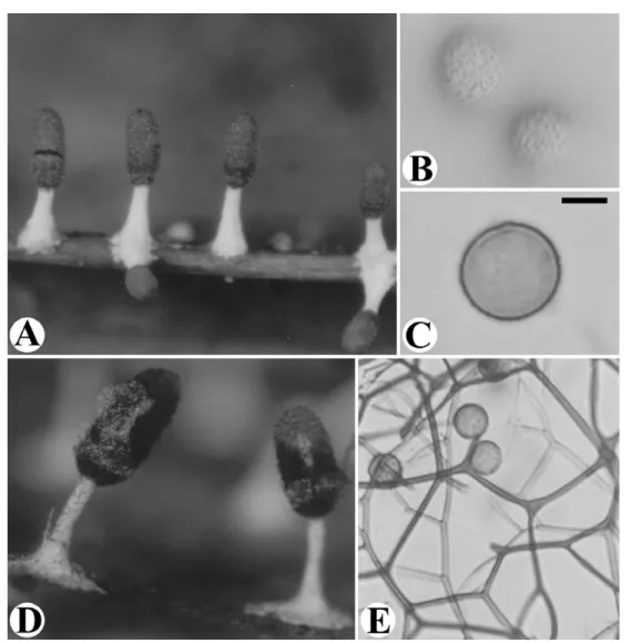
190. 1874. Fig. 1 Description and illustration: C.H. Liu, in Taiwania 27: 68, 70, 84 (1982).
Specimens examined: Taipei City: main campus of National Taiwan Univ., on wall of a plastic flower pot, CHL M394, Nov. 18, 1981; Peitou, Yangmingshan National Park, on fallen leaves of
Liquidambar formosana, CHL B2359, Dec. 16, 2000. Nantou Co.:
Yuchih Hsiang, Lianhua Pond, on decaying leaves, CHL M381, Oct. 25, 1981.
It is a very striking species and easily recognized in the field by the white calcareous stalk, the dark-colored, and metallic shinning conical sporangia.
Diachea subsessilis Peck, Annual Rep. New York State
Mus. 31: 41. 1879.
Description and illustration: C.H. Liu and Y.F. Chen, in Taiwania 44: 369-371 (1999).
Key to the species of Didymium from Taiwan 1a. Fructification sessile ….……….………... 2 1b. Fructification stipitate ……….………….. 8 2a. Peridium white and smooth as egg-shell ………..……….… 3
2b. Limecrystalsofperidium,looselyscatteredorcompacted,with
rough surface layer not as egg-shell ………..………...…… 4
3a. Capillitial threads rigidand profuse, quite long withtransverse
connections between two neighboring threads; spores 9-11 m in diameter ………..…………...…………..… Didymium listeri 3b. Capillitial threads not as above, usually short and sparse; spores
11.0-12.5 m in diameter …...… Didymium difforme 4a. With large vesicles intermixed with spore ……….... 5 4b. Vesicles absent ……….…. 6 5a. Plasmodiocarps branching and anastomosing to form an intricate
net; columella rising to about half of the height of plasmodiocarps; spores subglobose or polygonaled with ridges on the surface ……...……….…Didymium flexuosum 5b. Plasmodiocarps broadly effused, depressed; columella absent …... ………...………. Didymium serpula 6a. Spores 12-16 m in diameter ……….……Didymium leptotrichum 6b. Spores smaller than 11 m in diameter ……….……...…….7
Taiwania Vol. 56, No. 4 7a. Plasmodiocarps forming a perforated layer; peridium with large,
yellowish lime crystals ……..……...……Didymium perforatum 7b. Plasmodiocarps not forming a perforated layer; peridium with
small, colorless lime crystals ……..……..……. Didymium anellus
8a. Stalk calcareous ………..……….. 9
8b. Stalk not calcareous ……… 12
9a. Stalk smooth, filled with amorphous lime;peridium areolate; columella dark brownish, clavate ……..…... Didymium floccosum 9b. Stalk rough, with lime crystals ……..………... 10
10a. Peridium cartilaginous, orange yellow, with thinner pale lines of dehiscence, loosely covered by white or pale ochraceous lime crystals; spores not forming clusteredwarts … Didymium leoninum 10b. Spores with conspicuous clusters of warts ……... 11
11a. Sporangia discoid; columella absent; stalk tapering above …... ………...……….. Didymium lenticulare 11b. Sporangia globose to subglobose, with a small umbilicus below; columella present, white; stalk stout, the top extending into the sporangia …...……….... Didymium squamulosum 12a. Sporangia discoid, umbilicate below; spores 7.0-8.0 μm in diameter ...…Didymium clavus 12b. Sporangia not discoid ……….……... 13
13a. Columella whitish ………...………….. 14
13b. Columella brownish ……….………. 17
14a. Sporangia usually erect ……….……… 15
14b. Sporangia nodding ………...…. 16
15a. Stalk narrowing upward; columella globose; spores 8.5-10.0 μm in diameter …………...………...….... Didymium iridis 15b. Stalk uniform in width; columella stalked as goblet in shape; spores 10.0-11.5 μm in diameter .….... Didymium megalosporum 16a. Columella globose …………...…... Didymium verrucosporum 16b. Columella discoid or depressed globose, up to 0.30 mm in diameter ………. Didymium bahiense 17a. Spores larger than 10 μm in diameter ……… ……….... Didymium melanospermum 17b. Spores smaller ………...……… 18
18a. Collumella large, globose, calcareous, up to 0.2 mm in diameter ………..…Didymium minus 18b. Collumella smaller ……….……..……. 19
19a.Collumella small, conical, up to 0.11 mm in height ………..…… ………...………….……….. Didymium floccoides 19b. Collumella not as above ………...………… 20 20a. Peridium dark; columella dark; stalk usually black ………….….. ………..………... Didymium nigripes 20b. Peridium pale; columella paler, yellowish; stalk reddish
brown ………....…Didymium ovoideum
Didymium anellus Morgan, J. Cincinnati Soc. Nat.
Hist. 16: 148. 1894.
Description and illustration: S.M. Wang et al., in Biol. Bull. Nat. Taiwan Normal Uni. 16: 8 (1981).
Didymium bahiense Gottsb., Nova Hedwigia 15: 365.
1968.
Description and illustration: C.H. Chung and C.H. Liu, in Fung. Sci. 11: 123-124, 126 (1996a).
Specimens examined: Taipei City: farm of National Taiwan University, on leaves and fallen twigs, CHL B359, Apr. 3, 1984. Changhua City: main campus of National Changhua Normal University, on fallen leaves, J.H. Wu 46, Mar. 6, 1995. Nantou Co.: Huisun Forestry Station, on fallen leaves, CHL B498c, Apr. 1, 1985.
This species is difficult to distinguish from
Didymium megalosporum and Didymium eximium.
They all have white sporangia with long stalks, columella-like pseudocolumella and deep umbilici. To compare with the other two species, Didymium
bahiense have much tapering stalk and darker spores
which are warted or spinulose with distinct groups of larger warts instead of evenly warted in Didymium
megalosporum or minutely and evenly warted in Didymium eximium. In specimen CHL B359, lime
crystals in small swollen capillitial threads are observed. Our spore size is exactly same as that found in Tanzania (Härkönen and Saarimäki, 1991).
Didymium clavus (Alb. & Schwein.) Rabenh.,
Deutschl. Krypt.-Fl. 1: 280. 1844.
Description and illustration: C.H. Liu, in Taiwania 28: 103, 109, 112-113 (1983).
Specimens examined: Taipei City: Main campus of National Taiwan Univ., on bark of Bischofia javanica, CHL B94a, June 14, 1982; on bark, CHL B1266, Aug. 21, 1997. Tainan City on decayed wood, CHL B612, Aug. 4, 1986. Hualien County: Tayuling, Hohuanshan, on twigs, CHL B130, June 28, 1982.
The distinctive feature of this species is the white discoid sporangium with a thickened, basal umbilicus which is limeless and brown in color. It resembles
Diderma hemisphericum in size and shape. But the
stellate lime crystals covering the peridium make the sporangium of this species apparently not smooth in outer appearance, while in Diderma hemisphericum, it appears smooth as a white crust of lime granules.
Didymium difforme (Pers.) S. F. Gray, Nat. Arr. Brit.
Pl. 1: 571. 1821.
Description and illustration: C.H. Liu and Y.F. Chen, in Taiwania 43: 177-180 (1998).
Didymium flexuosum Yamashiro, J. Sci. Hiroshima
Univ., Ser. B. II, Bot. 3: 31. 1936.
Description and illustration: C.H. Liu and Y.F. Chen, in Taiwania 43: 178-179, 181 (1998).
Didymium floccoides Nann.-Bremek. & Y. Yamam.,
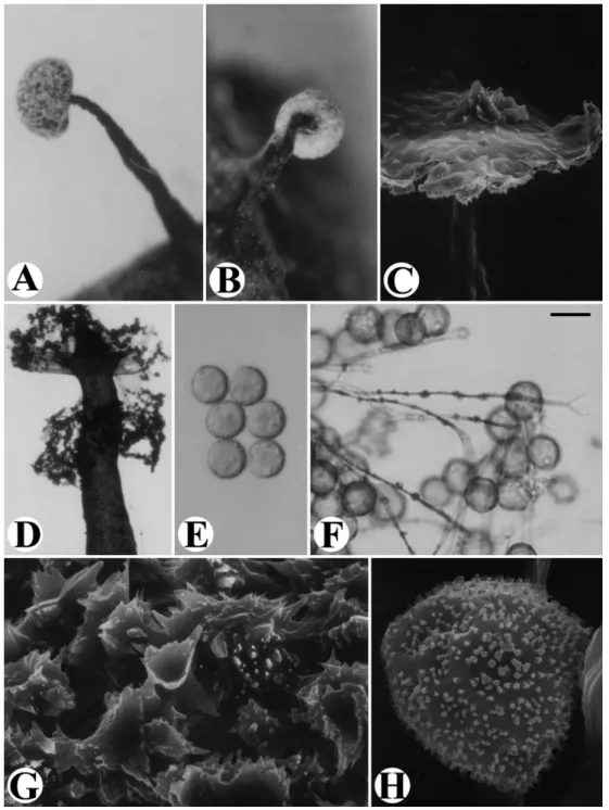
Proc. K. Ned. Akad. Wet., Ser. C, Biol. Med. Sci. 90: 323. 1987. Fig. 2 Fructification scattered, stipitate, (0.27-) 0.51-0.85 mm in total height. Sporangia hemispherical or flattened globose, white and rough except at the base around the stalk attachment, 0.18-0.38 mm in diameter. Stalk dark, blackish, wrinkled, erect or slightly curved, attenuate, limeless on the surface, filled with rounded debris matter, up to 2/3 or more of the total height. Peridium membranous, hyaline, densely covered by
Fig 1. Diachea leucopodia. A: Fruiting bodies. B: Spores, surface view. C: One spore, marginal view. D: Dehiscent fruiting bodies. E: Capillitial threads and spores. Scale bar: A = 350 μm; B-C = 4 μm; D = 230 μm; E = 14 μm.
white lime crystals. Hypothallus discoid, black brown. Columella dark brown, conical, small, up to 0.11 mm in height. Capillitium pale brown, slender, scarcely branching and anastomosing, with small nodules. Spores dark brown in mass, pale violaceous brown by transmitted light, globose, 7.5-9.0 (-10.0) m in diameter, mostly 7.5-8.0 m, minutely warted, warts often clustered. Plasmodium not observed.
Specimens examined: Taipei City: Peitou, Yangmingshan National Park, on fallen leaves, Jong 39, June 26, 2001 (moist-chamber culture: Mar. 18-June 26, 2001), Jong 67, July 18, 2001 (moist-chamber culture: June 26-July 18, 2001), Jong 71, Apr. 4, 2002 (moist-chamber culture: Feb. 15-Apr. 4, 2002).
This species is characterized by the white sporangia (hemispherical or flattened globose, rough), the long, wrinkled and dark stalk, and the small, conical columella. It differs from Didymium floccosum in the much smaller and shorter fruiting bodies, the non-umbilicate sporangia, and smaller spores with warts
not as distinct.
Didymium floccosum G.W. Martin, K.S. Thind &
Rehill, Mycologia 51: 160. 1959.
Description and illustration: C.H. Chung and C.H. Liu, in Taiwania 41: 175-178 (1996b).
Didymium iridis (Ditmar) Fr., Syst. mycol. 3: 120.
1829.
Description and illustration: C.H. Liu, in Taiwania 27: 69, 71, 74, 83 (1982).
Didymium lenticulare K.S. Thind & T.N. Lakh.,
Mycologia 60: 1083. 1968.
Description and illustration: C.H. Chung and C.H. Liu, in Taiwania 40: 375-379 (1995).
Taiwania Vol. 56, No. 4
Fig. 2. Didymium floccoides. A-B: Fruiting bodies. C: Columella, by SEM. D: Columella surrounded by remaining peridium. E: Spores. F: Capillitial threads and spores. G: Lime crystals on the outer surface of peridium, by SEM. H: Surface markings of spore, by SEM. Scale bar: A-B = 150 μm; C = 18.5 μm; D = 75 μm; E -F = 9.5 μm; G = 3.6 μm; H = 1.4 μm.
Didymium leoninum Berk. & Broome, J. Linn. Soc.,
Bot. 14: 83. 1873.
Description and illustration: C.H. Liu, in Taiwania 34: 6-8 (1989).
Didymium leptotrichum (Racib.) Massee, Monogr.
Myxogastr. 243. 1892. Fig. 3
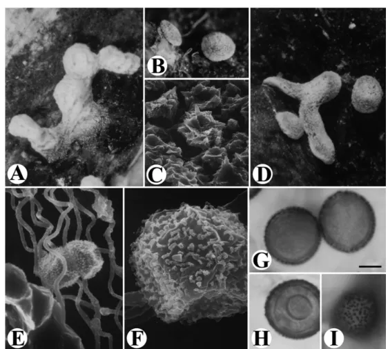
Fructification plasmodiocarpous, scattered to gregarious, 0.33-1.92 mm long, up to 4.15 mm in their longer dimension, accompanied by sessile, sporangiate forms. Sporangia usually flat-pulvinate, whitish, 0.27-0.44 mm in diameter, dehiscent circumscissile; plasmodiocarps curved, irregular or nodular, grayish white,appearing thickerinheightthan thesporangiate
Fig. 3. Didymium leptotrichum. A-B & D: Fruiting bodies. C: Lime crystals on the outer surface of peridium. E: Capillitial threads, by SEM. F: Surface markings of spore, by SEM. G-H: Spores, marginal view. I: Spore, surface view. Scale bar: A-B & D = 200 μm; C = 7.5 μm; E = 5 μm; F = 1.7 μm; G-I = 6 μm.
form. Peridium membranous, covered with a crumbly layer of minute lime crystals forming a rough crust, colorless in transmitted light. Hypothallus inconspicuous. Columella absent or represented by a thickened pale orange-brown base. Capillitium dense, threads slender, undulate, sparsely branched, with isolated swelling nodules, hyaline to pale brown. Spores black in mass, violaceous brown or reddish brown by transmitted light, coarsely and distinctly warted, the warts often arranged in an obscurely line pattern, globose or subglobose, 12-16 m in diameter. Plasmodium not observed.
Specimens examined: Taipei City: Peitou, Yangmingshan National Park, on moss, CHL B2280, CHL B2281, Sept. 16, 2000.
Our specimens with characteristics fulfill well with the descriptions for Didymium leptotrichum in the reference (Nannenga-Bremekamp, 1991). This species is almost identical with Didymium nivicolum Meylan in the flat plasmodiocarps with a rough and white crust on the peridium, the capillitial threads, and the dark, warted, large spores (Moreno et al., 2003).
Didymium nivicolum is a nivicolous species and
known from either the snow bank or alpine area (Mitchel et al., 1980; Ing, 1999; Moreno et al., 2003). As listed in the reference (Nannenga-Bremekamp, 1991), it was under Didymium leptotrichum as a synonym and stated “Whether Didymium nivicolum and
Didymium leptotrichum are really identical cannot be
certain till the type material has been examined”. We placed our specimen as Didymium leptotrichum since it has the priority and our collections are from lowland instead of alpine.
Didymium listeri Massee, Monograph of the
Myxogastres: 244. 1892.
Description and illustration: C.H. Liu and Y.F. Chen, in Taiwania 43: 179-180, 182 (1998).
Didymium megalosporum Berk. & M.A. Curtis,
Grevillea 2: 53. 1873.
Taiwania Vol. 56, No. 4
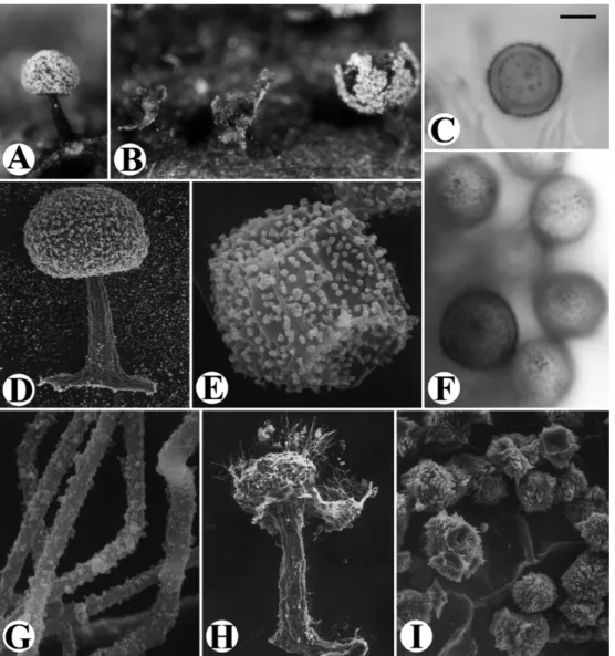
Fig. 4. Didymium minus. A: Fruiting body. B: Dehiscent sporangia. C: Spore, marginal view. D: Fruiting body, by SEM. E: Surface markings of spore, by SEM. F: Spores, surface view. G: Capillitial threads, by SEM. H: Dehiscent sporangium, showing columella, by SEM. I: Lime crystals on outer surface of peridium, by SEM. Scale bar: A-B = 250 μm; C, F = 4.5 μm; D = 120 μm; E, G = 1.4 μm; H = 85 μm; I = 10.5 μm.
Hsin-Chu Teacher’s College 7: 396-397 (1981). Specimens examined: Taipei City: farm of National Taiwan University, on fallen leaves, CHL B500, Apr. 11, 1985. Taipei County: Shiding, Wenshan Botanical Gardens of National Taiwan Univ., on fallen leaves and twigs, Yang 99-12 C5L1, Dec. 25, 1999.
The distinct characters of this species are the erect, white sporangium on a limeless brown stalk, the prominent stalked, whitish columella, and the large spores of 10-12 μm in diameter. Our specimens resemble Didymium nigripes in outer appearance of the white sporangia and the spore surface markings. They are different, however, in columella and capillitial threads. In Didymium nigripes, the columella are brown and subglobose, the capillitial threads bear dark thickenings, while in Didymium megalosporum the
columella are whitish and stalked as goblet in shape, the dark thickenings of the capillitial threads are not found.
Didymium melanospermum (Pers.) T. Macbr., N.
Amer. Slime-Moulds 88. 1899.
= Didymium melanospermum var. bicolor G. Lister, Monogr. Mycetozoa, 3rd Edi. (London): 115. 1925.
It was reported in a list by Nakazawa (1929), but no specimens were deposited in Taiwan.
Didymium minus (Lister) Morgan, J. Cincinnati Soc.
Nat. Hist. 16: 145. 1894. Fig. 4
0.42-0.80 (-1.0) mm in total height. Sporangia white, slightly depressed-globose, umbilicate below, (0.36-) 0.40-0.50 mm in diameter. Peridium membranous, covered with lime crystals, dehiscence irregular. Stalk erect or slightly curved, limeless, striate, brown to dull below, slightly narrowed and paler near the apex, 0.28-0.68 mm long. Columella large, globose, slightly depressed, rough, calcareous, brown, attaining to the center of the sporangium, 100-200 m in diameter. Capillitium of delicate, colorless to pale brown threads, scarcely branched and anastomosed, irregularly and densely marked by warts of various size on the surface under SEM. Hypothallus discoid, dark, membranous. Spores dark brown to nearly black in mass, brown to violaceous brown by transmitted light, globose, mostly 8.5 m, (7.5-) 8-10 m in diameter, minutely warted, the warts often in clusters on the surface. Plasmodium not observed.
Specimens examined: Taipei City: Peitou, Yangmingshan National Park, on fallen leaves, CHL B1423c, Apr. 1, 1998; Jong 19, June 28, 2001 (moist-chamber culture: June 4-28, 2001). Farm of National Taiwan University, on straw, CHL B254, Apr. 29, 1983. Nantou Co.: Huisun Forestry Station, on decaying twigs, CHL B498, Arp. 1, 1985.
The distinct characters of this species are the strongly umbilicate and stalked sporangia, and dark warted spores with warts often clustered on the surface. Our specimens with characters agree well in general with those of Didymium minus except that the surface of capillitial threads of ours is not smooth, a characteristic not found in the references (Martin and Alexopoulos, 1969; Nannenga-Bremekamp, 1991; Moreno et al., 2001).
Didymium nigripes (Link) Fr., Syst. Mycol. 3: 119.
1829.
Description and illustration: C.H. Liu, in Taiwania 27: 71-72, 74, 83 (1982).
Didymium ovoideum Nann.-Bremek., Med. Bot. Mus.
Herb. Utrecht 150: 780. 1958.
Description and illustration: C.H. Liu, in Taiwania 27: 71-72, 83 (1982).
Didymium perforatum Yamashiro, Journal of Science
of the Hiroshima University, B. II 3: 33. 1936. Description and illustration: C.H. Chung and C.H. Liu, in Taiwania 42: 278-279 (1997).
Specimen examined: Taipei City: campus of Affiliated High School of the National Taiwan Normal University, on dead leaves of
Eucalyptus robusta, C.-H. Chung M1507, Dec. 21, 1996.
The distinct characters of this species are the closely reticulate plasmodiocarps, the iridescent peridium sprinkledwithyellowish lime crystals,the darknetted
capillitium, and capillitial threads with nodose thickenings.
Didymium serpula Fr., Syst. Mycol. 3: 126. 1829.
Description and illustration: C.H. Liu, in Taiwania 27: 72-73, 75, 82 (1982).
Didymium squamulosum (Alb. & Schwein.) Fr., Symb.
Gasteromyc. 19. 1818.
Description and illustration: C.H. Liu, in Taiwania 27: 73, 75, 82 (1982).
Didymium verrucosporum A.L. Welden, Mycologia
46: 98. 1954.
Description and illustration: C.H. Liu, in Taiwania 27: 73, 76, 82 (1982).
LITERATURE CITED
Chung, C.-H. and C.-H. Liu. 1995. Didymium lenticulare
Thind & Lakhanpal (Physarales, Myxomycetes) New to Taiwan. Taiwania 40: 375-380.
Chung, C.-H. and C.-H. Liu. 1996a. Noets on Slime Molds
from Chunghua County, Taiwan (I). Fung. Sci. 11: 121-127.
Chung, C.-H. and C.-H. Liu. 1996b. Didymium floccosum
Martin, Thind & Rehill (Physarales, Myxomycetes) New to Taiwan. Taiwania 41: 175-179.
Chung, C.-H. and C.-H. Liu. 1997. Myxomycetes of Taiwan
VIII. Taiwania 42: 274-288.
Härkönen, M and T. Saarimäki. 1991. Tanzanian
Myxomycetes: first survey. Karstenia 31: 31-54.
Ing, B. 1999. The Myxomycete of Britain and Ireland.
Richmond Publication, Slough, UK, 374 pp.
Liu, C.-H. 1982. Myxomycetes of Taiwan III. Taiwania 27:
64-85.
Liu, C.-H. 1983. Myxomycetes of Taiwan IV: Corticolous
Myxomycetes. Taiwania 28: 89-116.
Liu, C.-H. 1989. Myxomycetes of Taiwan V: Two New
Records. Taiwania 34: 5-10.
Liu, C.-H. and Y.-F. Chen. 1998. Myxomycetes of Taiwan
X. Three new records of Didymium. Taiwania 43: 177-184.
Liu, C.-H. and Y.-F. Chen. 1999. Myxomycetes of
Taiwan—XII. New records and newly rediscovered species. Taiwania 44: 368-375.
Liu, C.-H., F.-H. Yang and J.-H. Chang. 2002.
Myxomycetes of Taiwan XIV. Three new records of Trichiales. Taiwania 47: 97-105.
Martin, G. W. and C. J. Alexopoulos. 1969. The
Myxomycetes. Univ. of Iowa Press, Iowa City, Iowa. 477 pp.
Mitchel, D. H. and S. W. Chapman. 1980. Notes on
Colorado fungi IV: Myxomycetes. Mycotaxon 10: 299-349.
Taiwania Vol. 56, No. 4
Moreno, G., C. Illana and M. Lizarraga. 2001. SEM studies
of the Myxomycetes from the Peninsula of Baja California (Mexico), III. Additions. Ann. Bot. Fennici 38: 225-247.
Moreno, G., A. Sanchez, A. Castillo, H. Singer and C. Illana. 2003. Nivicolous Myxomycetes from the Sierra
Nevada National Park (Spain). Mycotaxon 87: 223-242.
Nakazawa, R. 1929. A list of Formosan Mycetozoa. Trans.
Nat. Hist. Soc. Formosa 19: 16-30.
Nannenga-Bremekamp, N. E. 1991. A Guide to Temperate
Myxomycetes. Biopress Limited, Bristol, UK. 409 pp.
Shi, H. 1981. Myxomyetes in Yangmingshan Area, I. Bull.
Hsin-Chu Teacher’s College 7: 392-410.
Wang, S.-M., Y.-W. Wang and S. Huang. 1981. The
Revised Checklist of Myxomycetes in Taiwan. Biol. Bull. Natl. Taiwan Normal Univ. 16: 1-12.


