Microstructure and Magnetic Property of the Ferromagnetic
Fe-Pd-Rh Alloys
Yin-Chih Lin
1, Hwa-Teng Lee
2 1Department of Mold and Die Engineering, National Kaohsiung University of Applied Sciences
2Department of Mechanical Engineering, National Cheng Kung University
NSC Project No. : NSC 94-2212-E-151-008
Abstract
This study shows that an appearance of an intermediate martensitic structure exists between the fcc → L10 martensitic transformation in the aged
Fe-30Pd-4Rh alloys. The intermediate phase (L1m) has a
monoclinic structure with the lattice parameters of a = 3.193 Å, b = 3.684 Å, c = 3.141 Å,and β = 92.042°, as confirmed by TEM and X-ray diffraction. The crystal orientation relationships between the L1mand L10 can
be demonstrated as [101]L10//[100]L1m and
[112]L10//[11 1]L1m, respectively. This observation
suggests that the observed intermediate L1mmonoclinic
phase is tentatively the adaptive martensite.
The magnetization (M) versus magnetic field (H) M-H curve, measured at the temperatures of 50, 200, and 350 K of the alloys, first solution-treated (S. T.), then thermally aged at 450C for 100 hours, reveals an abrupt drop at the saturation remanence (Mr). This result indicates that two phases exist in the aged alloys, and these two phases have a different saturation remanence. The two phases, i.e., the adaptive L1m
monoclinic phase and ordered L10martensitic structure,
are confirmed by TEM and X-ray study.
Keywords:
Fe-Pd-Rh alloys, Adaptive L1m structure,TEM and X-ray diffraction, Magnetic property
1. Introduction
The adaptive structure is a metastable phase alternative to the normal martensitic phase. The adaptive phase cannot exist in the stress-free unconstrained state. It forms only in a constrained state when formation of a more stable normal martensite is suppressed by a large transition-induced elastic energy. A crystal lattice of the adaptive phase is derived from that of the normal martensite phase by crystal-plane shuffling. The main geometrical feature of the crystal lattice of the adaptive phase is that it is related to the parent phase lattice by an invariant plane crystal-lattice rearrangement. The smaller the twin surface energy and the greater the crystal-lattice mismatch, the easier it is to form an adaptive martensite. In the Fe-Pd alloy system, the adaptive martensite exists only within a narrow temperature range above the temperature of the martensitic transformation. In this case, the formation of
the adaptive structure can be perceived as the premartensitic phenomenon. The crystal-lattice structure of the adaptive martensite is very sensitive to the applied stress, and the appearance of the adaptive phase seems to occur just below the second-order transition. Therefore, the adaptive martensitic structure should generate extra diffraction spots related to accommodation shuffling of crystal plane. The incommensurate position of its diffraction spots is caused by random faulting of the periodic distribution of shuffling plane; such a fault, like the formation of stacking faults in the fcc lattice, leads to a shift of the diffraction spots from their regular positions. The value of the shift of the diffraction spots depends on the density of faults, and thus, is determined by the crystal-lattice mismatch. The incommensurate positions of the diffraction spots of the randomly faulted adaptive phase are not the same in all Brillouin zones [1,2].
The primary purpose of the present study is to provide insight into the phase transformation and magnetic property of the aged Fe-30Pd-4Rh ferromagnetic alloys by use of selected area diffraction pattern (SADP) of TEM, X-ray diffraction, and superconducting quantum interference device (SQUID). There is evidence of the intermediate L1m monoclinic
structure which should be formed between the fcc → L10 martensitic transformation. This observation
suggests that the observed intermediate L1m phase
seems to be the adaptive martensite. The crystal structure identification and the crystal orientation relationship between the adaptive L1m phase and the
normal L10martensite are confirmed by SADP of TEM,
while the lattice parameter of L1mand L10structures is
obtained by the calculation method using X-ray diffraction d-spacing in association with the measurement the SADP of TEM image. The result reveals that the intermediate L1m phase has a
monoclinic structure with the lattice parameters of a = 3.193 Å, b = 3.684 Å, c = 3.141 Å,and β = 92.042°, and the normal L10martensite has an ordered structure with
the lattice parameters of a = 3.876 Å, c = 3.684 Å, and c/a = 0.950, respectively. After the aged treatment, the magnetic property of the alloys reveals an abrupt drop at the saturation remanence (Mr). This phenomenon indicates that two phases exist in the aged alloys, and these two phases have a different saturation remanence. Further use of TEM and X-ray investigation reveals that the two phases are comprised of the adaptive L1m
monoclinic phase with normal L10martensitic structure.
The magnetic property of the above-mentioned character is coincident with the TEM and X-ray study.
2. Experimental procedure
The Fe-30Pd-4Rh (at%) alloys were melted by pure electrolytic iron (99.9%), pure palladium (99.95%), and pure rhodium (99.95%) in an arc vacuum furnace under a controlled protective argon atmosphere. The cast ingot was sealed in an evacuated quartz capsule and homogenized at 1050 C for 60 hours, then subsequently hot-and cold forged to a thickness of 2 mm. After forging, the specimens were sliced and sealed in an evacuated quartz capsule again, then were solution-treated at 950 C for 1.5 hours and quenched into water at room temperature. The aged treatment was performed in the temperature range of 400-550 C for various times. Thin foils for TEM were prepared by double jet electropolishing in a solution containing 82% acetic acid, 9% perchloric acid, and 9% methanol at a temperature in the range of –7C~10 C using a current density of 2 A/cm2 to 4 A/cm2. Transmission electron microscopy (TEM), with a double tilt stage, was performed in an analytical type high resolution electron microscope (Hitachi HF-2000) with a field emission gun operated at 200 kV, and a Philips CM 200 TEM operated at 200 kV, respectively. The X-ray diffraction patterns were detected at room temperature using an X-ray diffractometer (Siemens D5000 Karlsruhe) with Cu-Kradiation, and diffraction angles were in the 2 ranges from 35 to 140. The magnetic property measurements were carried out with a superconducting quantum interference device (SQUID) magnetometer. The magnetization versus magnetic field (M-H) curves for the samples were measured at 50, 200, and 350 K with the maximum applied field of 30000 Oe.
3. Results and discussion
3.1 γ
fccphase transformation into the adaptive
L1
mphase+L1
0structure observed by TEM
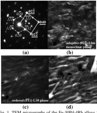
An essential TEM selected area diffraction pattern (SADP) with zone axis [101]L10//[100]L1m of the
Fe-30Pd-4Rh alloys S. T. at 950C for 1.5 h quenched in water then thermally aged at 450C for 100 h is shown in Fig. 1(a) (hkl denotes the ordered L10phase; hkl denotes
the adaptive L1mmonoclinic structure). On the basis of the
diffraction pattern analysis, an extra diffraction spot at the (0 22)L1m, (040)L1mposition can be seen from the SADP
micrograph that are related to accommodation shuffling of the crystal planes [1,2]. This effect is caused by random faulting of the periodic distribution of shuffling plane, which is similar to the formation of stacking faults in the fcc lattice in that the results will lead to a shift of the diffraction spots from their regular positions. The value of
the shift depends on the density of faults, and thus, is determined by the crystal-lattice mismatch. If there is a certain randomness in the fault distribution, then the position of the diffraction spots is incommensurate [1,2]. By calculating the X-ray diffraction d-spacing in association with SADP measurements, it is found that the adaptive L1m phase has a monoclinic structure with the
lattice parameters of a = 3.193 Å, b = 3.684 Å, c = 3.141 Å, and β= 92.042°, and the ordered L10 martensitic
structure has the lattice parameters of a = 3.876 Å, c = 3.684 Å, and c/a = 0.950. Figure 1(b) is a dark field (DF) image formed using adaptive (0 22)L1m monoclinic
reflection corresponding to Fig. 1(a). The DF image of Fig. 1(b) reveals that the intermediate L1mmonoclinic phases
are comprised of the periodic alternating antiphase boundaries (APBs) and microtwins. These planar faults strongly support the mechanism of coercivity in the aged Fe-Pd-Rh alloy system it has tended to favor APBs and microtwins pinning the magnetic domain wall that has been demonstrated as a possible source of magnetic hardening [3-6].
(a)
(b)
(c)
(d)
Fig. 1. TEM micrographs of the Fe-30Pd-4Rh alloys S. T. at 950C for 1.5 h quenched in water and thermally aged at 450 C for 100 h: (a) SADP of the zone axis [101]L10//[100]L1m (hkl denotes the ordered L10
martensitic structure; hkl denotes the adaptive L1m
monoclinic phase), (b) DF image of adaptive (0 22)L1m
monoclinic L1mreflection corresponding to (a), (c) DF
image of ordered (11 1)L10 L10 reflection
corresponding to (a), (d) BF image. The adaptive L1m
monoclinic structure has the lattice parameters of a = 3.193 Å, b = 3.684 Å, c = 3.141 Å, and β= 92.042°; The ordered L10 martensitic structure has the lattice
A dark field image using (11 1)L10 reflection,
corresponding to Fig. 1(a), is shown in Fig. 1(c). Figure 1(d) is a bright field (BF) image. The (0 22)L1m
adaptive L1mmonoclinic phase (gray contrast) shuffling
the ordered L10 martensitic (11 1)L10 crystal plane
(bright contrast) can be seen in the BF image.
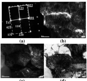
Figures 2(a)-(d) are a series of TEM micrographs taken from the alloys after S. T., then thermally aging at 550C for 110 h. Figure 2(a) is a typical SADP zone axis [112]L10//[11 1]L1m (hkl denotes the ordered
L10 phase; hkl denotes the monoclinic L1m structure).
Careful analysis of the SADP zone axis, reveals it as (11 1)L10//(101)L1m, in which the result is coincident
with the X-ray diffraction pattern as shown in Figs. 4(b)-(c). Note that in the SADP around the (022)L1m,
(121)L1m reflection, very small extra diffraction spots
can be seen in the micrograph, indicating that the L1m
→ L10phase transition is accompanied by a modulation
due to the composition fluctuations with shuffling of crystal planes [1-2,4-6]. Also, the intensity of the extra diffraction spot seems to gradually decrease compared to that of the specimen aged at 450 C for 100 h (Fig. 1(a)). Figure 2(b) is a DF image formed using (101)L1m
reflection that corresponds to Fig. 2(a). From the DF image of Fig. 2(b) analysis, we can see the tiny APBs morphology as well as tweed structures. Figure 2(c) is a
(a)
(b)
(c)
(d)
Fig. 2. TEM micrographs of the Fe-30Pd-4Rh alloys S. T. and thermally aged at 550C for 110 h: (a) SADP of the zone axis [112]L10//[11 1]L1m (hkl denotes the
ordered L10 phase; hkl denotes the monoclinic L1m
structure), (b) DF image for (101)L1m reflection
corresponding to (a), (c) DF image for (110)L10
reflection corresponding to (a), (d) BF image.
DF image form using (110)L10 reflection corresponding
to Fig. 2 (a) that the bright particles are the ordered L10
martensitic structures, in which we can also see the tiny
APBs. These APBs are the primary obstacles to the domain wall motion. The observed APBs microstructure in the L1m with L10structures strong illustration the
mechanism of coercivity in the Fe-Pd-Rh alloys is pinning controlled [3-6]. Figure 2(d) is a BF image. The tweed microstructures as well as the planar defect appear very sharply in the BF image. For further confirmation of the APBs and microtwins existing in the aged Fe-Pd-Rh alloys, a high magnification TEM micrograph including APBs and microtwins, indicated by arrow, is shown in Fig. 3(b). The APBs and microtwins pinning the magnetic domain are further demonstrated as a possible source of magnetic hardening of the alloys after S. T. and thermally aged treatment.
(a)
(b)
Fig. 3. TEM micrographs of the alloys after S. T. and thermally aged at 475C for 17 h showing APBs and microtwins. (a) BF, (b) DF.
3.2 X-ray diffraction pattern analysis
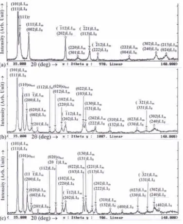
Figures 4(a)-(c) represent a series of X-ray diffraction patterns of the alloys S. T. and thermally aged at 450 C and 550 C for various times. The X-ray diffraction pattern of the alloys S. T. at 950C for 1.5 h and water quenched is shown in Fig. 4(a), in which two phases' (101)L1m and (111)L10 reflections are the main
diffraction peak. In the meanwhile, the (111)peak also appears, but there is no observablebctpeak in the X-ray
diffraction pattern. The X-ray experiment results exhibit that the addition of rhodium (Rh) element to the Fe-Pd alloy system will enhance the L1m and L10 phase
formation. When the alloy was aged at 450C for 100 h, many adaptive L1m monoclinic phases and ordered L10
martensitic structures appear in the X-ray diffraction patterns in addition to the (111)phase separation to the
(110)bct+L1mstructure as shown in Fig. 4(b). It is also
interesting to note that the X-ray diffraction peak for the plane (002)L10, (202)L10, (113)L10reflections corresponding
to the S. T. specimen of Fig. 4(a) had been transformed to
Fig. 4. X-ray diffraction patterns for the alloys solution treatment and after S. T. then thermally aged at 450C, 550C for given times: (a) 950 C for 1.5 h S. T. and quenched in water; (b) S. T. and aged at 450C for 100 h; (c) S. T. and aged at 550C for 110 h. (bctdenotes
the body centered tetragonal martensite, L1mdenotes
the adaptive L1mmonoclinic phase, L10 denotes the
ordered L10martensitic structure).
be the tetragonal splitting peak (200)L10;(002)L10,
(220)L10;(202)L10, and (131)L10;(113)L10reflections when
the specimen was aged at 550C for 110 h as shown in Fig. 4(c). These splitting peaks are the result of formation of the completely ordered tetragonal L10
martensitic structures attendant on the L1m→ L10phase
transformation reaction.
When compared with the L10 tetragonal splitting
peaks of Figs. 4(b) with (c), it discovers that the axial ratio c/a of the ordered L10martensite gradually decreased
as aging temperature was lowered to 450C. On the other hand, the degree of tetragonality develops when the specimen is aged at the lower temperature of 450C, in that case, it gives rise to the increase of the anisotropy and leads to enhance in the magnetic coercivity in the aged Fe-Pd-Rh alloys [7-12]. Careful analyses of Figs. 4(a)-(c) reveals a small shift in the diffraction angles demonstrating the lattice change due to the movement of the Pd-Rh atoms in association with the phase transformation. The γfcc phase separation into L1m+L10
phases confirmed by TEM is discussed in the previous Section 3.1. The two phases existing in the aged alloys, investigated by TEM and X-ray diffraction analyses, are exactly coincident.
3.3 Magnetic property
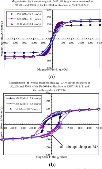
The mass magnetization (M) versus magnetic field (H) M-H curves, measured at temperatures 50, 200, and 350 K of the alloys S. T. at 950 C for 1.5 h and quenched in water, are shown in Fig. 5(a). The hysteresis loops reveal that the saturation magnetization values of the magnetic moment per unit mass are 137.4-99.5 (emu/g) for the S. T. sample measured at temperatures of 50-350 K. As shown in Fig. 5(b), we see the M-H curves measured at the same temperatures of the same alloys after S. T., then thermally aged at 450 C for 100 h. The hysteresis loops of Fig. 5(b) display a high saturation magnetization (Ms= 112.9-120.7 emu/g)
as well as a high coercivity Hc = 800-1000 Oe
throughout the temperatures about 50-350 K. Figure 5(b) is also revealing that the specimen aged at 450 C for 100 h possesses a superior coercivity than that of the S. T. specimen (Fig. 5(a)). This is due to much more ferromagnetic precipitates of the L1m and L10 phases
appearing in the latter specimen. The magnetic test suggests that the aged specimen increase in the saturation magnetization (Ms) and coercivity (Hc) can be
ascribed to the precipitation of the L1m and L10
structures. In addition, it is also interesting to note that the magnetization (M) versus magnetic field (H) M-H curve of the aged specimen exhibits an abrupt drop at saturation remanence (Mr) as indicated by arrow shown in Fig. 5(b). The characteristic magnetic property illustrates that two phases exist in the aged alloys, and the two phases have a different saturation remanence. The two phases existing in the aged alloys have been demonstrated by TEM and X-ray diffraction observation
and discussion in the above-Sections 3.1.-3.2. The characteristic magnetic property of the magnetic test exactly coincides with the microstructure analysis.
The magnetization behavior of the hysteresis loop of the alloys aged at 450C for 100 h has been shown in Fig. 5(b), in which some of the intrinsic magnetic properties and domain parameters of the aged Fe-Pd-Rh alloys are obtained from the experimental results and the equation data [3,13-14] as shown in the following: (1) anisotropy constant K1 = Ms×Ha/2 = 1.45×10⁷
ergs/cm3, (2) Ms (saturation magnetization) at R. T. =
970 emu/cc, (3) Ha(anisotropy field) = 30 kOe, (4) Tc
(Curie temperature) = 485C (758 K), (5) B (domain
wall thickness) =(A1/K1)1/2= 82 Å, (A1= 106ergs/cm,
A1: exchange parameter), (6)(domain wall energy) =
4(A1K1)1/2 = 15 ergs/cm2, (7) energy product (B×H)max theor= (4M
s/2)2= 37 MGOe.
(a)
(b)
Fig. 5. Magnetization (M) versus magnetic field (H) M-H curves measured at temperatures 50, 200, and 350 K of the Fe-30Pd-4Rh alloys: (a) solution-treated (S. T.) at 950C for 1.5 h and water quenched; (b) S. T. then thermally aged at 450C for 100 h.
4. Conclusions
1. On S. T. and quenched the alloys in water, the γfcc
phase separates into γfcc+L1m+L10 structures, then
by thermally aging the alloys, the γfcc+L1m+L10
phases decompose into bct+L1m+L10structures, as
verified by both the TEM and the X-ray diffraction pattern.
2. TEM and X-ray diffraction study confirms that an intermediate martensitic structure exists between the fcc → L10 martensitic transformation in the aged
Fe-Pd-Rh alloys. The intermediate phase (L1m) has a
monoclinic structure with the lattice parameters of a = 3.193 Å, b = 3.684 Å, c = 3.141 Å, and β = 92.042°. The observed intermediate L1mmonoclinic
phase suggests to be the adaptive martensitic structure.
3. TEM selected area diffraction pattern (SADP) reveals that the orientation relationships between the L10; L1mcan be demonstrated as [101]L10//[100]L1m
and [112]L10//[11 1]L1m, respectively.
4. The observed APBs and microtwins in the TEM image suggest that the primary mechanism of coercivity in the aged Fe-Pd-Rh alloy system is pinning controlled and the planar defects such as APBs and microtwins are the primary obstacles to domain wall motion.
5. The hysteresis loops of the aged alloys exhibit an abrupt drop at the saturation remanence (Mr) indicating two phases with a different saturation remanence existing in the aged alloys. The two phases, i.e., the adaptive L1mmonoclinic phase and
the ordered L10 martensitic structure are
demonstrated by TEM and X-ray diffraction study.
Acknowledgments
The authors would like to express their sincere appreciation to the National Science Council R.O.C. for supporting the work (under Grant-in-Aid for the project No: NSC-94-2212-E-151-008).
References
1. Khachaturyan, A. G., Shapiro, S. M. and Semenovskaya, S., Adaptive Phase Formation in Martensitic Transformation, PHYSICAL REVIEW B, Vol. 43, No. 13, pp. 10832-10843, 1991.
2. Shimizu, K. and Tadaki, T., Recent Studies on the Precise Crystal-Structural Analyses of Martensitic Transformations, Materials Trans. JIM, Vol. 33, No. 3, pp. 165-177, 1992.
3. Klemmer, T. J., Hoydick, D., Okumura, H., Zhang, B. and Soffa, W. A., Magnetic hardening and coercivity mechanisms in L10 ordered FePd
ferromagnets, Scripta Metallurgica et Materialia, Vol. 33, pp. 1793-1805, 1995.
Magnetization (M ) versus magnetic field (H ) M-H curves measured at 50, 200, and 350 K of the Fe-30Pd-xaRh alloy s as 950C/1.5h S. T.
-200 -150 -100 -50 0 50 100 150 200 -5000 -4000 -3000 -2000 -1000 0 1000 2000 3000 4000 5000
Magnetic Field, H /(Oe)
M ag n et iz at io n , M /( em u /g ) 350 K(Ms=99.5 emu/g) 200 K(Ms=124.7 emu/g) 50 K(Ms=137.4 emu/g)
Magnetization (M ) versus magnetic field (H ) M-H curves m easured at 50, 200, and 350 K of the Fe-30Pd-xaRh alloy s as 950C/1.5h S. T. and
therm ally aged at 450C/100h
-200 -150 -100 -50 0 50 100 150 200 -5000 -4000 -3000 -2000 -1000 0 1000 2000 3000 4000 5000
Magnetic Field, H /(Oe)
M ag n et iz at io n , M /( em u /g ) 350 K(Ms=112.9 emu/g) 200 K(Ms=118.5 emu/g) 50 K(Ms=120.7 emu/g) an abrupt drop at Mr
4. Klemmer, T. J., Liu, C., Shukla, N., Wu, X. W., Weller, D., Tanase, M., Laughlin, D. E. and Soffa, W. A., Combined reactions associated with L10
ordering, Journal of Magnetism and Magnetic Materials, Vol. 266, pp. 79-87, 2003.
5. Zhang, B. and Soffa, W. A., Magnetic domains and coercivity in polytwinned ferromagnets, Phys. Stat. Sol. (a), Vol. 131, pp. 707-725, 1992.
6. Sugiyama, M., Oshima, R. and Fujita, F. E., Mechanism of FCC-FCT thermoelastic martensite transformation in Fe-Pd alloys, Trans. of the Japan Institute of Metals, Vol. 27, No. 10, pp. 719-730, 1986.
7. Oshima, R., Successive martensitic transformations in Fe-Pd alloys, Scripta Metallurgica, Vol. 15, pp. 829-833, 1981.
8. Sohmura, T., Oshima, R. and Fujita, F. E., Thermoelastic FCC-FCT martensitic tramsformation in Fe-Pd alloy, Scripta Metallurgica, Vol. 14, pp. 855-856, 1980.
9. Muto, S., Oshima, R. and Fujita, F. E., Elastic softening and elastic strain energy consideration in the F.C.C.F.C.T. transformation of Fe-Pd alloys, Acta metall. mater., Vol. 38, No. 4, pp. 685-694, 1990.
10. Oshima, R., Sugiyama, M. and Fujita, F. E., Tweed structures associated with Fcc-Fct transformations in Fe-Pd alloys, Metall. Trans. A., Vol. 19A, pp. 803-810, 1988.
11. Sugiyama, M., Oshima, R. and Fujita, F. E., Martensitic transformation in the Fe-Pd alloy system, Trans. of the Japan Institute of Metals, Vol. 25, No. 9, pp. 585-592, 1984.
12. Oshima, R., Muto, S. and Fujita, F. E., Initiation of FCC-FCT Thermoelastic Martensite Transformation from Premartensitic State of Fe-30at% Pd Alloys, Materials Trans. JIM, Vol. 33, No. 3, pp. 197-202, 1992.
13. Livingston, J. D., The history of permanent magnet materials, JOM. February, pp. 30-34, 1990.
14. Cullity, B. D., : Introduction to Magnetic Materials, ed. by M. Cohen, Addison-Wesley, Reading, Massachusetts, USA, chap. 1-2, 1972.