行政院國家科學委員會專題研究計畫 成果報告
腦缺血變化於水腦症之影響: 腦缺血變化增生因子, VEGF
及 PLGF 於實驗性水腦症之分析及其重要性
計畫類別: 個別型計畫 計畫編號: NSC92-2314-B-002-254- 執行期間: 92 年 08 月 01 日至 93 年 12 月 31 日 執行單位: 國立臺灣大學醫學院外科 計畫主持人: 杜永光 共同主持人: 賴達明,廖漢文 報告類型: 精簡報告 處理方式: 本計畫可公開查詢中 華 民 國 94 年 9 月 6 日
中文摘要
常壓性水腦症(Normal pressure hydrocephalus, NPH)以及非阻塞性的慢 性水腦症是常見於老年人的神經系統疾病。有若干研究顯示病變的發生乃導致 於水腦造成腦室周圍組織的缺血。本研究是要探究血管生成因子在大鼠水腦模 式下是否會增加,以及其增加之位置分布與代表的意義。利用 22 隻大鼠水腦 模式,定量核糖核酸檢查發現 VEGF 受體及 PLGF 會在水腦生成時上升,而增 加的位置主要在基底核與海馬回等靠近腦室的地方,這也剛好符合臨床病例之 症狀(基底核異常引發步履不穩,而海馬回異常導致記憶力喪失)。免疫染色發 現在慢性水腦時 腦室周圍白質之血管內皮細胞 VEGF 受體表現增加。本實驗 因此證實缺血在水腦病變致病因素之意義,並且由於 PlGF 與病態炎症之血管 增生有關,或許炎症反應也是水腦病變致病因素之一。 英文摘要
Background and Purpose— In chronic hydrocephalus, a rolefor tissue hypoxia
resulting from cerebrovascular compressionis suggested. The purpose of this study
was to evaluate the geographic and temporal pattern of angiogenic factor elevation
after adultkaolin-induced hydrocephalus.
Methods— In 22 adult Sprague-Dawley rats, kaolin hydrocephaluswas induced and
quantitative reverse transcription polymerase chain reaction (QPCR) was performed at 1 week (short term) and 4 weeks (long term). Immunostaining of vascular endothelial growth factor receptor 1 (Flt-1) was also performed at 1 (short term), and 4 (long term) weeks.
Results— At 1 weeks, Placenta growth factor (PlGF) and Flt-1 wasincreased in
Striatal area and hippocampus but not cortical areas.Four weeks after hydrocephalus
induction, PlGF elevation persisted but Flt-1 elevation goes down. At 4 weeks, Flt-1
immunohistochemical changes in the periventricular area became most evident.
Conclusions— The observed Flt-1 and PlGF elevation supported the hypothesis that ischemia is the major mechnism of hydrocephalic damage. The proinflammatory cytokine PlGF may have significant role in the pathogenesis of chronic
Introduction
Recent investigations in experimental kaolin-induced hydrocephalushave shown
varying involvement of cholinergic, dopaminergic,and noradrenergic
neurotransmitter systems, indicating a rathercomplex neuronal disturbance[1-5].
Cerebral ischemia wassuggested as a potential cause for the observed neuronal
dysfunctionby xenon CT blood flow study or cerebral metabolism study using
magneticresonance spectroscopy[6-10].
Angiogenic factors has been found to be elevated in cerebral ischemic disorders. VEGF and its receptors, HIF-1α and recently PlGF were found to be elevated in astrocytes, neurons or endothelial cells following experimental focal cerebral
ischemia[11-15]. Their role of angiogenic factors in experimental hydrocephalus has never been evaluated.
In the present study we sought to assay the mRNA expression of VEGFR1 (Flt-1) and PlGF in different area of the brain following experimental Kaolin induced hydrocephalus. These data was confirmed at the protein level by the
immunohistochemistry of the Flt-1 expression.
Material and methods 一、水腦之生成及評估
本實驗以三週大之雄性 Sprague-Dawley rats 為對象。使用 Ketamine 90mg/kg 及 Xylocazine 10mg/kg 之混合液以肌肉注射之方式麻醉,在頸部及枕 骨交界處剃毛消毒後,以 27-gauge 之針頭進行大枕池(Cisterns magnum)之穿 刺,並緩慢注入 0.05ml 之無菌 Kaolin 懸浮液(250mg/ml)。 Sham operation 組則注入同量之生理食鹽水。動物由麻醉中恢復後送回動 物室,令其自由攝食及飲水,每週紀錄其習性、步伐及體重。 二、QPCR 每組四隻,在一週與四週時,將老鼠(包括打生理時鹽水的老鼠)在 pentobarbital 麻醉下犧牲,腦部在 5 分鐘內取出置於冰上,依廠商方法抽取全核糖核酸,再 取 2ug RNA 作反轉錄,接著用 cybergreen RTPCR 方法側 RNA 的量(Roche)。 相對 RNA 的量用 GAPDH 相對量來作校正。
二、以免疫組織化學法(Immunohistochemistry),測 VEGFR1 及 PLGF 於腦組織
內之分佈情形 1. 標本之準備
老鼠先以 5%之 Choloral hydrate 麻醉(0.008ml/gm, I.P.)後打開其胸腔, 於右心房處插入導管,先以 100ml 之生理食鹽水灌流,再以 4% paraformaldehyde solution in PBS(pH=7.35)灌流,固定腦組織。其後於 4°C 之環境下取出腦子,再以冷凍切片機將自 frontal tip 算起之後的 4-7mm(或相當於 foramen Monro 前後 3mm 之處)之腦組織切成 3mm 之冠狀向之厚塊,再切成 1mm 之切片。 切下之切片再於 4%之 paraformaldehyde solution in PBS 中進行一小時
之 postfixtion 後,於 30% sucrose/PBS solution 中在 4°C 溫度下進行 24
小時之 cryoprotection。這些 1mm 之切片再於液態氮中以 isopentane 緩 降溫冷凍後存於-70°C 之冰箱中。 2. 免疫組織染色 冷凍之 1mm 冠狀向薄片由冰箱中取出,再以冷凍切片機切成 10-16um 之薄片,所切出之系列薄片中,取出單數片進行本步驟研究,雙數片則 進行下一步驟中血管密度之研究。切片之後將這些單數片於 2.5% BSA,3000u/ml heparin in PBS 中浸泡 30 分鐘,再以 PBS 液洗三次,每 次 5 分鐘。
其後將切片分別置於 anti-PLGF polyclonal antibody(R & D Inc.)或 anti-VEGFR1
polyclonal antibody(Coldspring Harbor Co.)in antibody solution(包含 0.25% Triton
X, 1% BSA, 3000u/ml heparin, 及 10u/ml anti-Rnase in PBS)中 24 小時(4°C 下),
取出後,再以 PBS 洗三次,每次 5 分鐘,再將切片於室溫下置於
Peroxidase-conjugated antibody 中一小時,再以 PBS 洗三次。之後將切片置於 ABC Kit 6ul/ml A and B in PBS 中一小時,再以 PBS 溶液洗三次,最後以 DAB staining kit(Vector Labs-SK 4100)作染色。染色後之切片於顯微鏡下觀察其染 色情形,以決定 VEGF 及 PLGF 在腦內之分佈情形,同時攝影紀錄以與下一步 驟中,由偶數片所觀察得之腦內各部位之血管密度作比較。 四、腦組織內之血管密度 本實驗中有關腦部血管密度之分析,使用 factor 8 免疫切片染色法(Fong 1995, Ma 1998)。在前一步驟以冷凍切片所得之偶數薄片,以 10% normal horse serum 浸泡 2 小時,再以 PBS 洗三次,每次 10 分鐘。再將切片置於含有以 10%
normal horse serum 及 0.3% Triton X-100 作 1:50 稀釋 factor 8 之溶液中 16 小時(於
4°C 下),再置於含有 3% H2O2及 10% methanol 液中 20 分鐘,再以 PBST 洗三
次,每次 10 分鐘。以上切片取出後,再置於以 PBS 液(含 0.018% normal horse serum)作 1:200 稀釋之 biotinylated anti-rat IgG 液中,於室溫下留置 3 小時。
微血管會由 diaminobenzidine 染色。染色之切片以 computed image analyzer 下顯 微鏡下計算血管之密度。血管之密度分以下四級:1. no staining; 2. slight staining; 3. moderate staining; 4. dense staininig,以便進行統計分析。
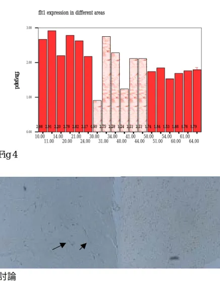
結果 一、水腦之生成 藉由 kaolin 注射,水腦生成率為 75%,圖一為大腦取下分析時所得之相片。 二、PlGF 之表現增加主要發生在基底核 由各區之 PlGF 表現,可知 PlGF 表現在基底核之位置持續增加、其他位置則 無明顯變化(fig 2)。 三、VEGF 受體之表現增加主要發生在基底核與海馬回。 Flt-1 mRNA 從第一週即增加,表現增加主要發生在基底核與海馬回 (fig 3), flt1 免疫染色可見正常時 flt-1 染不出來,而在四周之水腦腦室周圍血管內皮細 胞會被染出(fig 4)。圖四之箭頭指出血管內皮細胞之染出情形。 Fig 1 Fig2 0 0.5 1 1.5 2 2.5 3 3.5 Hippocampus Normal 1W 4W Striatum Normal 1W 4W Thalamus Normal striatum
Fig 3 10.00 11.0014.0020.0021.0024.0030.0031.0034.0040.0041.0044.0050.0051.0054.0060.0061.0064.00 0.00 1.00 2.00 3.00 f l t v g a p d D D D D D D D D D D D D D D D D D D 2.66 2.91 2.20 2.78 2.62 2.17 0.90 2.75 2.28 1.24 2.11 2.11 1.74 1.84 1.53 1.68 1.76 1.79
flt1 expression in different areas
Fig 4
討論
In this adult model of hydrocephalus, the cisternal injectionof kaolin causes an
inflammatory reaction and results in a progressive,but variable, dilation of the
ventricles[4]. As chronic hydrocephalusdevelops, stabilization of ventricular
enlargement and normalizationof intracranial pressure occurs. This model therefore
may be somewhat similar to some forms ofchronic hydrocephalus likepostmeningitic
hydrocephalus or the syndrome of normal pressure hydrocephalus.Our result showed
that 75% of our kaolin injected rat developed hydrocephalus on the time of sacrifice. No mortality was seen during the recovery period.
In the present investigation local changes in angiogenesis factor expression, as
measuredby quantitatively RT-PCR, were compared withtissue responses, as
demonstrated by immunohistochemistry,during the course of kaolin-induced
hydrocephalus. Both the RT-PCR response and the tissue responses displayed
significantregional and temporal alterations that were correlated in boththe
Striatal and Hippocampal Response in Kaolin-Induced Hydrocephalus
Very few studies have described damage to the striatal and hippocampal formationin
adult hydrocephalus[1, 2, 16].Most of these studies focus on the neuronal loss. Our
study is the first to demostrate angiogenic factor elevation in these two area of the brain[4]. In the human cerebral blood flow Mrspectroscope study, deep white matter is the primary site of decreased cerebral blood flow[6, 7]. Hippocampus, which is adjacent to the ventricle in rat, showed elevated Flt-1 expression in our study,
whichwas propably because of the ischemic drive. The striatal area was the other site of Flt-1 and PlGF overexpression in our study. This is also adjacent to the dilated cerebral ventricle. Striatum involved coordinated motor control. Through PET study and intracranial pressure manipulation, basal ganglia, thalamus and deep white matter was found to be the most vascular vulnerable area[1, 16].
The role of PlGF elevation in experimental hydrocephalus has never been reported. This is correlated with our study on elevated CSF PlGF in clinical hydrocephalus patients. Whether this is pathogenetic as PlGF is a proinflammatory factor or mearly a reflection of proangiogenic factor elevation remained to be studied[17, 18].
In conclusion, in the present descriptive study, we showed acorrelation of both the
short-term and the long-term neuronalresponses with a moderate but prolonged
angiogenic factor production.
參考資料
[1] Klinge P, Muhlendyck A, Lee S, Ludemann W, Groos S, Samii M, et al. Temporal and regional profile of neuronal and glial cellular injury after induction of kaolin hydrocephalus. Acta Neurochir Suppl 2002;81:275-7.
[2] Ding Y, McAllister JP, 2nd, Yao B, Yan N, Canady AI. Neuron tolerance during hydrocephalus. Neuroscience 2001;106(4):659-67.
[3] Del Bigio MR. Calcium-mediated proteolytic damage in white matter of hydrocephalic rats? J Neuropathol Exp Neurol 2000;59(11):946-54.
[4] Tashiro Y, Chakrabortty S, Drake JM, Hattori T. Progressive loss of glutamic acid decarboxylase, parvalbumin, and calbindin D28K immunoreactive neurons in the
cerebral cortex and hippocampus of adult rat with experimental hydrocephalus. J Neurosurg 1997;86(2):263-71.
[5] Tashiro Y, Drake JM, Chakrabortty S, Hattori T. Functional injury of cholinergic, GABAergic and dopaminergic systems in the basal ganglia of adult rat with
kaolin-induced hydrocephalus. Brain Res 1997;770(1-2):45-52.
[6] Braun KP, Gooskens RH, Vandertop WP, Tulleken CA, van der Grond J. 1H magnetic resonance spectroscopy in human hydrocephalus. J Magn Reson Imaging 2003;17(3):291-9.
[7] Bateman GA. Vascular compliance in normal pressure hydrocephalus. AJNR Am J Neuroradiol 2000;21(9):1574-85.
[8] Braun KP, van Eijsden P, Vandertop WP, de Graaf RA, Gooskens RH, Tulleken KA, et al. Cerebral metabolism in experimental hydrocephalus: an in vivo 1H and 31P magnetic resonance spectroscopy study. J Neurosurg 1999;91(4):660-8.
[9] Tanaka A, Kimura M, Nakayama Y, Yoshinaga S, Tomonaga M. Cerebral blood flow and autoregulation in normal pressure hydrocephalus. Neurosurgery
1997;40(6):1161-5; discussion 1165-7.
[10] Kimura M, Tanaka A, Yoshinaga S. Significance of periventricular
hemodynamics in normal pressure hydrocephalus. Neurosurgery 1992;30(5):701-4; discussion 704-5.
[11] Autiero M, Luttun A, Tjwa M, Carmeliet P. Placental growth factor and its receptor, vascular endothelial growth factor receptor-1: novel targets for stimulation of ischemic tissue revascularization and inhibition of angiogenic and inflammatory disorders. J Thromb Haemost 2003;1(7):1356-70.
[12] Autiero M, Waltenberger J, Communi D, Kranz A, Moons L, Lambrechts D, et al. Role of PlGF in the intra- and intermolecular cross talk between the VEGF receptors Flt1 and Flk1. Nat Med 2003;9(7):936-43.
[13] Beck H, Acker T, Puschel AW, Fujisawa H, Carmeliet P, Plate KH. Cell
type-specific expression of neuropilins in an MCA-occlusion model in mice suggests a potential role in post-ischemic brain remodeling. J Neuropathol Exp Neurol
[14] Ergenekon E, Gucuyener K, Erbas D, Aral S, Koc E, Atalay Y. Cerebrospinal fluid and serum vascular endothelial growth factor and nitric oxide levels in newborns with hypoxic ischemic encephalopathy. Brain Dev 2004;26(5):283-6.
[15] Hayashi T, Noshita N, Sugawara T, Chan PH. Temporal profile of angiogenesis and expression of related genes in the brain after ischemia. J Cereb Blood Flow Metab 2003;23(2):166-80.
[16] Klinge PM, Samii A, Muhlendyck A, Visnyei K, Meyer GJ, Walter GF, et al. Cerebral hypoperfusion and delayed hippocampal response after induction of adult kaolin hydrocephalus. Stroke 2003;34(1):193-9.
[17] Oura H, Bertoncini J, Velasco P, Brown LF, Carmeliet P, Detmar M. A critical role of placental growth factor in the induction of inflammation and edema formation. Blood 2003;101(2):560-7.
[18] Nagy JA, Dvorak AM, Dvorak HF. VEGF-A(164/165) and PlGF: roles in angiogenesis and arteriogenesis. Trends Cardiovasc Med 2003;13(5):169-75.