• 沒有找到結果。

Central Neurogenic Hyperventilation in A Conscious Patient with Chronic Brain Inflammation

N/A
N/A
Protected

Academic year: 2021

Share "Central Neurogenic Hyperventilation in A Conscious Patient with Chronic Brain Inflammation"

Copied!
6
0
0

加載中.... (立即查看全文)

全文

(1)

Central Neurogenic Hyperventilation in A Conscious Patient with

Chronic Brain Inflammation

Yi-Jen Chen

1

, Sheng-Feng Sung

2

, and Yung-Chu Hsu

2

1 Division of Pulmonary and Critical Care Medicine, Department of Internal Medicine;

2 Division of Neurology, Department of Internal Medicine, Ditmanson Medical Foundation, Chia-Yi Christian Hospital

Abstract

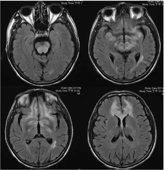
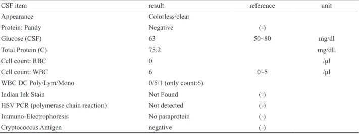
Central neurogenic hyperventilation (CNH) is a rare and easily forgotten diagnosis. A 53-year-old male patient presented with dyspnea for the past 1 month. His consciousness was clear and had bilateral upward gazing palsy. The arterial blood gas analysis showed severe respiratory alkalosis. Brain magnetic resonance imaging revealed symmetric hyperintense lesions involving the midbrain and the area surrounding the fourth ventricle. A brain biopsy showed gliosis and chronic inflammation. CNH results from an uninhibited respira- tory drive due to pons or medulla disorders, and a conscious patient can mislead the initial judgment of the physician. However, severe respiratory alkalosis and persistent nocturnal dyspnea should raise the clinical suspicion of CNH. Chronic brain inflammation with CNH has seldom been reported in the literature. This case provides another pathological possibility of CNH. (J Intern Med Taiwan 2013; 24: 328-333)

Key Words: Central neurogenic hyperventilation, Inflammation, Conscious

Introduction

Hyperventilation is defined as an increase in alveolar ventilation that results in excessive metabolic carbon dioxide expulsion. It may result in a decrease in arterial carbon dioxide tension to below the normal range. Many clinical conditions and diseases can lead to an excessive ventilatory drive, such as hypoxemia, pulmonary disorder, and metabolic disorder. Central neurogenic hyperventi- lation (CNH) is a rare respiratory syndrome which was first reported by Plum and Swanson in 1959.

1

It

is defined as respiratory alkalosis induced by lesions in the central nervous system without cardiac, pulmonary or other organic disease. Herein, we present the case of a conscious patient with CNH.

Case presentation

This 53-year-old male patient visited our emergency room (ER) with the chief complaint of progressive dyspnea in the recent one month. He denied drinking, smoking, or having previously undergone gastrointestinal surgery. The rapid respiratory rate was persistent even when he slept.

Reprint requests and correspondence:Dr. Yung-Chu Hsu

Address:Division of Neurology, Department of Internal Medicine, Ditmanson Medical Foundation Chia-Yi Christian Hospital, No. 539, Jhongsiao Rd., Chia-Yi City, Taiwan 60002

(2)

He worked in an industrial plastics factory and he had continued to work for the past month. He had visited a local clinic, however the symptoms had not improved at all. At our ER, his initial body tempera- ture was 36.8°C, with a pulse rate of 100 beats per minute, respiratory rate of 36 breaths per minute and blood pressure of 121/85 mmHg. His conscious- ness was clear. A physical examination did not demonstrate any remarkable findings, including clear breathing sounds and regular heart rhythm.

Neither audible cardiac murmurs nor leg edema were noted. The oxygen saturation was 99% in room air. A hemogram only revealed mild anemia, and the hemoglobin level was 13.0 g/dL. Biochemistry showed normal renal and liver function tests, and only mild hypokalemia was noted with a potassium level of 3.18 mmol/L. The initial chest radiograph showed neither significant lung lesions nor cardio- megaly. E lectrocardiography showed a normal sinus rhythm. The B-type natriuretic peptide

level was less than 5 pg/mL. Chest computed tomography revealed normal opacification of the pulmonary artery without evident filling defects and clear bilateral lung fields. Hyperventilation syndrome was initially suspected, however the symptom did not resolve after treatment for hyper- ventilation at the ER.

His respiratory rate was around 30 breaths per minute after hospitalization. Due to a normal cardiopulmonary function test, the hyperventilation was assumed to be caused by a neuropsychiatric disease such as generalized anxiety disorder. Thus alprazolam (0.5 mg) 1# tid was prescribed, however the patient became irritable. Alprazolam was thus discontinued and he subsequently calmed down. All of the series of arterial blood gas analysis showed persistent severe respiratory alkalosis and sufficient oxygen saturation (Table 1). After we excluded other etiologies of hyperventilation, CNH was highly suspected and a neurologist was consulted.

On neurological examination, the patient

was alert and his language function was normal.

His pupils were equal and reactive, however he had obvious upward vertical gaze palsy. No other brainstem signs existed. The muscle power of all four limbs was full with normal muscle tone, however deep tendon reflexes were brisk in all limbs. Plantar reflex showed a flexor response on both sides. His coordination system was relatively preserved. His neck was supple and there was no Kernig or Brudz- inski’s sign. Based on the neurological findings, we localized as a midbrain lesion.

Further brain magnetic resonance imaging was arranged which revealed far more extensive lesions: symmetric hyperintense FLAIR/T2 lesions involving bilateral periventricular white matter, frontotemporal regions, part of the basal ganglia, and midbrain (Fig. 1). The differential diagnosis from the neuroimaging findings included encephalitis, demyelinating disease, and infiltrative glioma. A cerebrospinal fluid study was within normal ranges, and the results of bacterial and viral detection tests were negative (Table 2). The level of folic acid and vitamin B12 were within normal ranges (folic acid:

7.23 ng/mL, vitamin B12: 692 pg/mL). Tracing back his history, there was no obvious evidence of malnutrition. Intoxication surveys including carbon monoxide and cocaine were all negative. Because the nature of the brain lesions was unknown, we suggested a brain biopsy. He was then transferred to another hospital for the biopsy and the final patho- logical report showed gliosis and chronic inflam- mation only. The patient did not come back to our hospital for further treatment. We could not contact with him or his family by the telephone. We are sorry that his outcome cannot be reported.

Discussion

Among many etiologies of hyperventilation, CNH is a so rare that easily forgotten diagnosis.

The consciousness of the patient with CNH is often

influenced by brain disorders. The fact that our

(3)

patient was able to work as usual and continue with his daily activities while he sought medical help is extremely rare, and made the initial diagnostic approach in the ER difficult. The arterial blood gas analysis revealed longstanding respiratory alkalosis with metabolic compensation. The white blood cell count of 6 /μl was only slightly increased, and would only have been significant had it been over 10 /μl.

Mildly elevated WBC counts with lymphocyte predomi- nance in cerebrospinal fluid analysis can appear with demyelinating processes, tumor, stroke, or rarely, parameningeal irritation (such as extensive sinusitis infiltrating the meninges). According to the symmetric lesions and no obvious parameningeal irritation focus

in the neuroimaging findings, a demyelinating process seemed most likely. Since this patient had a prolonged clinical course, a bacterial central nervous system infection was unlikely, so his pH and lactate levels were not measured.

The mechanism causing CNH is still poorly understood, and three have been proposed. First, and also the most traditional theory, was proposed by Plum and Swanson in 1959.

1

They suggested that there are three main respiratory centers in the brainstem: medullary center, pontine apneustic center, and pontine pneumotaxic center.

2

The medullary respiratory center mainly controls breathing impulses, while the other two pontine

Fig. 1. The FLAIR images of brain magnetic resonance imaging showed symmetric subcortical hyperintensity in bilateral

frontotemporal and basal ganglia, dorsal pontine, and midbrain.

(4)

especially primary central nervous system lymphoma, is the most common cause of CNH.

7,8

From the histological findings, our patient had a chronic immu- nological response to unknown stimuli. Chemical irritants in his work environment may have played a role. We proposed this final diagnosis and conclusion by carefully excluding other etiologies after the neuro- imaging diagnosis. Symmetric periventricular lesions on magnetic resonance imaging are usually caused by demyelination. The other two possible diagnoses were a central nervous system infection and tumor, however these lesions are not usually so symmetric. The clinical course and magnetic resonance imaging finding did not support the diagnosis of multiple sclerosis or paraneo- plastic syndrome. We therefore surveyed for uncommon demyelination diseases such as Wernicke’s encepha- lopathy, and toxic and drug related encephalopathy.

After these surveys and even a brain biopsy, only a toxin could explain the whole picture. We suspected that this centers adjust respiratory rhythm. Infiltrative

lesions, such as in our patient, damage the pons which may result in a disconnection syndrome of the above system.

3

The second hypothesis is that local lactate production in the central nervous system can stimulate medullary chemoreceptors.

4

The third theory, which is based on an animal model by Takahashi et al., also advocates that chemical stimu- lation may lead to CNH.

4

The authors found that a glutamate injection into lateral parabrachial neurons in the pons led to an increase in respiratory rate.

The characteristics of CNH are respiratory alkalosis with a decrease in arterial carbon dioxide tension (PaCO

2

) and normal arterial oxygen tension (PaO

2

) without evidence of pulmonary or cardiac abnor- malities. CNH may occur following stroke,

5

multiple sclerosis,

4

or brain tumors.

6

Tumor or inflamma- tory cytokine infiltration in the respiratory center may stimulate a tachypnea response.

6

Brain neoplasms, and

Table 1. The series of the arterial blood gas analysis

1

st

day 4

th

day 5

th

day 6

th

day (sleep)

PH 7.637 7.637 7.638 7.638

PCO

2

8.2 5.7 8.1 11.8

PO

2

126.7 157.5 154.4 116.7

HCO

3

8.6 5.9 8.5 12.4

BE -7.3 -9.5 -7.4 -4.3

O

2

Sat 99.1 99.4 99.4 99

Table 2. The results of cerebrospinal fluid examination

CSF item result reference unit

Appearance Colorless/clear

Protein: Pandy Negative (-)

Glucose (CSF) 63 50~80 mg/dl

Total Protein (C) 75.2 mg/dL

Cell count: RBC 0 /μl

Cell count: WBC 6 0~5 /μl

WBC DC Poly/Lym/Mono 0/5/1 (only count:6)

Indian Ink Stain Not Found (-)

HSV PCR (polymerase chain reaction) Not detected (-)

Immuno-Electrophoresis No paraprotein (-)

Cryptococcus Antigen negative (-)

(5)

patient had chronic central nervous system inflamma- tion due to chronic exposure to plastic elements, as the final biopsy report from the other hospital showed only gliosis and chronic inflammation. Unfortunately, we did not try steroid therapy, since central nervous system lymphoma is known to respond dramatically to steroids.

We regard this as a limitation to this case report.

There are very few reports about the rela- tionship between chronic brain inflammation and CNH, and this case provides further evidence of this relationship. Because of the insidious course and unusual presentation, it is a challange for physi- cians to make correct diagnosis at the ER. However, CNH should be added to the differential diagnosis of hyperventilation, particularly in the patients with severe respiratory alkalosis. Clinical alertness and careful neurological examinations, especially detail neuro-ophthalmic exam, may help to detect occult brain lesions.

References

1. Plum F, Swanson AG. Central neurogenic hyperventilation in man. Arch Neurol Psychiat 1959; 81: 535-49.

2. Chang CH, Kuo PH, Hsu CH, Yang PC. Persistent severe hypocapnia and alkalemia in a 40-year-old woman. Chest 2000; 118: 242-5.

3. Tarulli AW, Lim C, Bui JD, Saper CB, Alexander MP. Central neurogenic hyperventilation: a case report and discussion of pathophysiology. Arch Neurol 2005; 62: 1632-4.

4. Takahashi M, Tsunemi T, Miyayosi T, Mizusawa H. Revers- ible central neurogenic hyperventilation in an awake patient with multiple sclerosis. J Neurol 2007; 254: 1763-4.

5. Johnston SC, Singh V, Ralston HJ 3rd, Gold WM. Chronic dyspnea and hyperventilation in an awake patient with small subcortical infarcts. Neurology 2001; 57: 2131-3.

6. Tarulli AW, Lim C, Bui JD, Saper CB, Alexander MP. Central neurogenic hyperventilation: a case report and discussion of pathophysiology. Arch Neurol 2005; 62: 1632-4.

7. Nystad D, Salvesen R, Nielsen EW. Brain stem encephalitis with central neurogenic hyperventilation. J Neurol Neurosurg Psychiatry 2007; 78: 107-8.

8. Gaviani P, Gonzalez RG, Zhu JJ, Batchelor TT, Henson JW.

Central neurogenic hyperventilation and lactate production in

brainstem glioma. Neurology 2005; 64: 166-7.

(6)

慢性大腦發炎誘發之中樞神經性換氣過度

陳奕仁

1

  宋昇峰

2

  許永居

2

戴德森醫療財團法人嘉義基督教醫院  1 內科部胸腔暨重症科  2 神經內科

摘 要

內科急診常會遇見過度換氣症候群的病患,其發生的病因很多,中樞神經性病變因罕見 而不易列於起始的鑑別診斷中。於我們所報告的這位 53歲男性病患,當他因呼吸會喘求診 時,意識清楚且尚能工作。他的理學檢查顯示呼吸聲音清澈且心跳規則無雜音,動脈血分析 報告顯示極度呼吸性鹼中毒 PH: 7.637, PaCO

2

: 8.2 mmHg, PaO

2

:126.7 mmHg, HCO

3-

:8.6 mmol/L,

BE: -7.3 mmol/L and O

2

Sat: 99.1%. 胸部電腦斷層檢查未發現明顯病兆。神經學檢查發現其眼

睛向上看有困難,腦部核磁共振檢查顯示有大腦病變,經腦部切片檢查顯示其有慢性發炎狀

態。在處理過度換氣症候群的病患,對於極度呼吸性鹼中毒又有代謝性代償時,適當的神經

學檢查將有助於發現潛在的中樞性病兆。

數據

Fig. 1.  The FLAIR images of brain magnetic resonance imaging showed symmetric subcortical hyperintensity in bilateral  frontotemporal and basal ganglia, dorsal pontine, and midbrain.
Table 2. The results of cerebrospinal fluid examination

參考文獻

相關文件

The aim of this case report is to present the clinical and radiographic features of a 35-year-old female patient with autosomal dominant osteopetrosis type II who exhibited features

“Cowden’s syndrome affecting the mouth, gastrointestinal, and central nervous system: a case report and review of the literature,” Oral Surgery, Oral Medicine, Oral Pathology,

Accordingly, the present retrospective study analyzed bone loss as an objective clinical parameter for chronic periodontitis as a potential risk factor for the presence of OSCC in

Chronic traumatic ulcers are commonly found on the mucosa that is subjected to trauma from dentition such as buccal mucosa, lateral border of the tongue or

Chronic traumatic ulcers are commonly found on the mucosa that is subjected to trauma from dentition such as buccal mucosa, lateral border of the tongue or lips.. Lesions on

Histopathological examination of our case showed islands of basophilic mature cartilage in a dense fibrous connective tissue stroma with focal collection of chronic inflammatory

The growth in the number of vanco- mycin-induced thrombocytopenia cases presently seen may be associated with the increased use of the drug, especially in multiresistant patients

Introduction: Eagle’s syndrome is a rare condition that refers to chronic recurrent pain in the oropharynx, face, and neck due to elongation of the styloid process or calcification