• 沒有找到結果。

Long-term Survival of A Patient with Asymptomatic Left Ventricular Pseudoaneurysm after Acute Myocardial Infarction

N/A
N/A
Protected

Academic year: 2021

Share "Long-term Survival of A Patient with Asymptomatic Left Ventricular Pseudoaneurysm after Acute Myocardial Infarction"

Copied!
7
0
0

加載中.... (立即查看全文)

全文

(1)

Long-term Survival of A Patient with

Asymptomatic Left Ventricular Pseudoaneurysm after Acute Myocardial Infarction

Chun-Tai Mao, Ming-Feng Li, Yu-Cheng Kao, Wen-Jin Cherng, and Ming-Jui Hung

Department of Cardiology, Chang Gung Memorial Hospital, Chang Gung University College of Medicine, Keelung, Taiwan

Abstract

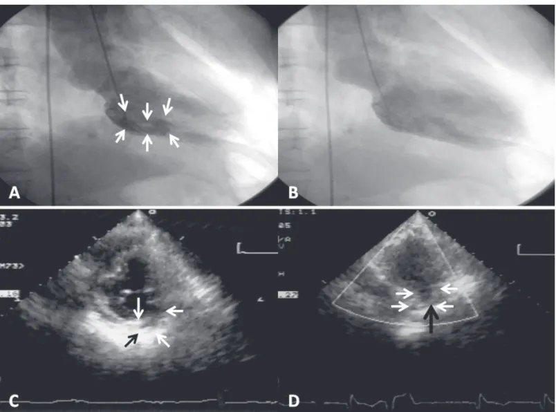
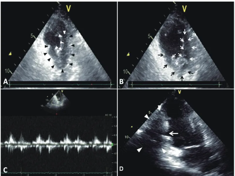
An 82 years old man developed a left ventricular pseudoaneurysm after acute myocardial infarction when he was 72 years old. Coronary angiography showed left main and triple-vessel coronary artery disease. On left ventriculography, a tubular-like pseudoaneurysm was demonstrated that originated from the basal inferoposterior wall of the left ventricle. He underwent coronary artery bypass surgery with no plication of the pseudoaneurysm because the surrounding tissues of pseudoaneurysm were all necrotic. The most recent follow-up transthoracic echocardiography revealed a hypokinetic basal inferior wall, impaired LV contraction with an ejection fraction of 44%, and an inferoposterior wall pseudoaneurysm. The patient was doing well more than 10 years after the myocardial infarction. The prognosis might be determined by the organized thrombi, aggressive pharmacologic treatment, and coronary artery bypass surgery. Although our patient has survived for more than 10 years with a nonsurgically treated post-infarction LV pseudoaneurysm, we could not provide an evidence to support that conservative therapy is enough for every patient with a post-infarction pseudoaneurysm. (J Intern Med Taiwan 2012; 23: 442-448)

Key Words: LV pseudoaneurysms, LV diverticulum, STEMI, CABG, Echo

Introduction

Left ventricular (LV) pseuodaneurysms develop when rupture of the free wall of the left ventricle is contained by pericardial adhesions or scar tissue

1

. Surgical resection of pseudoaneurysms is usually recommended because of the risk of spontaneous rupture

2

. However, few reports have been published on survival of patients with nonsurgically treated LV pseudoaneurysms

that developed as a result of acute myocardial infarction

3-6

. Herein, we present a patient who has survived for more than 10 years with a nonsurgically treated LV pseudoaneurysm that occurred after acute myocardial infarction.

Case report

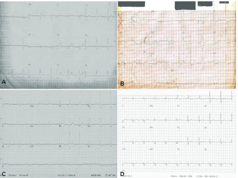
An 82-year-old man with a history of cigarette smoking, hypertension, and dyslipidemia developed an acute inferior myocardial infarction (Fig. 1 A &

Reprint requests and Correspondence:Dr. Ming-Jui Hung

Address:Department of Cardiology, Chang Gung Memorial Hospital, 222 Maijin Road, Keelung 20401, Taiwan

(2)

B) with no evidence of right ventricular infarction in February of 2002. The initial management was intravenous thrombolytic therapy. During the episode of acute myocardial infarction, the peak level of creatine kinase (6183 IU/L)-MB subform (12.2%) was 754 IU/L and the LV ejection fraction was 49% as measured by left ventriculography.

A tubular-like pseudoaneurysm originating from the basal inferoposterior wall of the left ventricle was noted (Fig. 2 A & B). The patient underwent coronary artery bypass surgery for his left main and triple-vessel coronary artery disease in March of 2002. Plication of the pseudoaneurysm was not performed by cardiac surgeon because the surrounding tissues of pseudoaneurysm were all

necrotic. No mechanical or electrical complications were noted during hospitalization. The patient made a good recovery and was discharged uneventfully.

He has been managed with atorvaststin, aspirin, isosorbide-5-mononitrate, carvedilol, amlodipine, and enalapril since 2002.

The patient underwent follow-up transthoracic echocardiography in 2003, 2005, and 2011. The size of the pseudoaneurysm in 2003 (Fig. 2 C) was similar to that in 2005 (Fig. 2 D). On parasternal short-axis view at the papillary muscle level, an acute angle between the contour of the aneurysm and the left ventricle was noted, suggesting an inferoposterior pseudoaneurysm. The most recent follow-up transthoracic echocardiography in 2011

Figure 1. A series of changes of 12-lead electrocardiograms in our patient. Before (A) and after (B) acute inferior

myocardial infarction electrocardiograms show a new Q-wave development (B) in inferior leads. Follow-up

electrocardiograms in 2007 (C) and 2011 (D) show serial changes (Q-wave and T-wave) after inferior

myocardial infarction.

(3)

revealed a hypokinetic basal inferior wall, impaired LV contraction with an ejection fraction of 44%, and an inferoposterior wall pseudoaneurysm (Fig.

3 A and B). Blood flow from the pseudoaneurysm to the LV cavity was noted (Fig. 3 C). On apical 2-chamber view, a saccular contour of the pseudoaneurysm with a narrow neck connecting it to the left ventricle was demonstrated (Fig. 3 D).

There was no pericardial effusion. The follow-up 12-lead electrocardiograms in 2007 (Fig. 1 C) and 2011 (Fig. 1 D) showed serial electrocardiographic changes of the present patient. Until June of 2012, the patient was doing well more than 10 years after the myocardial infarction without any evidence of

angina pectoris or heart failure.

Discussion

The development of LV pseudoaneurysms is a rare but potentially lethal complication of acute myocardial infarction, cardiac surgery, trauma, and infections. Most investigators consider surgery to be appropriate treatment for LV pseudoaneurysms

7

because of the risk of rupture when the condition is left untreated

8,9

. Up to now, post-infarction LV pseudoaneurysm is regarded as a surgical disease as recommended by ST-elevation myocardial infarction guideline

10

. However, some studies found that death was not attributable to cardiac rupture

Figure 2. Left ventriculograms (upper panels) and the follow-up transthoracic echocardiograms (lower panels) of our patient, who developed a basal inferoposterior pseudoaneurysm after inferior myocardial infarction. Systolic (A) and diastolic (B) frames of the contrast left ventriculograms show a tubular pseudoaneurysm (arrows) in the basal inferoposterior wall. The follow-up echocardiograms (short-axis views at the papillary muscle level) in 2003 (C) and 2005 (D) reveal little change in the size of the inferoposterior pseudoaneurysm (arrows).

Organized thrombi can be seen in panel D (black arrow).

(4)

in patients with pseudoaneurysms

4,6,11

. Yeo et al.

11

found that the all-cause mortality rates among patients with cardiac pseudoaneurysms were 31%

(13 of 42 patients) in patients who received surgical treatment and 60% (6 of 10 patients) in patients who received medical treatment during a median follow-up period of 4 years. Sakai et al.

12

did not recommend surgery if the pseudoaneurysm was connected to the LV wall with a narrow neck, or if it occurred in the presence of the postsurgical mitral valve. In addition, thrombus is frequently found in the pseudoaneurysm. Recently, some authors found that percutaneous closure of LV pseudoaneurysms

is a feasible alternative for high-risk surgical candidates

13

. However, further long-term follow-up studies are needed. The long-term survival of our patient might be related to the formation of organized thrombi within the pseudoaneurysm, which resulted in decreased LV pressure to its distal walls. Furthermore, aggressive pharmacologic treatment and coronary artery bypass surgery to decrease LV remodeling and preserve LV systolic performance might have played an important role in the long-term survival of this patient. The decline in the incidence of and mortality rate associated with cardiac rupture after acute myocardial

Figure 3. The most recent follow-up transthoracic echocardiograms. The parasternal short-axis views at the

end-systolic phase (A) and end-diastolic phase (B) show an tubular-like echo-free space posterior to the

inferoposterior wall (pseudoaneurysm, white arrows) without contractility (black and white arrowheads

in A) and with organized thrombi (black arrows in B). C. Doppler flow demonstrates blood flow entering

the pseudoaneurysm. D. Apical 2-chamber view shows a saccular contour (white arrowheads) of the

pseudoaneurysm with a narrow neck (arrow) connecting it to the left ventricle. LA = left atrium; LV = left

ventricle.

(5)

infarction over the last 30 years is associated with the increased use of reperfusion strategies and adjunct medical therapy

14

. The development of LV pseudoaneurysm is, therefore, rare

15

.

Recognition of LV pseudoaneurysm after acute myocardial infarction is still important because the presentation of LV pseudoaneurysm varies.

Pseudoaneurysm is most commonly detected using two-dimensional echocardiography. A narrow neck leading to the fundus is usually found. In contrast, a wide opening to the ventricular chamber is characteristic of true aneurysm. In addition, an acute angle between the contour of the aneurysm and the left ventricle, an abrupt transition from normal thickness of the myocardium to a thin-walled sac, a distinct discontinuity in the ventricular wall, and color Doppler studies demonstrating movement of blood from the suspected pseudoaneurysm to the LV cavity can be helpful in diagnosing pseudoaneu- rysms

4

. Unlike two-dimensional echocardiography, three-dimensional echocardiography provides good images of the LV posterior wall and is, therefore, an excellent tool for monitoring unruptured pseu- doaneurysms. In contrast to LV pseudoaneurysm, LV diverticula are rare congenital anomalies consisting of protrusion of endocardium and myocardium from the LV free wall

16

. Histopathologically, they are classified into fibrous or muscular. The former is non-contractile as it is constituted mostly from fibrous tissue. It most commonly originates from the apical or subvalvular positions. The muscular type includes all cardiac layers and usually emerges from the apex; therefore, it has a mechanical activity. This type is often associated with other congenital defects. Few case reports showed that diverticulae are not congenital but associated with hypertrophic cardiomyopathy

17,18

. Differentiation between these 2 entities is also important because LV diverticulum is often associated with serious complications, such as systemic thromboemblism, cardiac rupture, arrhythmia, and sudden death

19,20

.

Transthoracic echocardiography can show the origin, the neck (narrow or wide), the walls, and the contractility of the diverticula

21,22

. The multidetector computed tomography enables accurate evaluation of both LV myocardium and lumen

23

. Therefore, the multidetector computed tomography can establish the diagnosis of a LV pseudoaneurysm or diverticulum, which is the limitation of our case report. In our case, the outpouching echo-free space had no contractility and we believe that it was a case of pseudoaneurysm.

In conclusion, we present a patient who has survived for more than 10 years with a nonsurgically treated LV pseudoaneurysm that occurred after acute myocardial infarction. From this limited experience, however, we could not provide an evidence to support that conservative therapy is enough for every patient with a post-infarction pseudoaneurysm.

References

1. Dachman AH, Spindola-Franco H, Solomon N. Left ventricular pseudoaneurysm. Its recognition and significance.

JAMA 1981; 246: 1951-3.

2. Frances C, Romero A, Grady D. Left ventricular pseudoaneurysm. J Am Coll Cardiol 1998; 32: 557-61.

3. Hung MJ, Wang CH, Cherng WJ. Unruptured left ventricular pseudoaneurysm following myocardial infarction. Heart 1998; 80: 94-7.

4. Hung MJ, Cherng WJ, Kuo LT, et al. Echocardiographic characteristics of patients surviving nonsurgically treated left ventricular pseudoaneurysm after acute myocardial infarction. Am J Cardiol 2003; 91: 328-31.

5. Moreno R, Gordillo E, Zamorano J, et al. Long term outcome of patients with postinfarction left ventricular pseudoaneurysm. Heart 2003; 89: 1144-6.

6. Subban V, Sethuratnam R, Ajit MS. Asymptomatic large unruptured left ventricular pseudoaneurysm. Heart 2009; 95:

1139.

7. Brown SL, Gropler RJ, Harris KM. Distinguishing left ventricular aneurysm from pseudoaneurysm. A review of the literature. Chest 1997; 111: 1403-9.

8. Frances C, Romero A, Grady D. Left ventricular pseudoaneurysm. J Am Coll Cardiol 1998; 32: 557-61.

9. Davidson KH, Parisi AF, Harrington JJ, et al. Pseudoaneurysm of the left ventricle: an unusual echocardiographic presentation. Review of the literature. Ann Intern Med 1977;

86: 430-3.

10. Antman EM, Anbe DT, Armstrong PW, et al. ACC/AHA

guidelines for the management of patients with ST-elevation

(6)

myocardial infarction: a report of the American College of Cardiology/American Heart Association Task Force on Practice Guidelines (Committee to Revise the 1999 Guidelines for the Management of Patients with Acute Myocardial Infarction). Circulation 2004; 110: e82-292.

11. Yeo TC, Malouf JF, Oh JK, et al. Clinical profile and outcome in 52 patients with cardiac pseudoaneurysm. Ann Intern Med 1998; 128: 299-305.

12. Sakai K, Nakamura K, Ishizuka N, et al. Echocardiographic findings and clinical features of left ventricular pseudoaneurysm after mitral valve replacement. Am Heart J 1992; 124:

975-82.

13. Dudiy Y, Jelnin V, Einhorn BN, et al. Percutaneous closure of left ventricular pseudoaneurysm. Circ Cardiovasc Interv 2011; 4: 322-6.

14. Figueras J, Alcalde O, Barrabes JA, et al. Changes in hospital mortality rates in 425 patients with acute ST-elevation myocardial infarction and cardiac rupture over a 30-year period. Circulation 2008; 118: 2783-9.

15. Figueras J, Barrabes JA, Gruosso D, et al. Long-term course of stemi complicated by a moderate to severe pericardial effusion. Frequency of left ventricular pseudoaneurysm. Int J Cardiol 2012; 154: 212-4.

16. Yazici M, Demircan S, Durna K, Yasar E. Left ventricular diverticulum in two adult patients. Int Heart J 2005; 46:

161-5.

17. Maron BJ, Hauser RG, Roberts WC. Hypertrophic cardiomyopathy with left ventricular apical diverticulum.

Am J Cardiol 1996; 77: 1263-5.

18. Yamashita C, Nakamura H, Tobe S, Koterazawa T, Yamamoto S. Left ventricular diverticulum with hypertrophy of the left ventricular apex. Ann Thorac Surg 1992; 54:

761-3.

19. Sanada H, Shimizu M, Takeda R, et al. Two-dimensional echocardiographic and left ventriculographic evaluations of left ventricular diverticula. J Cardiol 1989; 19: 1107-16.

20. Speechly-Dick ME, Oliver RM, Slapak GI. Congenital left ventricular diverticula: a rare cause of sudden cardiac death.

Postgrad Med J 1992; 68: 378-80.

21. Iyisoy A, Kursaklioglu H, Celik T, Kose S, Genc C, Isik E.

A contractile left ventricular diverticulum. Int J Cardiovasc Imaging 2006; 22: 1-3.

22. Gonzales MR, Garg M. Images in cardiology: A rare finding of left ventricular diverticulum. Clin Cardiol 2005; 28: 308.

23. Ghersin E, Kerner A, Gruberg L, Bar-El Y, Abadi S, Engel A. Left ventricular pseudoaneurysm or diverticulum:

differential diagnosis and dynamic evaluation by catheter

left ventriculography and ECG-gated multidetector CT. Br J

Radiol 2007; 80: e209-11.

(7)

急性心肌梗塞併發無症狀的左心室偽瘤

—長期存活之病例報告

毛畯台  李明 峯  高瑜成  程文俊  洪明銳

基隆長庚醫院 心臟內科 長庚大學醫學院

摘 要

一位八十二歲男性於七十二歲時急性心肌梗塞後併發左心室偽瘤。冠狀動脈攝影顯示左 主冠狀動脈和三條冠狀動脈疾病;左心室攝影發現下後壁有一條狀偽瘤。他接受冠狀動脈繞 道手術但因偽瘤週邊組織皆壞死,所以無法以折疊術處理左心室偽瘤。最近的經胸心臟超音 波檢查顯示左心室下後壁運動低下、左心室收縮功能不良(射血分率為 44%)、左心室下後壁 有一偽瘤。此病患於心肌梗塞十年後仍存活良好。這預後可是能因為偽瘤內的組織化血栓、

積極的藥物治療和冠狀動脈繞道手術。雖然此個案以非外科治療後存活十年多,但我們無法

以此個案論證以後所有急性心肌梗塞後併發左心室偽瘤病患皆可只接受保守治療。

數據

Figure 1.  A series of changes of 12-lead electrocardiograms in our patient. Before (A) and after (B) acute inferior  myocardial infarction electrocardiograms show a new Q-wave development (B) in inferior leads
Figure 2.  Left ventriculograms (upper panels) and the follow-up transthoracic echocardiograms (lower panels) of our  patient, who developed a basal inferoposterior pseudoaneurysm after inferior myocardial infarction
Figure 3.  The  most  recent  follow-up  transthoracic  echocardiograms.  The  parasternal  short-axis  views  at  the  end-systolic phase (A) and end-diastolic phase (B) show an tubular-like echo-free space posterior to the  inferoposterior wall (pseudoan

參考文獻

相關文件

In this paper, we report a case of a patient who reported for bleeding gums to our department and was diagnosed with acute myeloid leukaemia incidentally on routine blood

The aim of this case report is to present the clinical and radiographic features of a 35-year-old female patient with autosomal dominant osteopetrosis type II who exhibited features

 In several long-term follow-up studies, MMP with exclusive oral involvement does not develop lesions in other sites during the course of the disease and also seems to be

Case Presentation: In this clinical case report, it is described a case of a 16-year-old male patient with an asymptomatic osteolytic lesion at first upper left molar apical level,

9,10,14–16 The Special Care Dentistry Association lists seven rationales for use of sedation and general anesthesia, which were similar rationales for treatment of the

Lingual thyroid is defined as an ectopic thyroid gland tissue located in the midline of the tongue base and it is uncommonly observed in clinical practice and is rare in children..

At the 11-month follow-up, the patient was still asymptomatic and the revitalization of the root canal was performed since the upper right maxillary incisor showed a distinct

Even though in this case the patient was treated for AML after the age of 5, the long-term side effects of the chemo and/or radiotherapy caused anomalies in all his permanent