Research Express@NCKU - Articles Digest
Research Express@NCKU Volume 10 Issue 9 - October 2, 2009 [ http://research.ncku.edu.tw/re/articles/e/20091002/4.html ]
Oct-3/4 expression reflects tumor progression
and regulates motility of bladder cancer cells
Ai-Li Shiau
1, Gia-Shing Shieh
2, Chao-Liang Wu
2,*1 Departments of Microbiology and Immunology, 2 Department of Biochemistry and Molecular
Biology, National Cheng Kung University, Tainan, Taiwan
wumolbio@mail.ncku.edu.tw *
Cancer Research 68: 6281–6291 AUG 1(2008)
C
ancer and embryonic stem cells exhibit similar behavior, including immortal, undifferentiated, andinvasive activities. Here we demonstrate that in clinical samples bladder tumors with intense expression of stem cell marker Oct-3/4 (also known as POU5F1) are associated with further disease progression, greater metastasis, and shorter cancer-related survival compared to those with moderate and low expressions. We used RT-PCR and immunohistochemical approaches to examine Oct-3/4 expression in a panel of clinical bladder TCC samples illustrating a portion of RT-PCR results in which various levels of Oct-3/4 mRNA were detected in four human primary bladder carcinoma specimens, but not in normal human bladder tissue (Fig. 1A). Further immunohistochemical staining of frozen sections confirms Oct-3/4 expression in superficial bladder tumor tissues, but not in normal tissues (Fig. 1B). H&E-stained bladder tumor with moderate Oct-3/4 expression served as a comparison (Fig. 1B, lower
right panel). Vast majority of Oct-3/4-positive tumor tissues suggest that Oct-3/4 expression in human
primary bladder carcinomas might induce a cascade of gene expression to promote tumorigenesis. We then systemically studied 57 patients with histologically confirmed superficial bladder TCC and 10 cases of non-neoplastic bladder tissues obtained by cystourethroscopic biopsies. Paraffin-embedded sections from tumor specimens were stained with H&E confirming the presence of at least 70% tumor cells. Overall, Oct-3/4 expression was seen in all tumor tissues but not in non-neoplastic bladder tissues. Of these tumor tissues, there were 25 (43.9%) with intense Oct-3/4 expression, 8 (14.0%) with moderate expression, and 24 (42.1%) with low expression. However, no significant association was found between Oct-3/4 expression and tumor grade, stage, morphology, and multiplicity. Univariate analysis revealed that multiplicity at diagnosis was the risk factor for tumor recurrence. Most importantly, tumors with intense Oct-3/4 expression were associated with further disease progression and greater metastasis compared with those with moderate or low expression (Fig. 1C). Furthermore, intense Oct-3/4
expression was also a risk factor for disease progression in bladder cancer subgroups, including grade II or III, as well as stage Ta-T1 or T2. Patients with low Oct-3/4 expression had longer cancer-related survival than those with high expression, as assessed by Kaplan-Meier survival analysis and log-rank test (Fig. 1C). The poor patient survival rate in Oct-3/4 high expression group may be attributed to greater disease progression and higher metastasis. Therefore, intensity of Oct-3/4 expression provides the prognostic information for patients with superficial bladder TCC. RT-PCR and immunoblot studies also demonstrate Oct-3/4 expression in human and murine bladder cancer cell lines, as well as murine HM-1 ES cells that served as the positive control for Oct-3/4 expression (Fig. 1D). In contrast, Oct-3/4
expression was not detectable in normal murine NMuMG epithelial cells. Of note, immortalized human epithelial (SV-HUC-1) and murine fibroblast (NIH3T3) cells expressed much smaller amounts of Oct-3/4 compared with bladder cancer cell lines. We also confirmed immunohistochemically Oct-Oct-3/4
Research Express@NCKU - Articles Digest
expression in MBT-2 and HM-1 cells, but not in NIH3T3 cells. Taken together, these results implicate Oct-3/4 as a novel tumor biological and prognostic marker for bladder cancer.
FIGURE 1. Oct-3/4 expression detected in human superficial bladder TCC predicts tumor progression.
A, examination of Oct-3/4 expression in four human bladder tumor tissues (PT1~PT4) and one normal
bladder tissue (NT) by RT-PCR. B, immunohistochemical detection of Oct-3/4 expression in three representative tumor sections (intense, moderate, and low) of human superficial bladder TCC, but not in normal bladder tissue (200× magnification). The upper right panel shows the magnified image (320× magnification) of the area indicated by the box in the upper middle panel, which reveals intranuclear staining of Oct-3/4 in tumor tissues. An Oct-3/4 moderate-stained section was also subjected to H&E-staining (lower right panel). Bars, 100 μm. C, correlation between Oct-3/4 expression and tumor progression (top left), metastasis (top right), and cancer-related survival (bottom left). D, detection of Oct-3/4 expression in bladder cancer cell lines as well as murine ES cells, as determined by RT-PCR and immunoblot analysis. Note that either none or very small amount of Oct-3/4 was detected in normal epithelial cells (SV-HUC-1 and NMuMG) and fibroblasts (NIH3T3). Expression of β-actin served as the loading control.
Oct-3/4 overexpression enhanced migration and invasion of bladder cancer cells in vitro, we then
Research Express@NCKU - Articles Digest
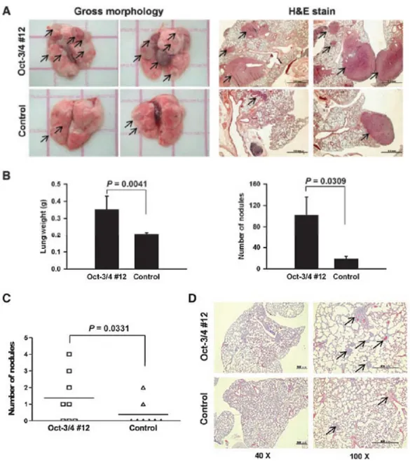
investigated in animal models whether the metastatic potential of bladder cancer cells correlated with Oct-3/4 expression. In the animal model of experimental pulmonary metastasis in which cancer cells were injected into mice via the tail vein, tumor nodules in the lung was detected more frequently in those inoculated with MBT-2/Oct-3/4 cells compared to those injected with the control cells, as revealed by gross examination and H&E staining (Fig. 2A). In particular, there was a significant increase in pulmonary metastatic colonization, both in lung weight as well as in the number and size of the tumor nodules (Fig. 2B). In the spontaneous metastatic model in which tumor cells were inoculated
subcutaneously into the mice, the number of visible metastatic nodules in the lung was greater in mice inoculated with MBT-2/Oct-3/4 cells than in those receiving control cells (Fig. 2C). Histological sections also revealed that mice inoculated with MBT-2/Oct-3/4 had more tumor lesions (Fig. 2D). Taken
together, these results indicate that Oct-3/4 overexpression promoted the metastatic behavior of bladder cancer cells in vivo.
FIGURE 2. Overexpression of Oct-3/4 promotes metastasis in animal models of experimental pulmonary and spontaneous metastases. A and B, in the mouse model of experimental pulmonary metastasis, C3H/HeN mice (n = 8) were inoculated with MBT-2/Oct-3/4 or control cells via the tail vein and killed 30 days after tumor cell inoculation. A, gross appearance of two representative lungs from each group of mice (left). The length of the small squire corresponds to 1 cm. Representative
Research Express@NCKU - Articles Digest
staining of the lungs of two mice from each group (right). The scale bars shown on 40× images correspond to 1 mm. Note that the numbers and sizes of tumor nodules indicated by arrows were increased in mice inoculated with MBT-2/Oct-3/4 cells. B, wet lung weight of each group of mice (left). Number of tumor nodules on the surface of the lung from each group of mice (right). columns, mean of eight determinations; bars, SEM. C and D, in the model of spontaneous metastasis, C3H/HeN mice (n = 8) were inoculated s.c. with MBT-2/Oct-3/4 or control cells and killed 60 days after tumor cell
inoculation. C, number of metastatic nodules on the surface of the lung from each group of mice. The mean values are indicated by horizontal bar. D, representative lung histology stained with H&E of each group of mice. The arrows denote tumor nodules, and the scale bars shown on 40× and 100× images correspond to 500 μm.
The expression of Oct-3/4 in bladder cancer promoted tumor progression and metastasis, which may accounted for by the activation of MMP-13, MMP-2, and MMP-9 expressions by Oct-3/4. Therefore, the expression of Oct-3/4 may contribute to the group of bladder cancer patients with poorer survival, and Oct-3/4 may act as a novel target for cancer therapy especially in cancers with high propensities for metastasis. In the syngeneic MBT-2 tumor model in immunocompetent mice, we also conclude that Ad5WS4, an oncolytic adenovirus driven by the Oct-3/4 promoter, may serve as a possible therapeutic strategy for bladder cancer with greater effectiveness and cancer-specific potentials.