國立臺灣大學醫學院暨工學院醫學工程學研究所 碩士論文
Institute of Biomedical Engineering College of Engineering
National Taiwan University Master Thesis
化學交聯水膠包覆人類血小板裂解液於促進血管新 生之探討
Developing a glyoxal-crosslinked hydrogel for sustained release of human platelet lysate to promote angiogenesis
陳光世
Guang-Shih Chen
指導教授:楊台鴻 博士 鄭乃禎 博士 Advisor: Tai-Horng Young, Ph.D.
Nai-Chen Cheng, M.D., Ph.D.
中華民國 107 年 7 月
July 2018
口試委員審定書
中文摘要
近年來,周邊性動脈阻塞性疾病的患者逐漸增加,但目前臨床上對於此疾病患者
的治療方式大多以支架、氣球將阻塞的動脈撐開或進行血管繞道手術,病情較嚴
重的患者更可能面臨到截肢的選擇。為了促進周邊性組織的血液循環,本研究將
生醫材料製作成水膠的形式,使用乙二醛將甲殼素與明膠相互交聯進而包覆血小
板裂解液,期望能於周邊性組織緩慢釋放生長因子並促進血管新生。為了要鑑定
此系統釋放包覆物的釋放機制,本研究包覆不同分子量大小的螢光異硫氰酸鹽右
旋醣酐及血小板裂解液進行分析,另一方面,於細胞實驗中我們發現血小板裂解
液不僅能夠促進人類臍動脈內皮細胞和纖維母細胞細胞株(HS68)的移動,亦能使
人類臍動脈內皮細胞形成管柱。此外,在受精卵及仿真皮膚系統中,我們發現血
小板裂解液結合生醫材料於促進血管新生方面具有相當大的潛能,未來值得繼續
研究。
Abstract
More and more people suffered from peripheral artery disease (PAD) due to insufficient
blood supply to the legs. Currently, the standard therapy for improving blood flow to
the affected extremity is either surgical or endovascular revascularization. However,
their therapeutic effects are sometimes limited and still many people with PAD may
require amputation. The purpose of our research is to combine biomaterials with human
platelet lysate (HPL) to increase the blood flow in the ischemia area. In our study, we
developed a chitosan-gelatin hydrogel crosslinked by glyoxal for sustained release HPL.
To investigate the release profile of this hydrogel system, we used hydrogel to
incorporate the FITC-dextran with different molecular weight and the release pattern
were determined. On the other hand, the angiogenic effect of HPL was studied in the
tube formation assay and transwell migration assay of human umbilical vein endothelial
cells (HUVEC). Moreover, supplementing culture medium with HPL induced the
migration of HS68 cells in an in vitro wound healing assay suggesting the potential of
HPL to facilitate wound closure. Chick chorioallantoic membrane (CAM) assay
revealed that HPL can stimulate angiogenesis in vivo. Given the results of in vitro and
in vivo experiments, we conclude that the hydrogel-based system of HPL has great
potential to increase the blood flow for the treatment of ischemic wounds.
Contents
口試委員審定書 ...i
中文摘要 ... ii
Abstract ... iii
Contents ... iv
List of figures ... vi
Chapter 1: Introduction ... 1
1.1 Peripheral artery disease (PAD) ... 1
1.2 Hydrogel ... 3
1.2.1 Hydrogel introduction ... 3
1.2.2 Chitosan ... 5
1.2.3 Gelatin ... 6
1.2.3 β-glycerophosphate (β-GP )& glyoxal ... 7
1.3 Human platelet lysate (HPL) ... 8
1.4 Motivation and Aims ... 10
1.5 Research Framework ... 11
Chapter 2: Materials and methods ... 12
Materials ... 12
Chemicals and Reagents ... 12
Cell Culture ... 13
Experimental Equipment ... 13
Methods ... 14
2.1 Preparation of chitosan/gelatin based (CS-GE) hydrogels ... 14
2.2 Rheological studies ... 15
2.4 Studies of the cargoes release pattern in hydrogels ... 16
2.5 Chemical cross-linker biocompatibility studies ... 17
2.6 The effect of HPL on cell proliferation ... 18
2.7 HS68 cells in vitro wound healing migration assay ... 19
2.8 HUVEC Transwell migration assay ... 20
2.9 HUVEC tube formation assay: ... 21
2.10 3D skin equivalent model ... 21
2.11 Chick Chorioallantoic Membrane Assay (CAM Assay) ... 22
2.12 Statistical analysis ... 23
3.5 Biocompatibility test of glyoxal ... 27
3.6 Human platelet lysate promote the cell activity of HUVEC/HS68 cells .... 27
3.7 HS68 cells wound healing migration assay ... 28
3.8 Human platelet lysate facilitate the migration of HUVEC ... 29
3.9 Human platelet lysate enhance tube formation in endothelial cells ... 29
3.10 3D human skin equivalent model ... 30
3.11 CAM assay ... 30
Chapter 4: Discussion ... 32
4.1 Glyoxal as chemical cross-linker in a hydrogel system ... 32
4.2 Rheological studies ... 33
4.3 HS68 cells migration ... 34
4.4 HUVEC migration ... 34
4.5 HUVEC tube formation ... 34
4.6 3D human skin equivalent model ... 35
4.7 CAM assay ... 36
Chapter 5: Conclusion ... 37
References: ... 38
Appendix: ... 45
List of figures
Fig. 1 Molecular structure of chitosan ... 6 Fig. 2 Typical gelatin molecules structure ... 7 Fig. 3 Molecular structure of (A.) β-GP (B.) glyoxal ... 8 Fig. 4 Gelification samples(2% chitosan, 1% gelatin and 7.125% β-GP) with
different concentrations of glyoxal. ... 45 Fig. 5 In vitro degradation test of CS-GE hydrogels. All the samples are
composed of 2% chitosan , 1% gelatin and 7.125% β-GP with different concentrations of glyoxal. ... 46 Fig. 6 Mechanical properties of different composition hydrogels. All the
samples are composed of 2% chitosan , 1% gelatin and 7.125% β-GP with different concentrations of glyoxal. Hydrogels are tested as
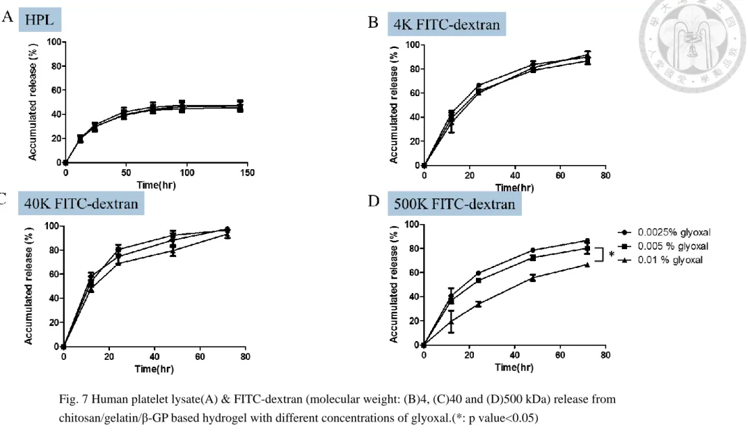
frequency sweeps at 37℃. ... 47 Fig. 7 Human platelet lysate(A) & FITC-dextran (molecular weight: (B)4,
(C)40 and (D)500 kDa) release from chitosan/gelatin/β-GP based
hydrogel with different concentrations of glyoxal.(*: p value<0.05) ... 48 Fig. 8 Cytotoxicity of glyoxal tested by alamar blue assay (A)Different
concentrations of glyoxal in medium incubates with HS68 cells for 24 hours (B)Chitosan/gelatin/β-GP based hydrogel in upper transwell and the cell activity of HS68 cells are measured on day 1, day3 and day5.(**:
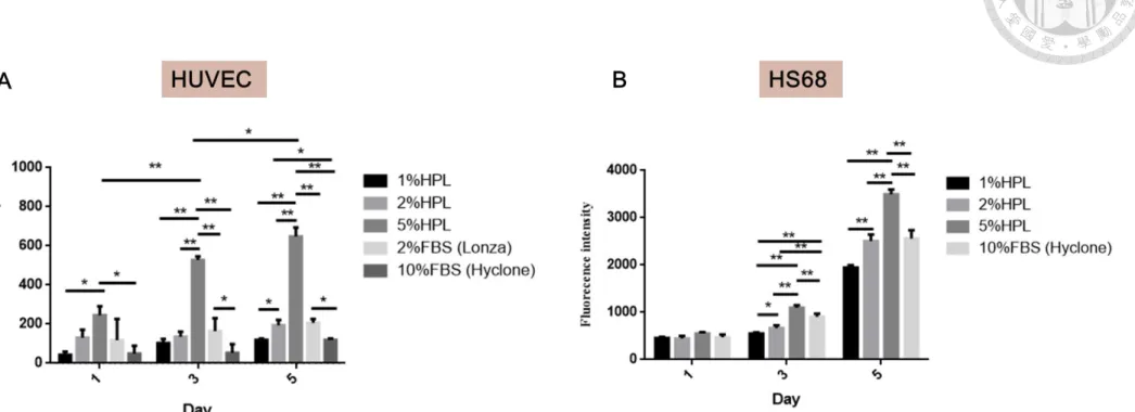
p value<0.01) ... 49 Fig. 9 Effect of HPL on cell activity tested by alamar blue assay (A) Different
amount of human platelet lysate in EBM incubates with HUVEC. The group 2% FBS(Lonza) without growth factor and 10% FBS(Hyclone) are as control. (B) Different amount of HPL in DMEM incubates with HS68 cells. The 10% FBS(Hyclone) are as control. (*: p value<0.05, **: p value<0.01) ... 50 Fig. 10 HS68 cells migration is observed in 24 hours (A)morphology of HS68
cells migration are photographed at 0 hour, 12hours and 24 hours.(B )Wound closure area (%) analyzed by image J.(*: p
value<0.05,**: p value<0.01 and scale bar=200μm) ... 51 Fig. 11 HPL embedded in chitosan/gelatin/β-GP based hydrogel with 0.005%
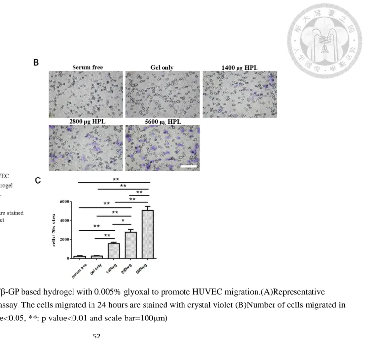
Fig. 12 Tube formation assay of HUVEC on different amount of
HPL.(A)Morphology of HUVEC on 2h, 4h, 6h and 24h (B)Number of junctions, master segments, nodes and meshes (HUVEC at 4 hours) are analyzed by image J (*: p value<0.05, **: p value<0.01 and scale
bar=100μm) ... 53 Fig. 13 Scheme of 3D skin equivalent model ... 54 Fig. 14 3D skin equivalent model photographed (A) 1.5 hr after seeding
HUVEC (B) after taking oring out (C) after adding hydrogel+HPL (scale bar=100μm) ... 55 Fig. 15 Timeline of CAM assay ... 56 Fig. 16 Result of in ovo test of CAM assay. (A) Control group was without
any treatment and the experiment group was Hydrogels combined with HPL (B) Microvascular vessels area was analyzed by image J. ... 57
Chapter 1: Introduction
1.1 Peripheral artery disease (PAD)
Peripheral artery disease (PAD) is characterized by ischemia in the lower extremities
due to narrowing of arteries with atherosclerotic plaque accumulation [1]. This
progressive atherosclerotic disorder could result in poor quality of life and high cost
of care. Nowadays, PAD affects 8–12 million individuals in North America and >200
million worldwide. By 2050, PAD incidence is predicted to double due to tobacco
use, an increase in type 2 diabetes, increasing rates of obesity in an aging population
and sedentary lifestyle [2, 3].
Critical limb ischemia (CLI), the most severe form of PAD associated with high
morbidity and mortality, lead to ischemia rest pain, non-healing ulcers and tissue
necrosis with gangrene [4]. The number of patients with PAD having CLI is estimated
at 1% to 3% and the annual number is estimated to be around 160,000 in the United
States [5, 6]. According to the Inter-Society Consensus for the Management of PAD,
25% of patients diagnosed with CLI will die within 1 year and an additional 30% will
receive amputation [7].
Currently, the standard therapy is either surgical or revascularization for those suitable
lesions while type D lesions are complex lesions. Types B and C lesions are
intermediate lesions. Endovascular therapy is the treatment of choice for type A lesions
and is the preferred treatment for type B lesions. Surgery is the treatment of choice for
type D lesions and is the preferred treatment of choice for low-risk patients with type C
lesions. In general, surgical therapy may be indicated in younger patients with longer
life expectancy. Alternatively, endovascular therapy is a more suitable treatment option
for elderly patients with comorbidities.
In some patients, revascularization therapy is not suitable since the atherosclerotic artery
occlusion is too extensive, and limb amputation remains the only treatment option.
Therefore, novel and more effective strategies such as stem cell therapy have emerged
as a promising alternative for treatment of disorders related to limb ischemia [9]. In this
study, we aim to develop a system including crosslinked hydrogels and human platelet
lysate to promote the angiogenesis in the ischemia tissue.
1.2 Hydrogel
1.2.1 Hydrogel introduction
Hydrogels are composed of three-dimensional, cross-linked networks of water-soluble
polymers and able to absorb from 10–20% (an arbitrary lower limit) up to thousands of
times their dry weight in water [10]. Biocompatibility is promoted by the high water
content of hydrogels and the physiochemical similarity of hydrogels to the native
extracellular matrix. Both natural polymers (e.g., hyaluronic acid, dextran sulfate,
chitosan, collagen) and synthetic polymers (e.g., polyethylene glycol(PEG)- polylactic
acid(PLA)-PEG) can be crosslinked to form hydrogels. The unique physical properties
of hydrogels have sparked interest in their use in drug/protein delivery system. Their
highly porous structure can easily be tuned by controlling the crosslinking degree in the
gel matrix and the affinity of the hydrogels for the aqueous environment in which they
are swollen [11]. In addition, their porosity also permits loading of drugs into the gel
matrix and subsequent drug release at a rate dependent on the diffusion coefficient of
the small molecule or macromolecule through the gel network.
Hydrogels can be formed physically or chemically. For those hydrogels whose networks are held together physically, they are called ‘physical’ hydrogels, or ‘reversible’
hydrogels formed by polyelectrolyte and multivalention of the opposite charge are also classified as ‘physical’ hydrogels. Calcium alginate is an example of this type of
hydrogel. For those hydrogels whose networks are covalently crosslinked, they are called ‘chemical’ or ‘permanent’ hydrogels. Like physical hydrogels, chemical
hydrogels are not homogeneous. They usually contain regions of low water swelling
and high crosslink density, called ‘clusters’, that are dispersed within regions of high
swelling, and low crosslink density. This may be due to hydrophobic aggregation of
crosslinking agents, leading to high crosslink density clusters [14]. Chemical hydrogels
can be formed in many ways, including using radiation, chemical cross-linkers and
multi-functional reactive compounds to crosslink polymer chains. Photosensitizer also
can be combined with monomers and multifunctional macromers to form hydrogels.
However, though hydrogels has several advantages, such as high porosity, high
biocompatibility and high deformability to conform to the shape of the surface to which
they are applied, they are limited in load-bearing applications due to low tensile
strength. It is clear that there are both significant advantages and disadvantages to the
use of hydrogels in tissue engineering, and the latter will need to be overcome so that
hydrogels will become practical and useful in this exciting field.
1.2.2 Chitosan
Chitosan is derived by partial deacetylation of chitin and is composed of a sugar backbone with comprising copolymers of glucosamine (β(1–4)-linked 2-amino-2-
deoxy-D-glucose) and N-acetylglucosamine (2-acetamido-2-deoxy-Dglucose) [15]. The
amino group in chitosan has a pKa value of ~6.5 [16], while the charge density is
dependent upon the solution pH and the degree of chitosan deacetylation. Because of
the presence of amine and hydroxyl groups, chitosan molecules can form hydrogen
bonds leading to the crystalline structure of the polymer [17]. It is reported that chitosan can be degraded via several enzymes such as β-N-acetylhexosaminidase, chitosanase,
chitinase and chitin deacetylase. In human body, chitosan can be biodegraded by
lysozyme, gastrointestinal enzymes and colon bacteria [18]. As a natural
polysaccharide, chitosan has many appealing characteristics including no adverse
reactions to human body, good biocompatibility and degradability, nontoxicity,
inhibition of inflammation, antibiosis and so on [19]. Due to its various advantages,
chitosan has received great attention in medical and pharmaceutical applications such as
cosmetic [20], cell encapsulation [21], drug delivery [22] and tissue engineering [23].
More importantly, chitosan has been demonstrated to accelerate wound healing due to
Fig. 1 Molecular structure of chitosan
1.2.3 Gelatin
As one of the natural polymers, gelatin is widely used in applications ranging from the
food industry to medicine and pharmaceutical processing [27, 28]. In tissue engineering
and regenerative medicine, increasing interest in the use of gelatin in this field stems
from its various desirable features such as biocompatibility, biodegradability, low cost
and ease of manipulation [29, 30]. Gelatin is a well-known biomaterial comprising
denatured and partially hydrolyzed native collagen. Compared with collagen, gelatin is
more prone to biodegradation and absorption [31, 32]. Moreover, the bioactive
sequences of collagen such as the arginine-glycine-aspartic acid (RGD) peptide for cell
attachment and matrix metalloproteinase (MMP)- sensitive degradation sites are
retained in the gelatin backbone [33]. Therefore, essential cellular functions, including
migration, proliferation and differentiation, can be facilitated via integrin-mediated cell
adhesion and cell-mediated enzymatic degradation [34, 35]. Typically, the extraction methods to obtain gelatin can be divided into “acid method” and “alkaline method”.
Gelatin obtained by the acid method is referred as “Type A” gelatin, whereas products
of the alkaline method is called “Type B" gelatin [36]. Gelatin structure is shown on
Fig. 2 .
Fig. 2 Typical gelatin molecules structure
1.2.3 β-glycerophosphate (β-GP )& glyoxal
Recently, more and more study reported that chitosan-based hydrogels are often
combined with β-glycerophosphate (β-GP) to constitute thermally responsive hydrogel
system. Due to the electrostatic attractions between the chitosan protonated amine
groups (-NH3+) and negatively charged phosphate molecules of β-GP (-HPO4- or -
PO42-), the hydrogel system composed of chitosan, gelatin and β-GP can stay in liquid
state without aggregation at low temperature [37, 38]. By adding β-GP as a weak base,
the pH of the solution is escalated close to physiological pH [39] so that the available –
NH on chitosan and gelatin chains may increases. Furthermore, the hydrophobic
system. Glyoxal can crosslink the available –NH2 on chitosan and gelatin chains to form
the networks and the mechanical properties of the hydrogel system is expected to be
better.
Fig. 3 Molecular structure of (A.) β-GP (B.) glyoxal
1.3 Human platelet lysate (HPL)
Platelets are intensively studied and used in various medical fields, such as plastic
surgery, dentistry and dermatology, since they mediate wound closure and healing [40,
41]. Platelets are mostly used in the form of platelet-rich plasma (PRP), where a higher
number of platelets are reconstituted in a small volume of plasma after centrifugation.
However, their main disadvantage is a short shelf-life, which hinders the use of PRP in
cell culture applications [42]. Therefore, it is necessary to develop methods for the
longer shelf-life. Abundant growth factors and cytokines that are stored in platelet
granules can be naturally released by thrombin activation [43, 44] and clotting, or
artificially released by freeze/thaw-mediated platelet lysis, sonication or chemical
treatment [45, 46].Compared to chemical activation of platelets to gain the releasate,
mechanical lysis to yield HPL appears preferable as being much easier, less time-
consuming, and less cost-effective. It further avoids the use of additional substances
such as thrombin which may cause side effects [47].
In recent years, many researches reported that HPL has the potential to replace fetal
bovine serum (FBS) for use in clinical applications [48, 49]. Some researches indicated
that HPL was feasible candidate to produce stem cells batches in compliance with the
good manufacturing practice (GMP) standards [50]. In order to prolong the retention
time of HPL in specific area, some scientists used electrospun fibers to control the
release of HPL [51]or made the HPL into hydrogels directly [52]. In this study, we
decided to use HPL from UltraGROTM due to its high quality. Moreover, the
components in HPL had been evaluated by ELISA in previous study [53]. Growth
factors stored in HPL include fibroblast growth factor (FGF), platelet-derived growth
factor (PDGF), transforming growth factor (TGF) and epidermal growth factor (EGF)
play an important role in cell communication and influence the rates of wound healing
[54-56].
1.4 Motivation and Aims
If directly placed HPL in the wounds directly, HPL can not retain in the wound area for
a long time. Therefore, we are aimed to combine HPL with hydrogels to prolong the
retention time of HPL in the wounds area. The objective of our study was to develop a
hydrogel vehicle and investigate the release profile of HPL. Moreover, the experiments
to prove the angiogenic effect of HPL are also conducted in this research.
1.5 Research Framework
Chapter 2: Materials and methods Materials
Chemicals and Reagents
Name Brand Model number
Chitosan Glutamate NovaMatrix, USA
PROTASAN UP G 213
Gelatin, from procine skin Sigma, USA G2625
β-glycerophosphate disodium salt
hydrate
Sigma, USA G9422
Glyoxal solution Sigma, USA 50649
Alamar Blue Invitrogen, USA DAL1100
Transwell cell culture inserts Corning Costar CLS3450
growth factor reduced Matrigel Corning 354230
Fluorescein isothiocyanate (FITC)- dextran (3~5 kDa)
Sigma FD4
Fluorescein isothiocyanate (FITC)- dextran (40 kDa)
Sigma FD40
Fluorescein isothiocyanate (FITC)- dextran (500 kDa)
Sigma FD500S
Collagen solution from bovine Sigma C4243
Collagenase type Ⅰ Gibco 17100017
BCA Protein Assay Kit Thermo 23225
Cell Culture
Name Brand
Human Umbilical Vein Endothelial Cells (HUVEC) Lonza Human Foreskin Fibroblast Cell line HS68 ATCC
Human Keratinocytes Cell line HaCaT ATCC
Dulbecco's Modified Eagle's Medium (DMEM-HG) Hyclone Endothelial Cell Growth medium (EGM-2) BulleKit Lonza
Human Platelet Lysate(HPL) UltraGRoTM, USA Endothelial Cell Growth medium 2(EGM-2) Promo Cell
Penicillin/Streptomycin (P/S) Biological, USA
Fetal Bovine Serum (FBS) Hyclone, USA
Phosphate-buffer saline (PBS) Omics Bio
Experimental Equipment
Name Brand Model number
Spark™ 10M multimode
TECAN Trading AG, Switzerland Spark™ 10M
Inverted Fluorescence Microscope
Leica, USA DMI 6000
Autoclave Mitutoyo, Taiwan EA-652
Refrigerator Marupin CR-530
μ-slide Ibidi 1803052
Vortex Scientific, USA G-560
Shaker bath Firstek scientific, USA B601
Centrifuge KUBOTA, USA 5910
Ultrapure water system MILLIPORE TANKPE060
Methods
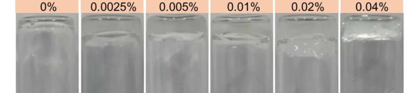
2.1 Preparation of chitosan/gelatin based (CS-GE) hydrogels
A solution of 4% (w/w) chitosan was prepared by dissolving the required amount of
chitosan powder in ddH2O at 60℃ overnight. 10% (w/w) gelatin solution was prepared
in the same way. Both of them were autoclaved before the test in this study. After 0.4mL
10% gelatin solution was blended into 4% 2mL chitosan solution and 0.6mL ddH2O
homogeneously, 1mL 28.5% (w/w) β-glycerophosphate (β-GP) solution was added
droply into the solution of chitosan and gelatin. The total volume was 4mL so that the
final concentration ratio of chitosan: gelatin: β-GP = 2 : 1 : 7.125 .The solution
composed of chitosan, gelatin and β-GP was called CS-GE mixture. The glyoxal
solution was diluted with ddH2O until the concentration reached 0.4 %. Different
volume of 0.4% glyoxal was added into CS-GE mixture homogeneously so that the
final concentration of glyoxal ranged from 0.0025% to 0.04%. In addition, gelification
due to the crosslinking between the amine group on chitosan and gelatin was estimated
through the vial tilting method [57].
2.2 Rheological studies
In order to determine the linear viscoelastic properties of the hydrogels, the small
amplitude oscillatory shear experiments were performed to measure the time-dependent
response of the samples. Hydrogels were made of CS-GE mixture (2% Chitosan,1%
gelatin and 7.125% β-GP) crosslinked with 0.0025%, 0.005% and 0.01% glyoxal. All the samples’ volume were 1 mL and shaped in 10 mm diameter, 5 mm height. The test
was under the condition that frequency sweeps in the range from 0.01 Hz up to 10 Hz at
37 ℃. Each rheological studies was repeated at least three times.
2.3 In vitro degradation test
The in vitro degradation test was studied by immersing the hydrogels into an enzymatic
solution and then monitoring their weight-losses in different time points. The enzymatic
solution consisted of 25U/mL collagenase in PBS. After 30 mm culture dishes were
with 3 mL enzymatic solution and incubated at 37℃. All the samples were removed
enzymatic solution at different time points and dried at 60℃. The remained hydrogels
were determined by following equation. (Wi= initial weight, Wx= weight at different
time points)
Hydrogels remained(%) = 1 −𝑊𝑖 − 𝑊𝑥
𝑊𝑖 × 100%
2.4 Studies of the cargoes release pattern in hydrogels A. Human platelet lysate (HPL) release
1000μg Human platelet lysate (HPL) determined by bicinchoninic acid assay (BCA
assay) was encapsulated in CS-GE mixture (2% Chitosan,1% gelatin and 7.125% β-GP)
crosslinked with 0.0025%, 0.005% and 0.01% glyoxal. PBS was added onto the
hydrogels incorporated HPL. On the specific time points of 12 hours, 24 hours, 48 hours
and 72 hours, PBS was completely moved and we added new PBS onto the hydrogels.
HPL released from hydrogels was quantified by BCA assay.
B. FITC-dextran release
To determine the release pattern of particles with different molecular weights, we
adopted a protocol modified from a previous study [58]. FITC-dextran with three
different molecular weight (4kDa, 40kDa and 500 kDa) was encapsulated in CS-GE
mixture (2% Chitosan,1% gelatin and 7.125% β-GP) crosslinked with 0.0025%, 0.005%
and 0.01% glyoxal. Then 100μL PBS was added onto the hydrogel incorporated FITC-
dextran. On the specific time points of 12 hours, 24 hours, 48 hours and 72 hours, PBS
was completely removed and then new PBS was added onto the hydrogels. FITC-
dextran release from hydrogels was measured by Spark™ 10M multimode microplate
reader (TECAN Trading AG, Switzerland) and determined by the calibration curves.
Each determination was performed a minimum of three times.
2.5 Chemical cross-linker biocompatibility studies
The Cytotoxicity of the chemical cross-linker glyoxal was tested in two ways.
A. Glyoxal solution in medium
HS68 cells (passage 27) were cultured in Dulbecco’s Modified Eagles
Medium(DMEM), 10%FBS and 1% penicillin/streptomycin and seeded in 48 well plate
with 15,000 cells per well. After the cells attached, medium was changed to DMEM,
10%FBS, 1% penicillin/streptomycin with different amounts of glyoxal from 0.00125%
up to 0.02%. alamar blue assay (excitation wavelength: 560nm, emission wavelength :
590nm) was used to quantify the cell activity after 24 hours. Data was reported as the
average of at least three samples for each group tested.
B. Hydrogel tested in Transwell assay
HS68 cells (passage 27) were cultured in DMEM, 10%FBS and 1%
crosslinked with 0.0025%, 0.005% and 0.01% glyoxal were added into 12 transwell
inserts (Corning) with 8.0 μm pore size. The transwell inserts were put into 12 well
plate and the cell activity was quantified by alamar blue assay (excitation wavelength:
560 nm, emission wavelength: 590 nm) at day 1, day 3 and day5.
2.6 The effect of HPL on cell proliferation
Alamar blue assay was used to evaluate the cell activity. The redox reaction that occur
in the mitochondria of the viable cells result in the production of resorufin, a compound
that is red in color and highly fluorescent. This product of redox reaction can be
measured spectrophotometrically.
The effect of HPL was shown in two ways.
A. Incubation with human umbilical vein endothelial cells (HUVEC)
HUVEC (Passage4) were seeded in 24-well culture plate with cell density: 1.0 x 10⁴
cells/well. When the cells attached to the 24-well culture plate after 24 hours, the
medium was changed to conditions of three different concentration of HPL (1%, 2%
and 5%) , 10% FBS (Hyclone) and 2% FBS (Lonza) in 0.5mL Endothelial cell Basal
Medium (EBM). For each sample, 0.5mL EBM containing 10% (v/v) alamar blue
reagent was added and incubated in 5% CO2, 37℃ atmosphere for 2 hours on day 1,
day 3 and day 5. In brief, 200μL of the solution was subsequently removed from the
wells and transferred in the transparent 96-well plate. The fluorescence intensity was
immediately measured with a standard spectrophotometer at fluorescence excitation
wavelength of 560 nm and an emission wavelength of 590 nm. The 10% FBS (Hyclone)
and 2% FBS (Lonza) group were as control and each group was tested at least three
wells.
B. Incubation with HS68 cells
HS68 cells (Passage28) were seeded in 24-well culture plate with cell density: 1.5 x 10⁴
cells/well. When the cells attached to the 24-well culture plate after 24 hours, the
medium was changed to conditions of three different concentration of HPL (1%, 2 %
and 5%) and 10% FBS (Hyclone) in 0.5mL DMEM. For each sample, 0.5mL DMEM
containing 10% (v/v) alamar Blue reagent was added and incubated in 5% CO2, 37℃
atmosphere for 2 hours on time of day 1, day 3 and day 5. In brief, 200μL of the
solution was subsequently removed from the wells and transferred in the transparent 96-
well plate. The fluorescence intensity was immediately measured with a standard
spectrophotometer at fluorescence excitation wavelength of 560 nm and an emission
wavelength of 590 nm. The 10% FBS group was as control and each group was tested at
least three well.
2.7 HS68 cells in vitro wound healing migration assay
(passage 28) cells were seeded in the 2 well culture-insert with cell density: 2 x 10⁴ cells
(in 70μL medium)/well. After the cells attachment, the cells were performed in
condition of serum, 1% HPL, 2% HPL, 5% HPL and 10% FBS in DMEM. The
migration of HS68 cells was observed 24 hours by Leica using the time lapse. Wound
closure area (%) was analyzed by image J (at least n=3).
2.8 HUVEC Transwell migration assay
Different amount of HPL (5600μg, 2800μg and 1400μg ) were blended into 250μL CS-
GE mixture (2% Chitosan&1% gelatin and 7.125% β-GP), then add into 3.25μL 0.4%
glyoxal solution mixed well and then it became hydrogel soon. The 24 well cell culture
plates were coated with 250 μL CS-GE hydrogel per well. Each group was immersed
with 700μL Endothelial cell Basal Medium (EBM) so that the HPL entrapped in the
hydrogels could sustained release into the medium to observe the effect of HPL.
Transwell inserts(Corning) with 8.0μm pore size were placed onto the top of the gels,
and 100μL of serum free media containing 5 x 10⁴ HUVEC was placed on top of the
insert. After 24 hours of incubation, all nonmigrant cells were removed from the upper
surface of the Transwell membrane with a cotton swab and migrant cells at the under
surface were fixed and stained with crystal violet. Images were obtained at 20x
magnification and the number of cells stained with crystal violet was counted by image
J (at least n=3). Values were reported as number of cells migrated per 20x field.
2.9 HUVEC tube formation assay:
To ascertain the angiogenic potential of HPL, we cultured HUVEC on growth factor
reduced Matrigel(Corning), which is considered a standard assay to analyze
vasculogenic capabilities. After 10μL Matrigel per well was coated on μ-slide (Ibidi) for
30min, HUVEC are resuspended in EBM supplemented under the condition of no
serum, 1%HPL, 2% HPL and 5% HPL. Finally, HUVEC were seeded 6 x 10⁴ cells/cm2
on Matrigel in 50uL medium per well. Tube-like structures are observed and captured
with a phase contrast microscopy on 2 hours, 4 hours, 6 hours and 24 hours after seeded
on Matrigel. Statistical analyzation of images on 4 hours (at least n=3) was proceeded
by the plug-in in image J, Angiogenesis Analyzer.
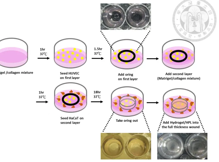
2.10 3D skin equivalent model
The protocol of this model was modified from a previous study [59] to evaluate the
effect of HPL entrapped in the CS-GE hydrogels to promote angiogenesis in three-
dimensional environment. First of all, 200μL growth factor reduced
Matrigel(Corning)/Collagen type 1 (Sigma) mixture (volume ratio= 2:1) was added into
24 well culture plate. After it gelled at 37℃ for 1h, HUVEC were seeded on top of this
layer with cell density of 105 cells per well in Endothelial cell Growth Medium (EGM) .
dropwise around the oring. HaCaT cells was seeded onto the second layer 1.25x10⁵
cells in 80μL DMEM. The system was incubated under the condition of 5% CO2 and 37
℃ for 18h. The wound in this three-dimensional skin equivalent model was formed by
taking the oring out. 50μL Hydrogels combined with 750 μg HPL was added to fill the
wound in this model. After 6 hours, tube formation of HUVEC was evaluated around
the oring.
2.11 Chick Chorioallantoic Membrane Assay (CAM Assay)
Fertile chicken eggs were obtained from the Animal Health Research Institute (Danshui
Dist., New Taipei City, Taiwan) and wiped clean. All the eggs were incubated under
constant 80% humidity at 37℃ in the hatch egg machine (HOONG SHENG, HY-01S).
The cultivation of eggs could be divided into ex ovo or in ovo. We decided to cultivate
the fertile eggs in ovo since in ovo cultivation could improve the survival of the
embryos [60]. On incubation day 7, we drilled a hole on the bottom and side of eggs
separately. The air chamber on the bottom of eggs was moved to the side of eggs by
suction and the observation window on the side of eggs was enlarged by using tweezers
to remove the pieces of eggshell. We placed an oring (diameter=8mm) onto chick
chorioallantoic membrane (CAM) and injected 100 μL the CS-GE hydrogels (final
concentration: 2% chitosan, 1% gelatin, 7.125% β-GP and 0.005% glyoxal) combined
with 1500 μg HPL into the oring. The angiogenesis of fertile chicken eggs undergoes
the peak growth period from day 7 to day 12 incubation and CAM architecture would
reached its mature morphology on day 13 and 14 [61]. The result was observed and
recorded at day 10 cause an angiogenic response occurs 72-96 hours after stimulation in
the form of an increased vessel density around the implant, with the vessels radially
converging toward the center like spokes in a wheel [62]. Besides, as there are many
blood vessels on both side of the CAM, and the only blood vessels that we want to
observe were the one that make direct contact with our materials. To decrease the
interferences from blood vessels underneath the CAM, milk was injected into the CAM
to block it out so that the view we observed would be clearer. Several qualitative,
quantitative, and semi-quantitative techniques, including blood vessel length, diameter,
density, vessel branch points, total area of the CAM, have been described for
assessment of angiogenesis [63]. We marked the 2 cm circle area around the oring and
analyzed the total blood vessels area around the materials by Image J (at least n=5).
2.12 Statistical analysis
The data were analyzed by one way ANOVA and Tukey’s multiple comparison test.
Probabilities of p value < 0.05 (*) and p value < 0.01 (**) were considered as
significant difference.
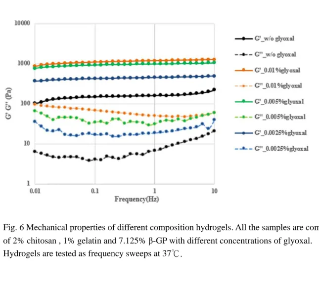
Chapter 3: Results 3.1 Rheological studies
Mechanical properties of CS-GE hydrogels crosslinked with different amount of glyoxal
are tested by rheometer (Fig.5). The shear storage modulus (G’) (reflecting the elastic behavior) and shear loss modulus (G”) (reflecting the viscous behavior) of the hydrogel
were compared between each group in this test. We could see that the value of G’ in
0.01% glyoxal group significantly higher than the 0.00125% glyoxal group and the control group. Nevertheless, the value of G’ in 0.01% glyoxal group was almost 1000
Pa, similar to the 0.005% glyoxal group. This study revealed that adequate glyoxal
concentration is essential for the fabrication of stiffer hydrogels with higher values of G’ as observed in the frequency sweeps performed on the hydrogels group [64].
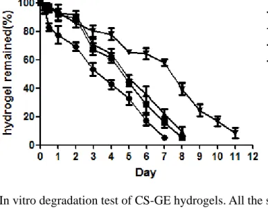
3.2 In vitro degradation test
In order to evaluate the degradation of CS-GE hydrogels, the weight loss of hydrogels
was measured under the collagenase digestion (Fig. 6). Hydrogels without glyoxal
would be degraded within seven days. The experimental groups with 0.0025% glyoxal
and 0.005% glyoxal degraded slower than the group without glyoxal. Hydrogels with
0.01% glyoxal degraded within twelve days.
3.3 Human platelet lysate release
The influence of different compositions of chitosan/ gelatin/ β-GP/ glyoxal hydrogels on
HPL release (Fig. 7A) was evaluated. All the experimental groups were loaded with the
same amounts of HPL and showed a burst release within 24 hours (the accumulated
release of HPL in each group was nearly 30%). From the result, we could see that each
group reacheda plateau in 72 hours. The accumulated release of HPL(72hours) from
hydrogels crosslinked with 0.0025%, 0.005%,0.01% glyoxal was 46.11±3.66,
44.47±2.84, 46.92±4.78%. There is no significant difference between the experimental
groups.
3.4 FITC-Dextran release
The influences of hydrogels with different compositions on the release profiles was
studied by the determination of the fluorescence intensity of FITC-dextran released
from hydrogels. When the hydrogels crosslinked with different amounts of glyoxal
(glyoxal final concentration: 0.0025%, 0.005% and 0.01%) were loaded with 4 kDa
FITC-dextran, all of the tested samples showed similar release profile (Fig. 7B). In the
first 12 hours, the accumulated release of 4 kDa FITC-dextran from hydrogels
crosslinked with 0.0025%, 0.005%, 0.01% glyoxal was 43.41±1.97, 39.22±2.29,
dextran. Fig. 7C showed the 40 kDa FITC-dextran release profile of hydrogels with
different compositions. The release profile of 40 kDa FITC-dextran was similar to 4
kDa FITC-dextran. The accumulated release of 40 kDa FITC-dextran from hydrogels
crosslinked with 0.0025%, 0.005%, 0.01% glyoxal was 58.20±5.17, 53.73±7.61,
48.05±4.75 % in 12 hours and nearly 90% in 72 hours.
However, the release profile of 500 kDa FITC-dextran was different from 4 kDa FITC-
dextran and 40 kDa FITC-dextran. As result shown on Fig. 7D, CS-GE hydrogels
composed of 0.0025%, 0.005% glyoxal released 40.63±6.4, 36.75±1.5 % 500 kDa
FITC-dextran in the first 12 hours. Nevertheless, the release of 500 kDa FITC-dextran
from hydrogel composed of 0.01% glyoxal in a relatively slow rates, it released 19.52±
9.1% 500 kDa FITC-dextran in the first 12 hours. From the result we could see that as
glyoxal content increased, the release rate of hydrogels decreased. Obviously, only the
hydrogel composed 0.01% glyoxal could significantly retarded the release rate of FITC-
dextran with a molecular weight of 500 kDa. After 72 hours, the accumulated release of
500 kDa FITC-dextran could reach almost 65%. Compared to the other experimental
groups, CS-GE hydrogels crosslinked with 0.01% glyoxal revealed greater potential to
sustained release cargoes.
3.5 Biocompatibility test of glyoxal
Cytotoxicity of glyoxal were evaluated by in two ways: incubated HS68 cells
with glyoxal directly and incubated HS68 cells with the free glyoxal didn’t crosslinked
the amine group by transwell assay. Fig. 8A showed the result that different
concentration of glyoxal from 0% to 0.02% in medium incubated with HS68 cells 24
hours directly. The fluorescence intensity of reducing alamar blue reagent in groups
(glyoxal concentration: 0.00125% and 0.0025%) had no significant difference to the
control group (glyoxal concentration: 0%). However, the value of 0.005% glyoxal group
was significantly lower than the control group and the value was almost zero in the
0.01% glyoxal group and 0.02% glyoxal group. Our result revealed that if glyoxal
concentration in medium higher than 0.005% may induce cytotoxicity. Fig. 8B showed
the result that the CS-GE hydrogels crosslinked with different concentration of glyoxal
from 0.0025% to 0.01% in transwell incubated with HS68 cells. The cell activity was
conducted by alamar blue assay on day 1, day 3 and day 5. We could see that no matter
on day 1, day 3 or day 5, all the experimental groups had no significant difference to the
10%FBS(Hyclone) group.
3.6 Human platelet lysate promote the cell activity of HUVEC/HS68 cells
assay to evaluate the cell activity on day 1, day 3 and day 5. As shown in Fig. 9A, we
could see that the fluorescence intensity in 5% HPL group was significantly stronger
than the other groups no matter on day 3 or day 5. The 2% FBS (Lonza) group without
growth factors and 10% FBS (Hyclone) were as control. Fig. 9B also showed that the
fluorescence intensity in 5% HPL group was significantly stronger than the other groups
no matter on day 3 or day 5. This result indicated that 5% HPL in medium was more
suitable for human foreskin fibroblast, HS68 cells, than the other groups.
3.7 HS68 cells wound healing migration assay
Fibroblasts migration played an important role in wound healing. After 12 hours
incubation HS68 cells in different conditions, our result showed that the speed of
migration in experimental group, 5% HPL, was faster than the other experimental
groups. Cells without serum were as control. At the time point of 24 hours, the gap
between cells on two sides was filled by the migrant cells only in the group of 5% HPL.
We used image J to calculate the wound closure area at the time point of 12 hours and
the result revealed that the wound closure area was 89.16±7.62% in the group of 5%
HPL. Nevertheless, under the circumstances of 2% HPL or lower, the wound closure
area was less than 50%.
3.8 Human platelet lysate facilitate the migration of HUVEC
To evaluate the effect of HPL, we used the transwell assay to count how many cells
migrated through the induction of growth factor in HPL. As our result on Fig. 11, we
could see that the group of 5600μg HPL had the most cells stained with crystal violet
after 24 hours of migration. Through the statistical analysis of image J, it showed that
the number of migrant cells in 5600μg HPL group was 5103±940 per 20x view and it
was significant higher than all the other groups. Our result indicated that the growth
factors in HPL could induce the migration of HUVEC.
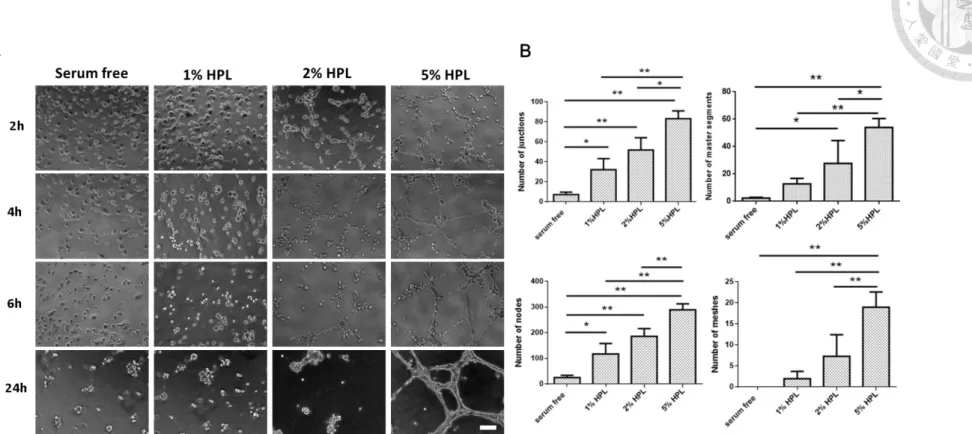
3.9 Human platelet lysate enhance tube formation in endothelial cells
We next examined whether human platelet lysate could promote the tube formation in
endothelial cells. In consistence to our previous work of HUVEC migration, we also
incubated the HUVEC under the condition of 1% HPL, 2% HPL and 5 % HPL to
monitor whether the endothelial cells formed tubes in 24 hours. Through the
observation of cells on time points of 2 hours, 4 hours, 6 hours and 24 hours, we could
see that the group of 5% HPL had the most significant effect on the promotion of
HUVEC tube formation. We used image J to analyzed the pictures at 4 hours. Several
indexes such as number of junctions, nodes ,master segments and meshes in 5%HPL
structures compared to serum free, 1%HPL and 2%HPL group.
3.10 3D human skin equivalent model
HPL was entrapped in CS-GE hydrogels to promote the angiogenesis. In this
experiment, we observed that HUVEC started to form tube-like structures after seeding
1.5 hours. The second layer was formed by Matrigel/collagen mixture after adding the
oring onto the first layer. When we took the oring out, we observed that HUVEC looked
circular inside the oring and formed tube-like structures outside the oring. HPL
combined with hydrogels was added to the wound in this model. After 6 hours treatment
of HPL and hydrogels, we found that HUVEC maintained the same tube-like structure.
3.11 CAM assay
Among many in vivo assays developed for the study of the angiogenesis, the chick
embryo chorioallanotoic membrane (CAM) assays stands for one of the most reliable
tools for the study of the effects of biological molecules on neovascularization [65, 66].
CAM assay was a closed system that allowed small quantities of therapeutic agents to
study angiogenesis. This assay has some advantages such as in vivo environment,
relatively low-cost and available to observe microvascular vessels directly [67].
Nevertheless, the eggs are immunodeficiency and sensitive to environmental factors. As
the scheme shown on Fig. 15, we opened a window on the side of eggs on day 7. After
the oring was placed onto the CAM, hydrogels combined with HPL was injected into
the oring. We observed the result three days after we put the biomaterials and
angiogenic molecules. The group without treatment was our negative control. In some
cases, we observed that the group of hydrogel and HPL revealed stronger angiogeneic
effect around the oring. However, we also found that the group without treatment make
no difference to the experimental in some cases. Through the image J analysis, we were
able to mark the area around the oring and calculate the blood vessels area in the
pictures. The vascular area of experimental group treated with CS-GE hydrogels and
HPL was 17.58±3.28 % and the group without treatment was 14.48±2.13 %. There was
no significant difference between our experimental group and control group.
Chapter 4: Discussion
4.1 Glyoxal as chemical cross-linker in a hydrogel system
A straightforward method for chitosan-based solution to form permanent hydrogel
networks is chemical cross-linking. Cross-linked chitosan networks can be prepared
using the available –NH2 and –OH chemical handles and cross-linkers that can form a
number of linkage chemistries such as aminecarboxylic acid bonding and Schiff base
formation [11, 68, 69]. In general, the networks of chitosan-based hydrogels can be
formed by using small molecule cross-linkers or enzymatically cross-linkers, polymer–
polymer reactions between activated functional groups and photosensitive agents [70].
For small molecular cross-linkers, including glutaraldehyde, formaldehyde,
diisocyanate, ethyleneglycol diglycidylether (EGDE), and others, it has been many
years using these chemical cross-linkers to reinforce the mechanical strength of
hydrogels [11, 68, 69, 71]. Though these hydrogels can offer desirable properties, the
main drawbacks of small molecule cross-linkers are potential toxicity and residual un-
reacted small molecular cross-linkers [72]. Previous study reported that Genipin, a
naturally derived chemical compound from the gardenia, showed great biocompatibility
and low cytotoxicity [73]. Nevertheless, the gelation time is at least one hour [74]which
is not suitable for clinical use and an anti-angiogenesis effect of genipin may decrease
the rate of wound healing [75]. Glutaraldehyde can crosslinked chitosan chains to form
hydrogels within one hour ; however, it is considered toxic for respiratory tract, eyes
and skin. For the convenience of clinical use, we use glyoxal as chemical cross-linker in
chitosan-based hydrogels so that the networks of hydrogels can form in short time.
Recently, glyoxal has been used as an alternative dialdehyde cross-linker in various
biomedical studies and applications and is considered safe [76, 77]. Moreover, previous
study revealed that glyoxal has been shown to be cytocompatible and support viability
of the cells [78]. This chemical cross-linker can be expected to be applied in tissue
engineering and protein/drug delivery.
4.2 Rheological studies
Compared with CS-GE hydrogels crosslinked with 0% glyoxal and 0.0025% glyoxal,
CS-GE hydrogels crosslinked with 0.005% glyoxal and 0.01% glyoxal revealed a higher
elasticity degree in their rheological behavior, and they could be classified as strong
hydrogels [79-81]. The strength of hydrogels mainly resulted from contemporary
presence of physical interactions between chitosan chains, gelatin chains and β-GP,
secondary bonds such as Van der Waals forces among polymers, and chemical
crosslinks between chitosan/gelatin chains and glyoxal [82]. Obviously, glyoxal
influenced the mechanical properties of the hydrogel by increasing the shear storage
4.3 HS68 cells migration
Previous research had found that HPL contained abundant of growth factors, such as
EGF, TGF-β1, PDGF-AB, PDGF-BB, and so on. Moreover, PDGF-BB had been
proved to participated in the migration of HS68 cells [83]. In present study we
examined the wound healing effect of HPL and it revealed that HPL could promote the
migration of HS68 cells in a dose-dependent manner. Based on previous researches, we
thought the reason may be abundant growth factor, PDGF-BB, in HPL.
4.4 HUVEC migration
As our result showed on Fig. 11, our study indicated that HPL have the power to
promote the migration of HUVEC. A previous study revealed that PDGF and vascular
endothelial growth factor (VEGF) are pivotal to the formation of capillary structures
[84]. While VEGF mainly regulates endothelial cells, PDGF signaling is crucial for
cells of the vascular wall, i.e., pericytes and smooth muscle cells [85]. In addition, HPL
comprises about PDGF-AB (about 1.5ng/mL), PDGF-BB (about 3.5ng/mL), and other
growth factors [53]. Due to the induction of PDGF-AB and PDGF-BB, the migration of
HUVEC are facilitated in our study.
4.5 HUVEC tube formation
Through the activation of compound that can stimulate angiogenesis, HUVEC may
differentiate into capillary-like structure [86]. The differentiation process involves
several steps in blood vessel formation, including cell adhesion, migration, alignment,
protease secretion, and tube-like structure formation [87]. Our result showed that tube
do not form after seeding HUVEC on ECM gel 6 hours and 24 hours under condition of
2% HPL or lower since the activation of compound that can stimulate angiogenesis is
not enough. According to previous study, it was reported that epidermal growth factor
(EGF) could stimulate the tube formation of HUVEC in a concentration-dependent
manner [88]. In our result, the 5% HPL group showed denser and stronger
tube-like structures no matter on 6 hours or 24 hours and we proposed that the reason
may be sufficient promotion effect exerted by EGF.
4.6 3D human skin equivalent model
Previous study indicated that Keratinocyte secrete angiogenic growth factors, such as
VEGF and PDGF[89]. Therefore, we seeded HaCaT cells on the second layer.
According to our result, it revealed that HUVEC already formed tube-like structures
before we add the hydrogels and HPL into the wound in this model. To solve the
problems we encountered, there are some modifications need to do. First, the
composition of medium in seeding HUVEC should be modified to slow down the rate
of tube formation. Second, we need to cut the time between seeding HaCaT cells and
4.7 CAM assay
Though we found that hydrogels combined with HPL could induce the abundant
formation of vascular network in some cases, there are still some limitations. Since we
opened a window on the side of eggs , the view we observed are limited. Moreover, the
disturbance from embryo was possible to result in the move of oring and hydrogels. The
view we observed was easy to be interfered with egg shells so that it’s hard for us to
capture the oring without egg shells. Since the eggs without treatment were able to form
the vascular network themselves, it was not an easy way for us to observe prominent
difference between the control and experimental group. Some modifications of this
assay can be expected in the future. We can try to inject lens culinaris agglutinin (LCA)
and use the microscope to visualize the microvasculature.
Chapter 5: Conclusion
In summary, we made the CS-GE hydrogels crosslinked with three different
concentration of gloxal (0.0025%, 0.005% and 0.01%). The rheology study showed that
CS-GE hydrogels crosslinked with 0.005% glyoxal and 0.01% glyoxal had better mechanical properties since the its elastic modulus (G’) was higher than the other
groups. Degradation behavior of hydrogels crosslinked with 0.01% glyoxal obviously
degrade slower than the other group. In the sustained release experiment, our result
suggested that CS-GE hydrogels crosslinked with different concentration of glyoxal had
similar release profile of HPL, 4 kDa FITC-dextran and 40 kDa FITC-dextran.
Nevertheless, in the 500 kDa FITC-dextran release experiment, CS-GE hydrogels
crosslinked with 0.01% glyoxal showed slower release rate than the other groups. In
addition, we also proved that HPL can not only stimulate the migration of HS68 cells
and HUVEC but promote the tube formation of HUVEC. Though we haven’t prove that
CS-GE hydrogels combined with HPL can stimulate angiogenesis in the CAM assay, we
still observed that eggs treated with hydrogels and HPL showed more complicated
vascular network in some cases. On the other hand, we also try to build a 3D skin
equivalent model to assess the angiogenic effect of HPL. Even there are some
References:
1. Fowkes, F.G.R., et al., Comparison of global estimates of prevalence and risk factors for peripheral artery disease in 2000 and 2010: a systematic review and analysis. The Lancet, 2013. 382(9901): p. 1329-1340.
2. Criqui, M.H. and V. Aboyans, Epidemiology of peripheral artery disease.
Circulation research, 2015. 116(9): p. 1509-1526.
3. Nehler, M.R., et al., Epidemiology of peripheral arterial disease and critical limb ischemia in an insured national population. Journal of vascular surgery, 2014.
60(3): p. 686-695. e2.
4. Parikh, P.P., Z.-J. Liu, and O.C. Velazquez, A Molecular and Clinical Review of Stem Cell Therapy in Critical Limb Ischemia. Stem Cells International, 2017.
2017.
5. Becker, F., et al., Chapter I: definitions, epidemiology, clinical presentation and prognosis. European Journal of Vascular and Endovascular Surgery, 2011. 42: p.
S4-S12.
6. Kalbaugh, C.A., et al., Peripheral Artery Disease Prevalence and Incidence Estimated From Both Outpatient and Inpatient Settings Among Medicare Fee‐
for‐Service Beneficiaries in the Atherosclerosis Risk in Communities (ARIC) Study.
Journal of the American Heart Association, 2017. 6(5): p. e003796.
7. Norgren, L., et al., Inter-society consensus for the management of peripheral arterial disease (TASC II). Journal of vascular surgery, 2007. 45(1): p. S5-S67.
8. Kasapis, C. and H.S. Gurm, Current approach to the diagnosis and treatment of femoral-popliteal arterial disease. A systematic review. Current cardiology reviews, 2009. 5(4): p. 296-311.
9. Inampudi, C., et al., Angiogenesis in peripheral arterial disease. Current opinion in pharmacology, 2018. 39: p. 60-67.
10. Hoffman, A.S., Hydrogels for biomedical applications. Advanced drug delivery reviews, 2012. 64: p. 18-23.
11. Hoare, T.R. and D.S. Kohane, Hydrogels in drug delivery: Progress and challenges. Polymer, 2008. 49(8): p. 1993-2007.
12. Campoccia, D., et al., Semisynthetic resorbable materials from hyaluronan esterification. Biomaterials, 1998. 19(23): p. 2101-2127.
13. Prestwich, G.D., et al., Controlled chemical modification of hyaluronic acid:
synthesis, applications, and biodegradation of hydrazide derivatives. Journal of Controlled Release, 1998. 53(1-3): p. 93-103.
14. Drumheller, P.D. and J.A. Hubbell, Densely crosslinked polymer networks of poly
surfaces. Journal of Biomedical Materials Research Part A, 1995. 29(2): p. 207- 215.
15. Elviri, L., et al., Controlled local drug delivery strategies from chitosan hydrogels for wound healing. Expert opinion on drug delivery, 2017. 14(7): p. 897-908.
16. Bhumkar, D.R. and V.B. Pokharkar, Studies on effect of pH on cross-linking of chitosan with sodium tripolyphosphate: a technical note. Aaps Pharmscitech, 2006. 7(2): p. E138-E143.
17. Mi, F.-L., et al., Synthesis and characterization of biodegradable TPP/genipin co- crosslinked chitosan gel beads. Polymer, 2003. 44(21): p. 6521-6530.
18. Kean, T. and M. Thanou, Biodegradation, biodistribution and toxicity of chitosan. Advanced drug delivery reviews, 2010. 62(1): p. 3-11.
19. Pellá, M.G., et al., Chitosan-based hydrogels: from preparation to biomedical applications. Carbohydrate polymers, 2018.
20. Kumar, M.R., et al., Chitosan chemistry and pharmaceutical perspectives.
Chemical reviews, 2004. 104(12): p. 6017-6084.
21. Hoemann, C., et al., Tissue engineering of cartilage using an injectable and adhesive chitosan-based cell-delivery vehicle. Osteoarthritis and cartilage, 2005.
13(4): p. 318-329.
22. Kempe, S., et al., Characterization of thermosensitive chitosan-based hydrogels by rheology and electron paramagnetic resonance spectroscopy. European Journal of Pharmaceutics and Biopharmaceutics, 2008. 68(1): p. 26-33.
23. Kim, I.-Y., et al., Chitosan and its derivatives for tissue engineering applications.
Biotechnology advances, 2008. 26(1): p. 1-21.
24. Azuma, K., et al., Chitin, chitosan, and its derivatives for wound healing: old and new materials. Journal of functional biomaterials, 2015. 6(1): p. 104-142.
25. Baldrick, P., The safety of chitosan as a pharmaceutical excipient. Regulatory toxicology and pharmacology, 2010. 56(3): p. 290-299.
26. Jayakumar, R., et al., Biomaterials based on chitin and chitosan in wound dressing applications. Biotechnology advances, 2011. 29(3): p. 322-337.
27. Djagny, K.B., Z. Wang, and S. Xu, Gelatin: a valuable protein for food and pharmaceutical industries. Critical reviews in food science and nutrition, 2001.
41(6): p. 481-492.
28. Gómez-Guillén, M., et al., Functional and bioactive properties of collagen and
hydrogels for bioengineered cell sheet carriers. Biomacromolecules, 2010. 11(5):
p. 1387-1397.
31. Matthyssen, S., et al., Corneal regeneration: A review of stromal replacements.
Acta biomaterialia, 2018.
32. Mimura, T., et al., Tissue engineering of corneal stroma with rabbit fibroblast precursors and gelatin hydrogels. Molecular vision, 2008. 14: p. 1819.
33. Vandooren, J., P.E. Van den Steen, and G. Opdenakker, Biochemistry and molecular biology of gelatinase B or matrix metalloproteinase-9 (MMP-9): the next decade. Critical reviews in biochemistry and molecular biology, 2013.
48(3): p. 222-272.
34. Heino, J., et al., Evolution of collagen-based adhesion systems. The international journal of biochemistry & cell biology, 2009. 41(2): p. 341-348.
35. Nichol, J.W., et al., Cell-laden microengineered gelatin methacrylate hydrogels.
Biomaterials, 2010. 31(21): p. 5536-5544.
36. Totre, J., D. Ickowicz, and A.J. Domb, Properties and hemostatic application of gelatin. Biodegradable Polymers in Clinical Use and Clinical Development, 2011:
p. 91-109.
37. G Tahrir, F., F. Ganji, and T. M Ahooyi, Injectable thermosensitive
chitosan/glycerophosphate-based hydrogels for tissue engineering and drug delivery applications: a review. Recent patents on drug delivery & formulation, 2015. 9(2): p. 107-120.
38. Kim, S., et al., A chitosan/β-glycerophosphate thermo-sensitive gel for the delivery of ellagic acid for the treatment of brain cancer. Biomaterials, 2010.
31(14): p. 4157-4166.
39. Jarry, C., et al., Effects of steam sterilization on thermogelling chitosan‐based gels. Journal of Biomedical Materials Research Part A, 2001. 58(1): p. 127-135.
40. Barsotti, M.C., et al., Effect of platelet lysate on human cells involved in different phases of wound healing. PLoS One, 2013. 8(12): p. e84753.
41. Dessels, C., M. Potgieter, and M.S. Pepper, Making the switch: alternatives to fetal bovine serum for adipose-derived stromal cell expansion. Frontiers in Cell and Developmental Biology, 2016. 4: p. 115.
42. Sovkova, V., et al., Platelet lysate as a serum replacement for skin cell culture on biomimetic PCL nanofibers. Platelets, 2017: p. 1-11.
43. Atashi, F., et al., Autologous platelet-rich plasma: a biological supplement to enhance adipose-derived mesenchymal stem cell expansion. Tissue Engineering Part C: Methods, 2014. 21(3): p. 253-262.
44. Li, H., et al., Autologous platelet-rich plasma promotes neurogenic
Journal of Neuroscience, 2013. 123(3): p. 184-190.
45. Chen, L.W., et al., The corneal epitheliotrophic abilities of lyophilized powder form human platelet lysates. PloS one, 2018. 13(3): p. e0194345.
46. Van Pham, P., et al., Activated platelet-rich plasma improves adipose-derived stem cell transplantation efficiency in injured articular cartilage. Stem cell research & therapy, 2013. 4(4): p. 91.
47. Bieback, K., Platelet lysate as replacement for fetal bovine serum in
mesenchymal stromal cell cultures. Transfusion Medicine and Hemotherapy, 2013. 40(5): p. 326-335.
48. Hofbauer, P., et al., Human platelet lysate is a feasible candidate to replace fetal calf serum as medium supplement for blood vascular and lymphatic endothelial cells. Cytotherapy, 2014. 16(9): p. 1238-1244.
49. Naskou, M.C., et al., Platelet lysate as a novel serum-free media supplement for the culture of equine bone marrow-derived mesenchymal stem cells. Stem cell research & therapy, 2018. 9(1): p. 75.
50. Saury, C., et al., Human serum and platelet lysate are appropriate xeno-free alternatives for clinical-grade production of human MuStem cell batches. Stem cell research & therapy, 2018. 9(1): p. 128.
51. Pignatelli, C., et al., Electrospun silk fibroin fibers for storage and controlled release of human platelet lysate. Acta biomaterialia, 2018. 73: p. 365-376.
52. Robinson, S.T., et al., A novel platelet lysate hydrogel for endothelial cell and mesenchymal stem cell-directed neovascularization. Acta biomaterialia, 2016.
36: p. 86-98.
53. Huang, C.-J., et al., Comparison of corneal epitheliotrophic capacities among human platelet lysates and other blood derivatives. PloS one, 2017. 12(2): p.
e0171008.
54. Golebiewska, E.M. and A.W. Poole, Platelet secretion: From haemostasis to wound healing and beyond. Blood reviews, 2015. 29(3): p. 153-162.
55. Mussano, F., et al., Cytokine, chemokine, and growth factor profile of platelet- rich plasma. Platelets, 2016. 27(5): p. 467-471.
56. Nurden, A.T., et al., Platelets and wound healing. Frontiers in bioscience: a journal and virtual library, 2008. 13: p. 3532-3548.
57. Ghobril, C. and M. Grinstaff, The chemistry and engineering of polymeric
59. Murphy, K.C., et al., Engineering fibrin hydrogels to promote the wound healing potential of mesenchymal stem cell spheroids. Acta biomaterialia, 2017. 64: p.
176-186.
60. Sys, G.M., et al., The in ovo CAM-assay as a xenograft model for sarcoma.
Journal of visualized experiments: JoVE, 2013(77).
61. Schlatter, P., et al., Quantitative study of intussusceptive capillary growth in the chorioallantoic membrane (CAM) of the chicken embryo. Microvascular
research, 1997. 54(1): p. 65-73.
62. Ribatti, D., The chick embryo chorioallantoic membrane (CAM). A multifaceted experimental model. Mechanisms of development, 2016. 141: p. 70-77.
63. Ribatti, D., et al., Erythropoietin is involved in angiogenesis in human primary melanoma. International journal of experimental pathology, 2010. 91(6): p.
495-499.
64. Pacelli, S., et al., Nanodiamond-based injectable hydrogel for sustained growth factor release: preparation, characterization and in vitro analysis. Acta
biomaterialia, 2017. 58: p. 479-491.
65. Nguyen, M., Y. Shing, and J. Folkman, Quantitation of angiogenesis and
antiangiogenesis in the chick embryo chorioallantoic membrane. Microvascular research, 1994. 47(1): p. 31-40.
66. Vázquez, F., et al., METH-1, a human ortholog of ADAMTS-1, and METH-2 are members of a new family of proteins with angio-inhibitory activity. Journal of Biological Chemistry, 1999. 274(33): p. 23349-23357.
67. Nowak-Sliwinska, P., T. Segura, and M.L. Iruela-Arispe, The chicken
chorioallantoic membrane model in biology, medicine and bioengineering.
Angiogenesis, 2014. 17(4): p. 779-804.
68. Berger, J., et al., Structure and interactions in covalently and ionically crosslinked chitosan hydrogels for biomedical applications. European Journal of
Pharmaceutics and Biopharmaceutics, 2004. 57(1): p. 19-34.
69. Hennink, W.E. and C.F. van Nostrum, Novel crosslinking methods to design hydrogels. Advanced drug delivery reviews, 2012. 64: p. 223-236.
70. Bhattarai, N., J. Gunn, and M. Zhang, Chitosan-based hydrogels for controlled, localized drug delivery. Advanced drug delivery reviews, 2010. 62(1): p. 83-99.
71. Kulkarni, A.R., et al., A Novel Method for the Synthesis of the PEG‐Crosslinked Chitosan with a pH‐Independent Swelling Behavior. Macromolecular bioscience, 2005. 5(10): p. 925-928.
72. Aminabhavi, T. and S. Dharupaneedi, Production of chitosan-based hydrogels for biomedical applications, in Chitosan Based Biomaterials Volume 1. 2017,