行政院國家科學委員會專題研究計畫 成果報告
中醫藥配合組織工程技術對促進大鼠坐骨神經再生之評估
計畫類別: 個別型計畫
計畫編號: NSC92-2614-B-039-024-
執行期間: 92 年 08 月 01 日至 93 年 07 月 31 日
執行單位: 中國醫藥大學中國醫學研究所
計畫主持人: 陳悅生
共同主持人: 姚俊旭
計畫參與人員: 王海亭,章如瑛,林麗如
報告類型: 精簡報告
處理方式: 本計畫可公開查詢
中 華 民 國 93 年 10 月 4 日
中醫藥配合組織工程技術對促進大鼠坐骨神經再生之評估
計畫編號:NSC 92-2614-B-039-024- 執行期限:2003.08.01 至 2004.07.31 主持人:陳悅生 中國醫藥大學中國醫學研究所 共同主持人:姚俊旭、劉百栓 中台醫護技術學院醫學工程暨材料研究所 計畫參與人員:王海亭、章如瑛、林麗如 中國醫藥大學中國醫學研究所 AbstractThe present study provides in vivo trials of silicone rubber chambers filled with different concentrations of bilobalide (0, 50, 100, 200, 400 µM) and Schwann cells (1.5 × 10⁵ cell/ml) in a 1:1 volumetric addition to bridge a 15 mm sciatic nerve defect in rat.
Keywords: Bilobalide; Schwann cells; Peripheral nerve regeneration
中文摘要 本結論報告在說明不同濃度銀杏葉 酯與史旺氏細胞混合物對受損傷坐骨神 經再生之影響。 關鍵詞:銀杏葉酯;史旺氏細胞;周邊神 經再生 Introduction
In nervous system, our group has successfully shown that the bilobalide pre-filled in a silicone rubber conduit can guide and promote peripheral nerve regeneration across a wide 15-mm gap (Chen et al., 2004). Bilobalide, extraction of the leaves of Ginkgo biloba, has been used
to treat cerebral ischemia for its capability to improve peripheral arterial occlusion (Kleijnen and Knipschild, 1992). It has been also reported that the bilobalide has neuroprotective effects and is efficient in neurodegenerative diseases, such as Alzheimer’s and Parkinson’s disease (Thompson, 1995). Besides the conduit material and the growth-promoting substance used in anastomosis, we also need to note the influence of neural cells on the processes of nerve regeneration. For example, it has been reported that Schwann cells seeded within nerve guidance channels can enhance axonal regeneration (Evans et al., 2002; Schlosshauer et al., 2003). The basal lamina of the Schwann cells can provide a substratum for the regenerating axons to adhere and grow (Madison et al., 1988). In this study, we therefore used a silicone rubber chamber filled with a mixture of bilobalide and Schwann cells for establishing a nerve bridge across a 15-mm gap for rat sciatic nerves. Morphological and histological techniques were used to evaluate the functional recovery of the nerve.
Materials and Methods
Schwann cell harvest
Schwann cells obtained from continuous cell lines are very different from cells isolated from primary cultures. Continuous cell lines exhibit a non-Schwann cell-like morphology and do not associate with neurons. Primary cultures were therefore used in the present study. Sprague Dawley rats, 5-7 days old, were anesthetized and then decapitated. The skin was cut back revealing muscle tissue by the hind limbs. The nerve was extracted and held on ice in Gey Balance Salt Solution (GBSS) with 6.5 mg of glucose per ml of GBSS. The nerve was cleaned to remove hair and excess tissue by grasping one end of the nerve and gently pulling it through the tips of the foreceps. The nerve was then cut into 1 mm length pieces.The sciatic nerve pieces were dissociated by rinsing the pieces off of the tissue culture dishes and placed them in a centrifuge tube. The medium was removed from the pieces, which were washed with 5 ml of Hank’s Balanced Salt Solution (HBSS), 3 times for 5 minutes each. Five millimeters of trypsin/collagenase solution was added to the nerve pieces and incubated for 1 hour at 37°C. The trypsin/collagenase solution was removed and the pieces were washed with HBSS (3 times for 5 minutes each). After removing the HBSS, 3 ml of medium was added. The pieces were then mechanically dissociated using a 1-ml pipette until the medium became cloudy. The cell suspension was centrifuged for 10 minutes at 1000 rpm
and the supernatant was removed. The cell suspension was then placed into a 35 mm poly-L-lysine-coated plate with 7 ml of medium. The medium was composed of 90% DMEM, 10% FBS, 5 µl gentamicin per ml medium. Schwann cells were then purified by removing fibroblasts from the cultures.
Schwann cell purification
After cells had adhered to the bottom of the dish, Ara-C (Sigma), which is a metabolic poison to stop proliferation of contaminating fibroblasts, was added at a final concentration of 10-5 M. Ara-C was removed by washing after 3 days and replaced with DMEM containing 10% FCS, 20 µg/ml bovine pituitary extracts (Sigma) and 10 µM forskolin (Sigma). Cells were incubated at 37°C and 5% CO2 until confluent. Schwann cells were confirmed with the use of S-100 staining and culture purity was greater than or equal to 90%. Cells were not passaged more than 2 times because subculturing the cells more than twice could lead to immortalization of the Schwann cells. Finally, Schwann cell numbers were determined by trypan blue exclusion using a hemacytometer. Counts were done in triplicate and averages for total cell numbers and viability were calculated.
Surgical preparation of animals
Fifty adult Sprague-Dawley rats underwent placement of silicone chambers. The animals were anesthetized with an inhalational anesthetic technique (AErrane®, Baxter, USA). Following the skin incision,
fascia and muscle groups were separated using blunt dissection, and the right sciatic nerve was severed into proximal and distal segments. The proximal stump was then secured with a single 9-0 nylon suture through the epineurium and the outer wall of the silicone rubber chamber (1.47 mm ID, 1.96 mm OD; Helix Medical, Inc., Carpinteria, CA). Animals were divided into 5 groups. In group A (n = 10), the chambers were filled with 1.5 × 10⁵ Schwann cell/ml and 50 µM bilobalide (Sigma) in a 1:1 volumetric addition. The volume for each filling was approximately 25.5 µl. Similarly, chambers in groups B (n = 10), C (n = 10), and D (n = 10) were filled with the Schwann cells (1.5 × 10⁵ cell/ml) and 100, 200, and 400 µM bilobalide in a 1:1 volumetric addition, respectively. Finally in group E (n = 10), the chambers were filled with a mixture of the Schwann cells (1.5 × 10⁵ cell/ml) and normal saline in a 1:1 volumetric addition. After loading the fillings, the distal stump was then secured into the other end of the chamber. Both the proximal and distal stumps were secured to a depth of 1 mm into the chamber, leaving a 15-mm gap between the stumps. The muscle layer was re-approximated with 4-0 chromic gut sutures, and the skin was closed with 2-0 silk sutures. All animals were housed in temperature (22°C) and humidity (45%) controlled rooms with 12-hour light cycles, and they had access to food and water ad libitum. All chambers remained in place for 8 weeks, at which time the nerves were re-exposed and the chambers examined for the presence of regenerated nerve across the
15-mm gap.
Histological techniques
Sciatic nerve sections were taken from the middle regions of the regenerated nerve in the chamber. After the fixation, the nerve tissue was post-fixed in 0.5% osmium tetroxide, dehydrated, and embedded in spurs. The tissue was then cut to 5-µm thickness by using a microtome with a dry glass knife, stained with toluidine blue. All tissue samples were observed under optical microscopy. An image analyzer system (Image-Pro Lite, Media Cybernetics, USA) coupled to the microscope then observed neural components in each nerve section at magnifications between 40x and 400x.
Results
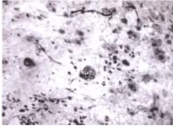
Spindle-shaped cellular morphology of Schwann cells cultured on the culture plate was viable and there was no sign of infection (Figure 1). After incubating with the anti-S100 antibody, nuclei of the Schwann cells were stained dark blown. The colorful profile of the dark granules of reaction-product made it easy to discriminate the labeled Schwann cells from the fibroblasts (Figure 2).
Gross examination of the silicone rubber chambers at 8 weeks revealed successful regeneration in the groups receiving the mixture of Schwann cells and bilobalide at 50, 100, 200 and 400 µM, with 44% (4 of 9, one died during experiment), 50% (5 of 10), 30% (3 of 10), and 60% (6 of 10) of the animals exhibiting a regenerated
nerve cable across the 15-mm gap, respectively. In comparison, 50% (5 of 10) of the animals in the group with Schwann cells only showed such regenerated nerve cables.
Figure 3 shows a representative cross section of regenerated nerve specimen. The epineurial and perineurial regions of the regenerated nerves consisted mainly of a collagenous connective tissue matrix in which circumferential cells resembling perineurial cells and fibroblasts were seen. In addition, the nerve fibers were loosely distributed in the endoneurium. Nuclei of Schwann cells were interspersed among these nerve fibers. Axons in the endoneurium were easily defined by their surrounding myelin sheaths stained dark blue by the toluidine blue. However, we also noted a number of regenerated nerve cables in the conduits with or without the fillings displayed a less mature ultrastructural organization. Specifically, those immature structures had no axons in the endoneurium, which was largely occupied by blood vessels (Figure 4). Some of the immature nerve cables had cores completely composed of a fibrin matrix, which is only seen in the early stage of regeneration.
Discussion and Conclusion
In our previous study, we found that pre-filled bilobalide in the regenerative chamber could provide a promoting effect on the regeneration of dissected rat sciatic nerves (Chen et al., 2004). In this study, we added Schwann cells into the regenerative
chamber containing the bilobalide to see if the mixture could further accelerate the growth of injured rat sciatic nerve. Like the results seen in our aforementioned study (Chen et al., 2004), we found the mixture of Schwann cells and bilobalide in the regenerative chamber could potentiate the formation of nerve cables successfully grew across the 15-mm gaps, which could never happen in a regenerative chamber with no grow-promoting fillings (Madison et al., 1988). This result is not surprising since the Schwann cells have been shown a promising supplement for use in entubulization repair of nerve defects (Mosahebi et al., 2001, 2002). The main reason is that Schwann cells can produce several growth factors, such as insulin-like growth factor-I (Hansson et al., 1986), ciliary neurotrophic factor (Rende et al., 1992), and brain-derived neurotrophic factor (Meyer et al., 1992), which have shown positive effects on regenerating axons. Similarly, the neuroprotection effect exerted by bilobalide could also simultaneously attenuate the degeneration and promote the regeneration of the injured nerve (Ahlemeyer et al., 2001). Though these results are encouraging, nevertheless, the added bilobalide in the conduits seemed could not further strengthen the supporting role of Schwann cells for regenerating nerves. We found that either the successful rate of nerve across the gap or the microstructure of the regenerated nerve in the groups with the mixture of Schwann cells and bilobalide were not significantly improved as compared to the group with Schwann cells only. This result
suggests that the bilobalide may not activate the Schwann cells in the conduits and therefore could not further invigorate their growth-promoting effects on regenerating nerves.
To sum up, the results of this study show that nerves have the capacity for regenerating across a large gap given an appropriate environment, namely a nerve guide filled with Schwann cells and bilobalide. Although the bilobalide did not significantly promote Schwann cell activation, the techniques we used in this study provided a method to incorporate traditional Chinese medicine and western biomedical science to nerve regeneration.
References
Ahlemeyer, B., V. Junker, R. Hühne and J. Krieglstein. Neuroprotective effects of NV-31, a bilobalide-derived compound: evidence for an antioxidative mechanism. Brain Res. 890: 338-342, 2001.
Chen, Y.S., C.H. Wu, C.H. Yao and C.T. Chen. Ginsenoside Rb1 enhances peripheral nerve regeneration across wide gaps in silicone rubber chambers. Int. J. Artif. Organs. 25: 1103-1108, 2002.
Chen, Y.S., C.J. Liu, C.Y. Cheng and C.H. Yao. Effect of bilobalide on peripheral nerve regeneration. Biomaterials 25: 509-514, 2004.
Evans, G.R.D., K. Brandt, S. Katz, P. Chauvin, L. Otto, M. Bogle, B. Wang, R.K. Meszlenyi, L. Lu, A.G. Mikos and
C.W. Patrick Jr. Bioactive poly(L-lactic acid) conduits seeded with Schwann cells for peripheral nerve regeneration. Biomaterials 23: 841-848, 2002.
Hansson, H.A., L.B. Dahlin, N. Danielsen, L. Fryklund, A.K. Nachemson, P. Polleryd, B. Rozell, A. Skottner, S. Stemme and G. Lundborg. Evidence indicating trophic importance of IGF-I in regenerating peripheral nerves. Acta Physiol. Scand. 126: 609-614, 1986. Kleijnen, J. and P. Knipschild. Ginkgo
biloba. Lancet 340: 1136-1139, 1992. Lin, F.H., G.C. Dong, K.S. Chen, G.J. Jiang,
C.W. Huang and J.S. Sun. Immobilization of Chinese herbal medicine onto the surface-modified
calcium hydrogenphosphate. Biomaterials 24: 2413-2422, 2003.
Madison, R., C.F. Da Silva and P. Dikkes. Entubulation repair with protein additives increases the maximum nerve gap distance successfully bridged with tubular prostheses. Brain Res. 447: 325-334, 1988.
Meyer, M., I. Matsuoka, C. Wetmore, L. Olson and H. Thoenen. Enhanced synthesis of brain-derived neurotrophic factor in the lesioned peripheral nerve: different mechanisms are responsible for the regulation of BDNF and NGF mRNA. J. Cell Biol. 119: 45-54, 1992. Mosahebi, A., B. Woodward, M. Wiberg, R.
Martin and G. Terenghi. Retroviral labeling of Schwann cells: In vitro characterization and in vivo transplantation to improve peripheral nerve regeneration. Glia 34: 8-17, 2001.
Mosahebi, A., P. Fuller, M. Wiberg and G. Terenghi. Effect of allogeneic Schwann cell transplantation on peripheral nerve regeneration. Exp. Neurol. 173: 213-223, 2002.
Rende, M., D. Muir, E. Ruoslahti, T. Hagg, S. Varon and M. Manthorpe. Immunolocalization of ciliary neuronotrophic factor in adult rat sciatic nerve. Glia 5:25-32, 1992.
Schlosshauer, B., E. Müller, B. Schröder, H. Planck and H.W. Müller. Rat Schwann cells in bioresorbable nerve guides to promote and accelerate axonal regeneration. Brain Res. 963: 321-326, 2003.
Thompson, C.B. Apoptosis in the pathogenesis and treatment of disease. Science 267: 1456-1162, 1995.
Yao, C.H., C.C. Tsai, Y.S. Chen, C.J. Chang, B.S. Liu, C.C. Lin and Y.H. Tsuang. Fabrication and evaluation of a new composite composed of tricalcium phosphate, gelatin and chi-li-saan as a bone substitute. Am. J. Chin. Med. 30: 471-482, 2002.
Figure 1: The optical microscopic examination of the Schwann cells
Figure 2: Schwann cells cultures immunostained with anti-S100
Figure 3: Light micrograph of a representative cross section of regenerated nerve specimen
Figure 4: Some of the regenerated nerve sections were immature with a microstructure nearly completely composed of fibrin strands and blood vessels only