Sweeping technique combined with micellar electrokinetic
chromatography for the simultaneous determination of
flunitrazepam and its major metabolites
Chiung-Wei Huang, Hsiu-Ping Jen, Ren-De Wang, You-Zung Hsieh
∗Department of Applied Chemistry, National Chiao Tung University, Hsinchu, Taiwan Received 23 July 2005; received in revised form 15 January 2006; accepted 17 January 2006
Available online 17 February 2006
Abstract
A sweeping technique, in conjunction with micellar electrokinetic chromatography, for the simultaneous determination of flunitrazepam and its major metabolites, 7-aminoflunitrazepam and N-desmethylflunitrazepam, is described. The optimized conditions for the sweeping and separation were a pH 9.5 buffer, 25 mM borate, 50 mM cetyltrimethylammonium bromide, 30% MeOH (v/v), and a 151-mm injection length. The calibration functions were all linear with the coefficient of determination (r2) exceeding 0.996 for the three target compounds. Using the sweeping procedure,
the limits of detection were determined to be 13.4, 5.6, and 12.0 ng/mL for flunitrazepam, 7-aminoflunitrazepam, and N-desmethylflunitrazepam, respectively, and the sensitivity enhancement for each compound was within the range of 110–200 fold. The RSDs for the retention time and the peak area were less than 4.10%. The optimized sweeping method was also used to examine a spiked urine sample. We conclude that sweeping with micellar electrokinetic chromatography has considerable potential use in clinical and forensic analyses of flunitrazepam and its metabolites. © 2006 Elsevier B.V. All rights reserved.
Keywords: Sweeping; Micellar electrokinetic chromatography; Flunitrazepam; Cetyltrimethylammonium bromide
1. Introduction
Flunitrazepam (Rohypnol), a nitro-containing benzodi-azepine, is used as a hypnotic and anesthetic induction agent. It is administered orally or by intravenous injection at doses of 2 mg. It has physiological effects similar to those of other benzodiazepines and has a potency that is ca. 10 times that of benzodiazepine. The illicit use of flunitrazepam usually involves a combination of other drugs, although it may be used alone. It has been used illegally in Asia since the early 1980s. In Taiwan, it appears to be used most frequently in conjunction with alco-hol, with which it seems to have a synergistic effect, producing disinhibition and amnesia. This has given flunitrazepam, espe-cially tasteless and odorless solutions, the reputation of being a “date-rape” drug.
Flunitrazepam can be detected in blood, plasma, and urine [1,2]. Because of its low dosage, biotransformation through
∗Corresponding author. Tel.: +886 3 5731785; fax: +886 3 5723764.
E-mail address: [email protected] (Y.-Z. Hsieh).
N-demethylation, and the high volume of distribution, fluni-trazepam and its metabolites occur at low blood levels after therapeutic administration [3]. Fig. 1 shows the pathway for flunitrazepam metabolism. Two major metabolites of flunitrazepam – 7-aminoflunitrazepam and N-desmethylflunit-razepam – can be detected when flunitN-desmethylflunit-razepam is injected or mixed into drinks[4].
Because of the rapid growth in the extent of abuse of flu-nitrazepam, a simple and consistent method is needed for its determination. Some analytical techniques for detecting fluni-trazepam have been reported, including the use of immunoassays [5], high-performance liquid chromatography (HPLC) [6,7], and gas chromatography/mass spectrometry (GC/MS) [8,9]. From the perspective of qualitative analysis, GC/MS provides additional spectral information as well as excellent sensitivity. Although GC/MS is capable of providing reliable data that can usually be used as scientific proof in a court of law, the method has disadvantages in that it involves time-consuming derivatiza-tion prior to the GC/MS analysis.
In recent years, capillary electrophoresis (CE) has expanded its scope and range in both instrumentation and applications 0021-9673/$ – see front matter © 2006 Elsevier B.V. All rights reserved.
Fig. 1. The major metabolic pathway for the detoxification of flunitrazepam in humans.
[10]. CE has proven to be a powerful analytical tool for sepa-rating charged species in diverse samples because of its many advantageous features, which include high column efficiency, rapid analysis times, and small sample volumes. However, the benefits derived by the high separation efficiency of CE can be overshadowed by its low UV detection sensitivity. Thus, using on-line sample preconcentration to overcome the poor sensitivity of CE has been the focus of a number of investigations[11,12]. For example, Quirino and Terabe[13]reported that neutral com-pounds could be concentrated effectively, when the technique of micellar electrokinetic chromatography (MEKC) combined with stacking was utilized. They later reported a sweeping method that can pick and accumulate neutral or charged ana-lytes into a narrow zone by the pseudostationary phase in MEKC [14–16].
In this paper, we report on an approach involving the use of a sweeping technique combined with MEKC for the simul-taneous determination of flunitrazepam and its major metabo-lites, 7-aminoflunitrazepam and N-desmethylflunitrazepam. The effects of the buffer pH, buffer concentration, cationic surfac-tant, organic modifier, and injection length on the analysis are described. We optimized the sweeping MEKC conditions to enhance the detection sensitivity with satisfactory resolution. We also employed the optimized sweeping MEKC method in an examination of a spiked urine sample.
2. Materials and methods
2.1. Apparatus
CE analysis was performed on a Beckman P/ACE MDQ CE system equipped with a photodiode-array detector (Fullerton, CA, USA). A personal computer, controlled by Beckman Coulter MDQ 32 Karat software was used for data collection. A 60 cm (50 cm to the detector)× 50 m I.D. fused-silica capillary tube (Polymicro Technologies, Phoenix, AZ, USA) was used. The capillary column was assembled in a car-tridge format. The temperature of the capillary tube during electrophoresis was maintained at 25◦C. The electrophoresis separation was performed at an applied voltage of −25 kV. Sample was pressure-injected at 0.5 psi with an extended time. The UV absorption detector was set at 240 nm for sweeping MEKC.
2.2. Chemicals
Flunitrazepam, 7-aminoflunitrazepam, and N-desmethyl-flunitrazepam were purchased from Radian International (Austin, TX, USA). Sodium tetraborate was obtained from Sigma (St. Louis, MO, USA). Cetyltrimethylammonium bro-mide (CTAB) was purchased from Merck (Hohenbrunn,
Ger-many). All other chemicals were analytical grade. Water was purified using a Milli-Q water system (Millipore, Bedford, MA, USA) and filtered through a 0.22-m filter.
2.3. Procedure
New capillaries were conditioned prior to separation by wash-ing with methanol, water, 1 M NaOH, and water for 10 min each. The capillary was flushed between runs with 0.1 M NaOH, methanol, and water for 3 min each. For the sweeping MEKC procedure, the stock solutions were diluted with a buffer solution that did not contain CTAB surfactants. The borate background solution (BGS) contained an appropriate amount of CTAB and methanol. The BGS was first passed through the capillary for 3 min and the sample solution was then pressure-injected into the capillary. Finally, voltages were applied at negative polar-ity. Other experimental conditions are described in the Section 3.
The SPE cartridges (Oasis MCX, 3 mL/60 mg) were condi-tioned with 2 mL methanol and 2 mL H2O. A 3-mL urine sample was spiked with 30-L of a standard solution (10 g/mL), then mixed with 60L pH 5.0 phosphate buffer (1 M) and passed through the cartridge. The cartridge was washed with 2 mL HCl (0.1 M) and 2 mL methanol, and was then dried under a vacuum for 10 min. The analytes were eluted with 3 mL dichloromethane/2-propanol/ammonium hydroxide solu-tion (78:20:2, v/v/v). This organic solusolu-tion was then evaporated to dryness. The residue was dissolved in 300L buffer and was used directly for MEKC.
3. Results and discussion
3.1. Effects of separation conditions for flunitrazepam and its major metabolites
Flunitrazepam and its metabolites (Fig. 1) are hydropho-bic substances, with a neutral charge in slightly or strongly basic environments, which could interact with micelles. Thus, a sweeping technique using CTAB was employed to achieve on-line sample concentration[15,16]. After the voltage was applied with a negative polarity from inlet (cathode), the EOF, under the influence of the cationic CTAB surfactant, moved toward the outlet (anode). Because the velocity of the EOF was higher than that of the CTAB micelle, the analytes stacked at the boundary by the CTAB micelle and moved toward the anode.
When performing the analysis using MEKC, the pH and concentration of the buffer solution were adjusted so as to obtain adequate separation. The migration times of the analytes increased with increasing pH value from pH 9.5 to pH 10.5 with a similar sensitivity enhancement. The peaks also broadened and their heights decreased for electrolyte concentrations lower than 25 mM. The migration time of the analytes was delayed with increasing electrolyte concentration. Taking all of these phe-nomena into consideration, we conclude that the pH 9.5 buffer with 25 mM electrolyte is the most suitable for the separation.
In the basic buffer solution, flunitrazepam and its metabolites acted as neutral analytes, and migrated with the electroosmotic flow. When they interacted with the positively charged CTAB micelles, however, the decrease in their apparent electrophoretic
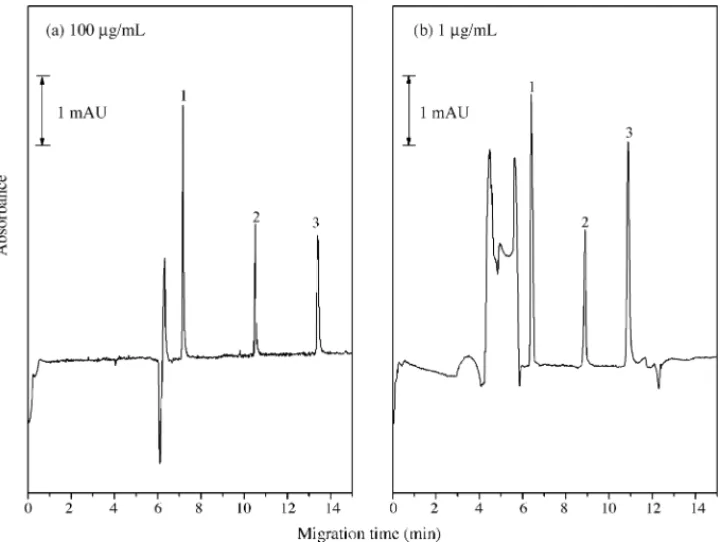
Fig. 2. Normal MEKC and sweeping MEKC analysis of flunitrazepam and its major metabolites. (a) MEKC analysis. Analyte concentration: 100g/mL; injection length: 1.51 mm. (b) Sweeping MEKC analysis. Analyte concentration: 1g/mL; injection length: 151 mm. Conditions: capillary, 60 cm long (50 cm to detector), 50m I.D.; buffer solution: 25 mM borate buffer (pH 9.5), 50 mM CTAB, 30% CH3OH (v/v), conductivity 7.28 mS/cm; sample matrix: 25 mM borate buffer (pH 9.5);
Table 1
Calibration lines, coefficient of determination (r2), limits of detection (LODs), migration times, and values of RSD for flunitrazepam, 7-aminoflunitrazepam, and
N-desmethylflunitrazepam using the MEKC and sweeping MEKC techniques
Flunitrazepam 7-Aminoflunitrazepam N-desmethyl-flunitrazepam MEKC
Calibration linea y = 101x + 224 y = 130x− 121 y = 71.5x− 228
Coefficient of determination r2= 0.997 r2= 0.999 r2= 0.997
LOD (S/N = 3,g/mL) 1.87 0.52 1.74
Migration time (min) 10.54 7.17 13.10
RSD (%; n = 5)
I. Migration time 0.55 0.25 0.62
II. Peak area 2.85 2.04 1.48
Sweeping MEKC
Calibration lineb y = 11.5x− 682 y = 28.2x− 853 y = 24.7x− 3.10 × 10³
Coefficient of determination r2= 0.996 r2= 0.999 r2= 0.998
LOD (S/N = 3, ng/mL) 13.4 5.6 12.0
Migration time (min) 8.48 6.21 10.24
RSD (%; n = 5)
I. Migration time 0.39 0.28 0.51
II. Peak area 1.14 2.01 4.10
SEheightc 110 140 200
aCalibration line (10–200g/mL): peak area (arbitrary units) = slope × concentration (g/mL) + y-intercept. b Calibration line (50–1000 ng/mL): peak area (arbitrary units) = slope× concentration (ng/mL) + y-intercept. cSE
height= (peak height obtained with sweeping MEKC/peak height obtained with MEKC)·dilution factor.
velocities caused these analytes to become focused. When the CTAB concentration was increased from 10 to 50 mM, separa-tion and peak height improved, suggesting that the sweep effect became more efficient. Nevertheless, when the CTAB concen-trations exceeded 50 mM, the separations became poor. These results suggest that the use of 50 mM CTAB provides the best condition for the separation.
Increasing the percentage of methanol in the buffer had a dramatic influence on the analyte migration time and the peak focusing effect. The results showed that adding an organic sol-vent to the buffer modified the polarity of the BGS, which further changed the EOF. It also improved the resolution by modifying the partition of the analytes between the solution phase and the micelle phase. The experimental results indicate that adding 30% methanol to the buffer solution provided the best condition for the separation.
In general, prolonging the sample injection length in sweep-ing MEKC is advantageous, in terms of achievsweep-ing better sensi-tivity for a separation. A long sample zone, however, increases the sweeping time and may have a negative influence on the effi-ciency of the sweeping procedure. Using the optimal conditions discussed above, we found that an injection length of 151 mm is suitable for the complete separation of all the peaks.
3.2. Comparing normal MEKC and sweeping MEKC
Fig. 2depicts the results of normal MEKC and the sweeping MEKC separation of flunitrazepam, 7-aminoflunitrazepam, and N-desmethylflunitrazepam under optimized conditions.Fig. 2a was obtained when the sample solution was the same as the running buffer, but did not contain micelles. The concentration of each analyte was 100g/mL.Fig. 2b was obtained in a manner
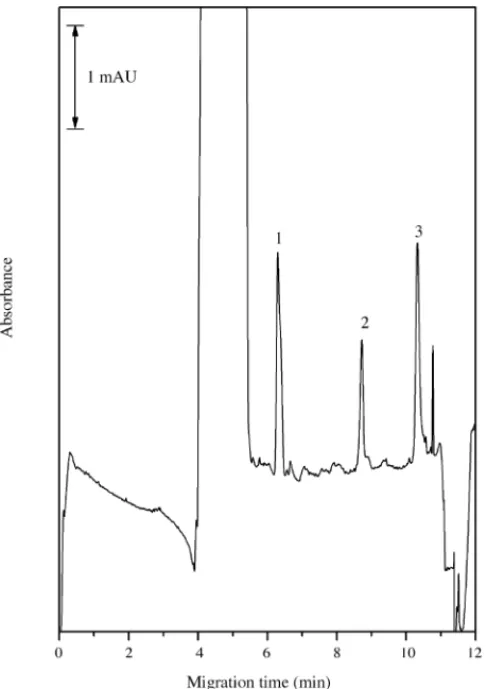
Fig. 3. Sweeping MEKC electropherogram of a spiked urine sample. Analyte concentration: 0.3g spiked in a 3-mL urine sample before SPE extraction. The other conditions are the same as inFig. 2.
similar to that ofFig. 2a, but the injection length was 151 mm and the sample concentration was diluted 100-fold. The sensitivity enhancement in terms of peak heights (SEheight) for the three analytes was calculated. Flunitrazepam, 7-aminoflunitrazepam, and N-desmethylflunitrazepam had ca. 110-, 140-, and 200-fold enhancements in their detection sensitivities, respectively.
Table 1 presents the calibration lines, coefficient of deter-mination (r2), limits of detection (LODs), migration times, and RSDs for the three analytes using MEKC and sweeping MEKC techniques. For analyses conducted using the normal MEKC procedure, the LODs were in the lowg/mL range. When the sweeping MEKC procedure was used, the LODs were less than 13.4 ng/mL. Table 1 also presents the reproducibility of the migration times and peak area. The RSD for the migration time was less than 0.62% for either separation procedure. The RSD of the peak area was also less than 4.10%. According to these results, both processes are acceptable separation methods, but the sweeping MEKC procedure is superior to MEKC in detec-tion sensitivity.
Fig. 3 illustrates the use of the sweeping MEKC method in analyzing a urine sample spiked with flunitrazepam and its metabolites. The separation of these analytes in urine was ade-quate. The total separation time was less than 12 min. Thus, sweeping MEKC can be used as a rapid screening method for the analysis of flunitrazepam and its major metabolites.
In conclusion, we report that a sweeping technique com-bined with MEKC permits the simultaneous determination of flunitrazepam and its major metabolites through a process that is easily performed, and does not require a derivatization step. The optimized parameters for the sweeping MEKC method were: running buffer, 25 mM borate buffer (pH 9.5); CTAB, 50 mM; organic modifier, 30% MeOH (v/v); injection
length, 151 mm. The LODs ranged from 5.6 to 13.4 ng/mL. Accordingly, sweeping in conjunction with MEKC represents an alternative approach with enhanced sensitivity for analyzing flunitrazepam and its major metabolites.
Acknowledgement
This study was supported by grant (NSC 93-2113-M-009-022) from the National Science Council of Taiwan.
References
[1] D. Borrey, E. Meyer, W. Lambert, C. Van Peteghem, A.P. De Leenheer, J. Chromatogr. B 765 (2001) 187.
[2] M. Kollroser, C. Schober, J. Pharm. Biomed. Anal. 28 (2002) 1173.
[3] H.G. Boxenbaum, H.N. Posmanter, T. Macasieb, K.A. Geitner, R.E. Weinfeld, J.D. Moore, A. Darragh, D.A. O’Kelly, L. Weissmann, S.A. Kaplan, J. Pharmacokinet. Biopharm. 6 (1978) 283.
[4] H. Sch¨utz, Benzodiazepines II, Springer, Berlin, 1989. [5] W. Huang, D.E. Moody, J. Anal. Toxicol. 19 (1995) 333.
[6] M.D. Robertson, O.H. Drummer, J. Chromatogr. B 667 (1995) 179. [7] W. He, N. Parissis, J. Pharm. Biomed. Anal. 16 (1997) 707.
[8] M.A. ElSohly, S. Feng, S.J. Salamone, R. Brenneisen, J. Anal. Toxicol. 23 (1999) 486.
[9] D. Borrey, E. Meyer, W. Lambert, S. Van Calenbergh, C. Van Peteghem, A.P. De Leenheer, J. Chromatogr. A 910 (2001) 105.
[10] S.F.H. Li, Capillary Electrophoresis, Principle, Practice, and Applica-tions, Elsevier, Amsterdam, 1992.
[11] J.L. Beckers, P. Boˇcek, Electrophoresis 21 (2000) 2747. [12] P. Britz-McKibbin, S. Terabe, J. Chromatogr. A 1000 (2003) 917. [13] J.P. Quirino, S. Terabe, Anal. Chem. 70 (1998) 149.
[14] J.P. Quirino, S. Terabe, Science 282 (1998) 465.
[15] J.-B. Kim, J.P. Quirino, K. Otsuka, S. Terabe, J. Chromatogr. A 916 (2001) 123.