IINNTTRROODDUUCCTTIIOONN
Liver fibrosis is a common consequence of many liver diseases, including viral hepatitis,
alcoholic hepatitis, primary biliary cirrhosis, primary sclerosing cholangitis and biliary atresia. Hepatic regeneration is an integral process in the liver’s response to chronic injury, and has mainly been studied in the setting of acute liver regeneration following partial hepatectomy [1]. Proliferating cell nuclear antigen (PCNA) Received : 6 September 2007. Revised : 13 November 2007.
Accepted : 11 December 2007.
Correspondence to : Jung-Chou Chen, School of Post-baccalaureate Chinese Medicine, China Medical University, 91 Hsueh-Shih Road, Taichung 404, Taiwan.
Xiao-Chai-Hu-Tang Attenuated the
Expression of Proliferating Cell Nuclear
Antigen and Epidermal Growth Factor in
a Rat Model of Hepatic Fibrosis
Ming-Ho Chen1,2,5, Jung-Chou Chen1,5, Chin-Chuan Tsai5,6, Wen-Chuang Wang3, Hsiao-Yen Hsieh4
1
Research Institute of Chinese Medicine, 5School of Post-baccalaureate Chinese Medicine, China Medical University, Taichung; 2Department of Chinese Medicine, 3Department of Pathology, 4
Department of Medical Research, Chiayi Christian Hospital, Chiayi; 6Department of Biological Science & Technology, I-Shou University, Kaohsiung, Taiwan.
B
Baacckkggrroouunndd//PPuurrppoossee.. To study the effect of Xiao-Chai-Hu-Tang on the expression of
proliferating cell nuclear antigen (PCNA) and to determine the relationship between the expression levels of PCNA and epidermal growth factor in a rat model of bile duct ligation-induced biliary fibrosis.
M
Meetthhooddss.. In this study, we used immunohistochemical staining to detect the expression of
proliferating cell nuclear antigen in hepatocytes and bile duct epithelium in bile duct-ligated rats with or without Xiao-Chai-Hu-Tang treatment for six weeks. Reverse transcription polymerase chain reaction was used to detect the level of epidermal growth factor mRNA expression.
R
Reessuullttss.. Treatment with Xiao-Chai-Hu-Tang for six weeks reduced the expression levels of
proliferating cell nuclear antigen and epidermal growth factor in hepatocytes and in bile duct epithelium. Expression of epidermal growth factor significantly correlated with the expression of proliferating cell nuclear antigen.
C
Coonncclluussiioonn.. Bile duct ligation significantly induced the proliferation of hepatocytes and bile
duct cells. Proliferation correlated with the increased expression of epidermal growth factor mRNA. Our data suggest that Xiao-Chai-Hu-Tang inhibits mRNA expression of epidermal growth factor, thereby reducing further hepatocellular proliferation in rats with biliary fibrosis. ( Mid Taiwan J Med 2008;13:65-74 )
K
Keeyy wwoorrddss
bile duct ligation, epidermal growth factor, liver fibrosis, proliferating cell nuclear antigen, Xiao-Chai-Hu-Tang
labeling index has been used as a proliferation index in the evaluation of liver regeneration in rats [2]. Beatriz et al. found that the development of fibrogenesis after bile duct ligation was characterized by the marked early proliferation of bile duct cells and hepatocytes [3].
Epidermal growth factor (EGF) is one of the earliest signals to appear after partial hepatectomy in rats [4]. Chronic hepatic regeneration constitutes an important part of the cirrhotic process. The development of cirrhosis has been reported to be associated with a progressive increase in EGF mRNA expression in bile duct ligation models [5].
The bile duct-ligated rat model has been examined extensively, and is used to induce progressive fibrosis and the development of secondary biliary cirrhosis. This model is also used to study proliferation of hepatocytes by assessing proliferating cell nuclear antigen expression, a process known to be related to increased expression levels of epidermal growth factor receptor in the nucleus [6]. Napoli et al. have shown that EGF is up-regulated in the rat bile duct ligation model, and that its increase is associated with progressive tissue remodeling. Tissue remodeling in this model is dominated by bile duct proliferation and hepatocyte regeneration [5].
Xiao-Chai-Hu-Tang (Sho-saiko-to, TJ-9, in Japanese) is an herbal medicine commonly prescribed to treat chronic hepatitis and liver cirrhosis in China and Japan. Sakae et al. have proved that Xiao-Chai-Hu-Tang exerts hepatoprotective effects on CCl4-induced liver injury [7]. In our previous study, we found that TJ-9 significantly reduces cholestasis and liver fibrosis in bile duct-ligated rats [8]. Furthermore, many researches have demonstrated the preventive and therapeutic effect of TJ-9 on experimental hepatic fibrosis. For example, Sakaida et al. reported that TJ-9 prevents hepatic fibrosis, reduces the expression of type (III) procollagen mRNA and inhibits the activation of Ito cells [9]. Shimizu et al. showed that TJ-9 has an antifibrotic effect on reducing collagen type (I)
synthesis and inhibiting the activation of Ito cells via the suppression of oxidative stress in hepatocytes and Ito cells [10].
The purpose of the current study was to investigate whether Xiao-Chai-Hu- Tang reduces the expression of PCNA and EGF in a rat model of bile duct ligation. We also determined the correlation between the expression of PCNA and that of EGF.
M
MAATTEERRIIAALLSS AANNDD MMEETTHHOODDSS Animals
Fifty male Wistar albino rats weighing approximately 200 to 250 g were purchased from the National Animal Center, kept on a standard rat diet with free access to tap water, and maintained on a 12-hour light-dark cycle. All animals received humane care. The study protocol was approved by the Chiayi Christian Hospital Ethics Committee on Animal Experiments.
Chemicals
The RNA extraction miniprep system was purchased from Viogene-Biotek Co. (Taiwan). Im Prom- TM
-reverse transcriptase was purchased from Promega Co. (USA). EGF and GAPDH primers were obtained from MdBio Inc. (Taiwan). Anti-PCNA antibody was purchased from Protech Technology Enterprise Co. (USA).
Preparation of Xiao-Chai-Hu-Tang extract
Xiao-Chai-Hu-Tang (Sho-saiko-to) extract powder was kindly provided by Ko-Da Pharmaceutical co., Taiwan. All of the herbs were authenticated by Dr. Shih-Chang Lee, China Medical University, Taiwan. Briefly, 1000 g of Xiao-Chai-Hu-Tang, comprising 333 g of Bupleurum chinese DC root, 133 g of Scutellaria baicalensis Georgi root, 133 g of Panax ginseng C.A. Meyer root, 67 g of Glycyrrhiza uralensis Fischer root, 67 g of Zingiber officinale Roscoe rhizome, 133 g of Pinellia ternata thunb. Breit. tuber, and 133 g of Zizyphus jujuba Mill fruit were decocted with 7 liters of boiling water in a stainless steel oven for 1 hour. The decoction was filtered and decocted again for another 50 minutes. The filtrate was then concentrated under reduced pressure (60 to 80 mm Hg) at 55 C by a
rotary vacuum evaporator followed by freeze drying at 45 C. The yield was 25.47% and it was diluted to a stock solution of 50 mg/mL.
Bile duct ligation model in rats
After an accommodation period of two weeks, the rats were randomly assigned to one of three groups: (A) sham operation (n = 10), (B) bile duct ligation (BDL) (n = 20), (C) bile duct ligation and treatment with Xiao-Chai-Hu-Tang at a dose of 0.5 g/kg via intragastric gavage (n = 20). Rats were anaesthetized with 0.1 mL/100g Zoletil (tietamine and zolezetam, Virbac, France) via intraperitoneal injection. A midline abdominal incision was made and the common bile duct was identified, doubly ligated with 3-0 black silk, and transected between two ligatures. Sterile saline (2 mL) was instilled into the peritoneum at the end of surgery, and the abdomen was closed in two layers with 3-0 black silk [11]. Rats in the sham group underwent an operation identical to that performed in the BDL group, except that the bile duct was not ligated or transected. Rats in the Xiao-Chai-Hu-Tang-treated group underwent ligation of the bile duct and were treated with dry powder 0.5 g/kg by intragastric gavage everyday except Sunday for 6 weeks. After 42 days, all of the animals were sacrificed under Zoletil anesthesia, and blood was collected from the carotid artery. Livers and spleens were removed at the same time. Half of the organs were fixed in 10% formalin and sectioned into 4 µm slices for histological examination, and half were immediately snap-frozen in liquid nitrogen for extraction of total RNA.
Immunohistochemical staining for PCNA
Immunohistochemical staining for PCNA on the paraffin-embedded liver tissue was performed with anti-PCNA antibody as previously described [2,12]. Briefly, 4 µm thick sections of the formalin-fixed, paraffin-embedded materials were mounted on glass slides. After deparaffinization, the sections were incubated in 0.3% methanolic hydrogen peroxide for 30 minutes to block endogenous peroxidase activity. Nonspecific protein binding was inhibited by treatment with 10% goat serum before exposure
to the primary antibody. The sections were then incubated with monoclonal IgG antibody against PCNA diluted at 1: 200. The detection of the binding of primary reagents was achieved by an avidin-biotin-peroxide complex kit. Diaminobenzidine was used to visualize peroxidase deposition at the antigenic sites. These sections were then counterstained with methyl green. Definitive reddish-brown staining of the nucleus confirmed PCNA positivity. Finally, PCNA labeling indexes were determined by random evaluation of at least 500 hepatocytes and bile duct epithelial cells at a magnification of
400.
RT-PCR of epidermal growth factor
Total RNA was extracted by the total RNA extraction miniprep system (Viogene, Viogene-Biotek co., Taiwan, ROC). After that, cDNA preparation and polymerase chain reaction (PCR) were performed as previously described [13,14]. EGF primers were Sense: 5’-AGA AGA GGA AAG GCA AGG GGT TAG G-3’and Antisense: 5’-CCT GAA CAT GAG AAG TCC CAC GAT G-3’.
An initial denaturation at 95 C for 5 min, 35 cycles at 95 C for 40 sec, 54 C for 1 min, 72 C for 1.5 min, and then 1 cycle of extension at 72 C for 7 min were performed using a DNA thermocycler (Applied Biosystems Gene Amp PCR System 2400). The PCR products were stored at 20 C and DNA was separated by electrophoresis on a 5% acrylamide gel and 1xTBE running buffer. The products were then stained with ethidium bromide and visualized using an UV transilluminator. Finally, we used a Kodak Digital Science ID 3.02 and DC40/DC120 Camera to analyze the PCR products. The target mRNA signals were normalized to GAPDH mRNA signals and expressed as relative abundance.
Statistical analysis
Results are expressed as mean SD, and the data obtained were evaluated by one way analysis of variance (ANOVA) as appropriate. Differences in the intensity of PCNA and EGF between the bile duct ligation group and the other
groups were evaluated by the Student’s t test. The level of significance was set at p < 0.05 for each analysis. The correlation between the intensity of PCNA and that of EGF was evaluated by Pearson correlation.
R
REESSUULLTTSS
Effect of Xiao-Chai-Hu-Tag on the expression of PCNA protein
Within the first week, one rat in the sham group, six rats in the BDL group and seven Xiao-Chai-Hu-Tang-treated rats died due to infection following the operation. These fifteen rats were excluded from the study. Therefore, the final study population comprised nine rats in the sham group, nine rats in the BDL group and seven Xiao-Chai-Hu-Tang-treated rats.
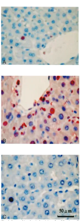
Microscopic examination of the liver sections from the sham group revealed nonspecific morphologic change of liver parenchyma, as well as trace amounts of reddish-brown positive PCNA labeling of hepatocytes and bile duct epithelial cells (Figs. 1A, 2A). Liver sections in the BDL group revealed massive portal bile duct cell proliferation and bridging fibrosis (Fig. 2B), and scant inflammation and necrosis or apoptosis in the hepatocytes (Fig. 1B). Liver sections in the Xiao-Chai-Hu-Tang-treated group revealed significantly reduced bile duct cell proliferation and fibrosis (Fig. 2C).
The PCNA indexes of hepatocytes and bile duct epithelial cells were significantly higher in specimens in the BDL group than in specimens in the sham group (p < 0.001), (Table). In contrast, the PCNA indexes of hepatocytes and bile duct epithelial cells were significantly lower in the Xiao-Chai-Hu-Tang-treated group than in the BDL group (p < 0.01).
Effect of Xiao-Chai-Hu-Tang on the expression of EGF mRNA
The mRNA expression of EGF was determined by RT-PCR. The expression was markedly enhanced in the BDL group (Fig. 3). As expected, a 280 bp band representing EGF mRNA was clearly shown. No equivalent band was seen in the negative control. The target mRNA signals were expressed as relative
Fig. 1. Immunohistochemical staining of proliferating cell nuclear antigen (PCNA) in hepatocytes in bile duct-ligated rats. A: Sham operation. B: Bile duct ligation for 6 weeks without treatment. C: Bile duct ligation and treatment with 0.5 g/kg Xiao-Chai-Hu-Tang for 6 weeks. The reddish-brown staining of the nucleus was confirmed to be PCNA-positive (original magnification 400).
C
B
A
abundance (Table). The relative abundance of EGF transcripts in the Xiao-Chai-Hu-Tang-treated group was significantly lower than that in the BDL group (0.48 0.19 vs 0.84 0.13, p < 0.001).
Correlation between PCNA expression and EGF expression
Pearson correlation comparison revealed a significant correlation between the expressions of PCNA protein and EGF mRNA in rat hepatocytes (r = 0.751, p < 0.01, Fig. 4). Similarly, Pearson correlation also showed a significant correlation between the expressions of PCNA protein and EGF mRNA in rat bile duct epithelial cells (r = 0.584, p < 0.01, Fig. 4).
Fig. 2. Immunohistochemical staining of proliferating cell nuclear antigen (PCNA) in bile duct epithelial cells in bile duct-ligated rats. A: Sham operation. B: Bile duct ligation for 6 weeks without treatment. C: Bile duct ligation and treatment with 0.5 g/kg Xiao-Chai-Hu-Tang for 6 weeks. The reddish-brown staining of the nucleus was confirmed to be PCNA-positive (original magnification 400).
C
B
A
Fig. 3. A: The mRNA expression of epidermal growth factor (EGF) was evaluated by RT-PCR analysis. Total RNA was extracted from liver tissue after 6 weeks of bile duct ligation (BDL). Lane M = DNA markers; Lane A = sham-operated rat; Lane B = BDL without treatment; Lane C = BDL and Xiao-Chai-Hu-Tang at a dose of 0.5g/kg BW/day. GAPDH as the internal control; (-) as the negative control. B: Densitometric results of EGF mRNA level in RT-PCR. Signals were normalized to GAPDH and expressed in arbitrary units. ***p < 0.001 vs BDL alone.
B
A
D
DIISSCCUUSSSSIIOONN
Bile duct ligation (BDL) in rats results in progressive fibrosis in the virtual absence of marked inflammation and necrosis [15], resembling that of human biliary liver fibrosis. Therefore, BDL allows for the detection of antifibrotic effects that are not obscured by radical scavenging or anti-inflammatory properties of therapeutic agents [16].
PCNA is a highly conserved 36 kDa nuclear protein that is highly modulated during the cell cycle [17]. PCNA expression begins in the late G1 phase and becomes maximal in the S phase. It is, therefore, used as a proliferation index in the evaluation of liver regeneration [18]. Measuring PCNA expression can also be used as an alternative to tritiated thymidine labeling for marking S phase cells. Furthermore, its expression correlates with the incorporation rate of bromodeoxyuridine in hepatocytes [19]. PCNA
labeling indexes have been shown to be higher in damaged liver (nonfulminant and fulminant hepatitis) than in normal liver [2]. Chiyiiwa et al showed that PCNA expression was higher in the cirrhotic liver before partial hepatectomy [20]. Our results showed that there was a marked increase in both PCNA positive hepatocytes and bile duct epithelial cells in the BDL group. Zimmermann found the same phenomenon [6].
Beatriz et al. has proposed that there may be a two-stage process of fibroblast modulation [3]. The first stage, a very early event associated with ductular reaction, is characterized by periductular fibroblast proliferation and modulation of myofibroblasts [21]. The second stage involves mainly the hepatic stellate cells, which, after being activated, express desmin and α-smooth muscle actin, and participate in portal and septal fibrosis [22]. Our results show that Xiao-Chai-Hu-Tang significantly reduced the Fig. 4. A: The correlations between the expression of PCNA and EGF mRNA in hepatocytes. A: Sham operation. B: Bile duct ligation for 6 weeks without treatment. C: Bile duct ligation and treatment with 0.5 g/kg Xiao-Chai-Hu-Tang for 6 weeks. r = 0.751, p < 0.01. B: The correlations between the expression of PCNA and EGF mRNA in bile duct epithelial cells. r = 0.584, p < 0.01.
A
B
Table. Effect of Xiao-Chai-Hu-Tang on proliferating cell nuclear antigen (PCNA) and epidermal growth factor (EGF) mRNA expression in bile duct-ligated rats
Sham op BDL BDL+TJ-9 0.7 0.4 11.0 3.5 3.7 5.6* PCNA (H) Group PCNA (B) 0.25 0.11 0.84 0.13 0.48 0.19 EGF 0.2 0.3 100.7 43.1 32.3 55.3*
Results are expressed as mean SD, and the data obtained were evaluated by one way analysis of variance (ANOVA) as appropriate. The differences in the intensity of PCNA and EGF between the bile duct ligation group and the other groups were evaluated by the Student’s t-test. *p < 0.01, p < 0.001 (vs BDL). Sham op = sham operation; BDL = bile duct ligation; BDL+TJ-9 = bile duct ligation and Xiao-Chai-Hu-Tang at a dose of 0.5 g/kg body weight/day. H = hepatocytes; B = bile duct epithelial cells.
PCNA indexes of both hepatocytes and bile duct cells. Therefore, we presume that Xiao-Chai-Hu-Tang inhibits the proliferation of periductular fibroblasts in the early stage of fibrogenesis by reducing the PCNA indexes.
Epidermal growth factor (EGF) is a potent mitogen in numerous cell types, including hepatocytes. EGF is secreted into bile to enhance the proliferation of bile duct epithelial cells and hepatic stellate cells [23,24]. Oguey et al reported that EGF receptor expression was involved in the maintenance of hepatocellular masses in a bile duct ligation model of cirrhosis [25]. EGF mRNA has been shown to increase 10-fold within 15 minutes after partial hepatectomy in rats, before returning to baseline levels by 24 hours [4]. After 3 weeks of bile duct ligation, there was a 25-fold increase in EGF mRNA expression [5].
Napoli et al found that EGF, platelet derived growth factor (PDGF) and transforming growth factor (TGF-β) were up-regulated in a BDL model of cirrhosis, and that their increases were associated with progressive tissue remodeling [5]. This tissue remodeling was dominated by bile duct proliferation with some hepatocyte regeneration. Therefore, EGF may play a role in the proliferation of bile duct epithelial cells and hepatic stellate cells (Ito cells) [26]. According to our results, EGF mRNA was 40% lower in rats treated with Xiao-Chai-Hu-Tang than in rats that underwent bile duct ligation but did not receive the drug. We presume that Xiao-Chai-Hu-Tang inhibited further bile duct proliferation by reducing the mRNA expression of EGF.
After 6 weeks of bile duct ligation, the PCNA labeling indexes in hepatocytes and bile duct cells correlated well with the level of EGF mRNA expression. Zimmermann et al also showed that the percentage of EGF receptor positive nuclei was closely related to the percentage of PCNA positive hepatocytes [6]. These findings support our results.
Shimizu et al. has confirmed that the antifibrotic effect of Xiao-Chai-Hu-Tang is associated with the regulation of extracellular
matrix proteins, including type I collagen, α-smooth muscle actin expression, retinoid disappearance and hepatic stellate cell proliferation [27]. Several growth factors, including TGF-β1, EGF and PDGF, may contribute to the fibrogenic response and trigger the transformation of hepatic stellate cells in vivo [28]. A preliminary report concluded that Xiao-Chai-Hu-Tnag inhibited PDGF-induced proliferation of hepatic stellate cells [29]. Xiao-Chai-Hu-Tang also has been shown to inhibit TGF-β1 mRNA expression in the human Ito cell line LI90, and to inhibit TGF-β1 protein secretion through the suppression of a specific region of the TGF-β1 promotor [30].
In this study, bile duct ligation in rats induced significant hepatocellular proliferation, including hepatocytes and bile duct cells. Furthermore, this proliferation correlated with the increased expression of EGF mRNA. We conclude that Xiao-Chai-Hu-Tang inhibits hepatocellular proliferation in the early stage of fibrogenesis by reducing the expression of EGF mRNA in rats with biliary fibrosis.
A
ACCKKNNOOWWLLEEDDGGMMEENNTTSS
This research was supported by a grant from the Chiayi Christian Hospital, Taiwan. The authors would like to express their thanks to Chan-Jan Chen (the superintendent of Chiayi Christian Hospital), and the Molecular Biology Department of National Chung-Cheng University for their generous support and technical assistance.
R
REEFFEERREENNCCEESS
1. Fausto N, Laird AD, Webber EM. Liver regeneration. 2. Role of growth factors and cytokines in hepatic regeneration. [Review] FASEB J 1995;9:1527-36. 2. Wolf HK, Michalopoulos GK. Hepatocyte
regeneration in acute fulminant and nonfulminant hepatitis: a study of proliferating cell nuclear antigen expression. Hepatology 1992;15:707-13. 3. Tuchweber B, Desmouliere A, Bochaton-Piallat ML,
et al. Proliferation and phenotypic modulation of portal fibroblasts in the early stage of cholestatic fibrosis in the rat. Lab Invest 1996;74:265-78.
4. Mullhaupt B, Feren A, Fodor E, et al. Liver regeneration of epidermal growth factor RNA. Rapid increases in immediate-early phase of liver regeneration. J Biol Chem 1994;269:19667-70. 5. Napoli J, Prentice D, Niinami C, et al. Sequential
increases in the intrahepatic expression of epidermal growth factor, basic fibroblast growth factor, and transforming growth factor beta in a bile duct ligated rat model of cirrhosis. Hepatology 1997;26:624-33. 6. Zimmermann H, Ganz P, Zimmermann A, et al. The
overexpression of proliferating cell nuclear antigen in biliary cirrhosis in the rat and its relationship with epidermal growth factor receptor. J Hepatol 1995;23: 459-64.
7. Sakae A, Masakane H, Yukio O, et al. Treatment of chronic liver injury in mice by oral administration of Xiao-Chai-Hu-Tang. J Ethnopharmacol 1989;25:181-7.
8. Chen MH, Chen JC, Tsai CC, et al. Sho-saiko-to prevents liver fibrosis induced by bile duct ligation in rats. Am J Chin Med 2004;32:195-207.
9. Sakaida I, Matsumura Y, Akiyama S, et al. Herbal medicine sho-saiko-to (TJ-9) prevents liver fibrosis and enzyme-altered lesions in rats liver cirrhosis induced by a choline-deficient L-amino acid-defined diet. J Hepatol 1998;28:298-306.
10. Shimizu I, Ma YR, Mizobuchi Y, et al. Effect of Sho-saiko-to, a Japanese herbal medicine, on hepatic fibrosis in rats. Hepatology 1999;29:149-60.
11. Plebani M, Panozzo MP, Basso D, et al. Cytokines and the progression of liver damage in experimental bile duct ligation. Clin Exp Pharmacol Physiol 1999; 26:358-63.
12. Hsu SM, Raine L, Ranger H. Use of avidin-biotin peroxidase complex (ABC) in immunoperoxidase techniques: a comparison between ABC and unlabeled antibody (PAP) procedure. J Histochem Cytochem 1981;29:557-80.
13. Iredale JP, Benyon RC, Arthur MJ, et al. Tissue inhibitor of metalloproteinase-1 messenger RNA expression is enhanced relative to interstitial collagenase messenger RNA in experimental liver injury and fibrosis. Hepatology 1996;24:176-84. 14. Lee HS, Huang GT, Miau LH, et al. Expression of
matrix metalloproteinases in spontaneous regression of liver fibrosis. Hepatogastroenterology 2001;48: 1114-7.
15. Gerling B, Becker M, Waldschmidt J, et al. Elevated serum aminoterminal procollagen type-III-peptide parallels collagen accumulation in rats with secondary biliary fibrosis. J Hepatol 1996;25:79-84.
16. Boigk G, Stroedter L, Herbst H, et al. Silymarin retard collagen accumulation in early and advanced biliary fibrosis secondary to complete bile duct obliteration in rats. Hepatology 1997;26:643-9.
17. Bravo R, Fey SJ, Bellatin J, et al. Identification of a nuclear and of a cytoplasmic polypeptide whose relative proportions are sensitive to change in the rate of cell proliferation. Exp Cell Res 1981;136:311-9. 18. Celis JE, Bravo R, Larsen PM, et al. Cyclin: a nuclear
protein whose level correlates directly with the proliferative state of normal as well as transformed cells. Leuk Res 1984;8:143-57.
19. Gerland P, Defraef C. Cyclin/PCNA immunostaining as an alternative to tritiated thymidine pulse labeling for marking S phase cells in paraffin sections from animal and human tissues. Cell Tissue Kinet 1989; 22:383-92.
20. Chijiwa K, Nakano K, Kameoka N, et al. Proliferating cell nuclear antigen, plasma fibronection, and liver regeneration rate after seventy percent hepatectomy in normal and cirrhotic rats. Surgery 1994;116:544-9. 21. Abdel-Aziz G, Rescan PY, Clement B, et al. Cellular
sources of matrix proteins in experimentally induced cholestatic rat liver. J Pathol 1991;164:167-74. 22. Rockey DC, Boyles JK, Gabbiani G, et al. Rat hepatic
lipocytes express smooth muscle actin upon activation in vivo and in culture. J Submicrosc Cytol Pathol 1992;24:193-203.
23. Marti U, Burwen SJ, Jones AL. Biological effects of epidermal growth factor, with emphasis on the gastrointestinal tract and liver: an update. [Review]
Hepatology 1989;9:126-38.
24. Bachem MG, Reiss U, Gressner AM. Liver fat storing cell proliferation is stimulated by epidermal growth factor/transforming growth factor alpha and inhibited by transforming growth factor beta. Biochem Biophys
Res Commun 1989;162:708-14.
25. Oguey D, Marti U, Reichen J. Epidermal growth factor receptor in chronic bile duct obstructed rats: implications for maintenance of hepatocellular mass.
Eur J Cell Biol 1992;59:187-95.
26.St. Hilare RJ, Hradek GT, Jones AL. Hepatic sequestration and biliary secretion of epidermal growth factor: evidence for a high-capacity uptake system. Proc Natl Acad Sci USA 1983;80:3797-801.
27.Shimizu I. Sho-saiko-to: Japanese herbs medicine for protection against hepatic fibrosis and carcinoma. [Review] J Gastroenterol Hepatol 2000;15(Suppl):84-90.
28. Pinzani M, Gesualdo L, Sabbah GM, et al. Effects of platelet-derived growth factor and other polypeptide mitogens on DNA synthesis and growth of cultured rat liver fat-storing cells. J Clin Invest 1989;84:1786-93. 29. Kayano K, Sakaida I, Uchida K. Inhibitory effect of
the herbal medicine Sho-saiko-to (TJ-9) on cell
proliferation and procollagen gene expression in cultured rat hepatic stellate cells. J Hepatol 1998;29: 642-9.
30. Zhao G, Ozaki I, Mizuta T, et al. Analysis of components and mechanisms of herbal medicine Sho-saiko-to (TJ-9) on TGF-beta expression in human Ito cell line LI90. J Gastroenterol Hepatol 2000;15 (Suppl):F34.
1,2,5 1,5 5,6 3 4 1 5 2 3 4 6 // mRNA mRNA (p < 0.05 ) mRNA (p < 0.01 ) mRNA mRNA 2008;13:65-74 404 91 2007 9 6 2007 11 13 2007 12 11