行政院國家科學委員會專題研究計畫 成果報告
電化學改質鈦植體與表面接枝之製程及性質研究
計畫類別: 個別型計畫
計畫編號: NSC93-2320-B-040-041-
執行期間: 93 年 08 月 01 日至 94 年 07 月 31 日 執行單位: 中山醫學大學口腔材料科學研究所
計畫主持人: 丁信智 共同主持人: 陳震漢
計畫參與人員: 鄭人華 簡伯仁
報告類型: 精簡報告
報告附件: 出席國際會議研究心得報告及發表論文
處理方式: 本計畫涉及專利或其他智慧財產權,2 年後可公開查詢
中 華 民 國 94 年 10 月 25 日
行政院國家科學委員會專題研究計畫成果報告
電化學改質鈦植體與表面接枝之製程及性質研究
Processing and properties of electrochemically modified and surface grafted titanium implants
計畫編號:NSC 93-2320-B-040-041 執行期限:93 年 8 月 1 日至 94 年 7 月 31 日
主持人:丁信智 中山醫學大學口腔材料研究所 ([email protected]) 共同主持人:陳震漢 中興大學獸醫微生物學研究所
計畫參與人員:鄭人華 簡伯仁 中山醫學大學口腔材料研究所
一、中文摘要鈦及鈦合金具有高機械性質、質輕、耐 疲勞、抗腐蝕性及優異的生物相容性,使鈦 合金表面擁有生物活性的能力,一直是生醫 材料界汲汲追求的目標之一。本研究利用簡 單電化學方法,進行鈦金屬表面改質,進一 步利用共架偶合連接矽烷及接枝幾丁聚醣分 子。使用掃描式電子顯微鏡、紅外線光譜儀 及薄膜 X 光繞射儀評估電化學處理後之鈦金 屬性質,鍵結強度及開路電位分析幾丁聚醣 接枝之介面穩定性。結果指出純鈦金屬經簡 單的電化學處理後,出現 anatase 相,接枝幾 丁聚醣分子後表面形態由顆粒狀轉為平滑結 構,開路電位分析顯示電位趨向穩定值。
關 鍵 詞 : 鈦、接枝、表面修飾、幾丁聚醣、
生物活性
Abstract
The intriguing Ti metal has excellent biocompatible, superior mechanical properties and better resistance to corrosion. The aim at making titanium bioactive is pursuited. In this study, simple electrochemical treatment method was used to perform surface modification of Ti, followed by chitosan grafting via silane coupling.
SEM, thin film XRD, and FTIR were used to characterize the treated Ti metals, tensile test and OCP for chitosan-grafted samples. Experimental results indicated the appearance of the anatase phase on electrochemically treated surface.
Morghology was changed to much smooth structure after chitosan grafting to APTS.
Moreover, the OCP of chitosan-grafting titanium samples shifted towards a steady state.
Keywords: titanium, grafting, surface
二、緣由與目的
The efforts of development of bioactive implants for load-bearing applications have been made in the field of biomedical engineering. The most promising metallic system for implant applications was found to be titanium and titanium alloys because of its excellent biocompatibility, superior mechanical properties and better corrosion resistance.
Unfortunately, like most metals, titanium exhibits poor bioactive properties and fails to bond to the bone tissues [1,2]. Many attempts have been made for improvement and optimization of Ti implant surface by creating a bioceramic coating on its surface or modifying the surface [3-10]. The primary goal is to combine the excellent mechanical properties of the metal with the bioactive properties of the ability to bind bone tissue.
The plasma-sprayed HA-coated implants have widely used for load-bearing replacement because hydroxyapatite (HA) has a good osteoconductivity [10]; however, long-term clinical application is been concerned. Thus, there has been increasing interest in chemical surface modifications of titanium implant, including alkali-etching treatments beveloped by Kokubo’s group [10]. According to Kokubo’s finds, titanium metal and its alloys had an apatite-forming ability that is attributed to the sodium titanate amorphous formed on the metal surface when subjected to NaOH and heat treatment. In a recent study, they preparation a bioactive titanium metal via anodic oxidation treatment [8], which the surface modification layer consisted of anatase and rutile phases, giving rise to much higher
titanate.
Chitosan is a polysaccharide obtained by deacetylation of natural chitin. Due to its various very desirable properties, e.g., low cost, no antigenicity, chemical inertness, low toxicity, high hydrophilicity, and good film forming properties [4,5], it has attracted much research in biomedical and drug-delivery applications [11- 12].
Our principal objective is to develop bioactive coatings based on these composites. In particular, combining titanium with a bioactive chitosan is expected to improve the bioactivity and resistance to corrosion. More importantly, the evolution in mechanical properties due to elasticity of chitosan can provide progressive load transfer from implant to the bone during the healing, thereby eliminating stress shielding.
三、實驗方法
3.1. Pretreatment of titanium
The commercially available 1-mm thick Ti plates were used as the substrates. Titanium surface was mechanically polished with diamond. Prior to treatment, the polished substrate surface was etched in HNO3 for 30 min at room temperature, followed by ultrasonic cleaning in acetone for 10 min and rinsing with distilled water for next 10 min and air-drying.
After which, the substrates were immersed in the treatment solution of H2O2 and performed at different applied voltages with CHI660A electrochemical work station (CH Instrument, Austin, Texas). The H2O2 treatment alone was as control. Then the substrates were washed with distilled water and vacuum-dryed. Some samples further were heat treated at 600oC for 1 hr.
3.2. Chitosan grafting
The first step of the modification was to graft the 3-aminopropyltriethoxysilane (APTS) onto electrochemically treated titanium surfaces by spin coating. Following this, polymeric chitosan solution was cross-linked onto APTS-grafted surface using GA reagent. After the reaction, the modified titanium samples were rinsed with distilled water and air dried at room temperature.
3.3. Evaluation
The surface topography of the samples was observed using field emission SEM. Phases of the titanium plates were analyzed by a thin- film X-ray diffractometer operated at 40 kV and 100 mA. A Fourier transform infrared (FTIR) spectroscopy in reflection absorption mode with a spectral resolution of 1 cm-1, was used to characterize the various functional groups on the coating surface. Bond strength of the chitosan-grafted samples was used to represent the present pull-out test results using an EZ-Test machine at a loading rate of 0.5 mm/min. The maximum fracture force can be recorded and averaged to obtain the mean value and standard derivation. Twelve samples at least were measured for each group. The stability of the chitosan-grafted surfaces was also checked by electrochemical experiments by means of open circuit potential (OCP)-time in Hank’s Balanced Salt Solution (HBSS).
四、結果與討論
4.1.1 Characterization of as-treated samples Nonstoichiometric, amorphous, insoluble titanium oxide layer of about 10 nm in thickness forms spontaneously on the Ti or Ti- alloy surface upon contact with air. Figure 1 shows the XRD patterns of the TiO2 layers on treated surfaces, indicating the dominant phase of anatase (2θ =25.4). In addition, the H2O2 pretreatment of titanium under volatage application results in around 100 nm thick film and large amounts of surface hydroxyl groups (FTIR) that is the keypoint in this study. Based on Yang et al. [8] a certain amount of titania of anatase and/or rutile structures on the oxidized titanium surfaces was required for the apatite formation. Besides, an oxide layer is a prerequisite for siloxane film formation on the metal surface. Some factors that influence the silane coupling agent selection are the type and concentration of hydroxyl groups on the substrate and the hydrolytic stability of the bonds formed.
We can predict that there was no distinct difference in morphology between untreated
and electrochemically treated titanium surface because of usage of a small voltage (Figure 2).
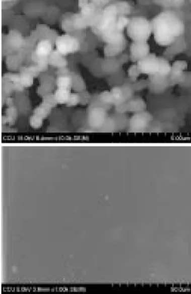
The aminosilane agent of APTS is considered as a candidate for modification on the surface of anodized Ti plates directly, for the advantages of the biocompatibility as well as high density of surface functional group. APTS is a heterofuctional molecule, with a silicon- containing function group at one end, an amino group at the other end, an alkaline chain in between. The Ti metal surface was covered with an oxide layer to allow coupling of biomolecules such as APTS through silanization. Figure 3(a) show the morphology of electrochemically treated Ti attaching APTS. It can be seen that APTS particles of approxumately 5 nm uniformly sprayed onto the Ti surface. More particularly, APTS with NH2 allows for connecting to other targeting biomolecules such as chitosan, as discussed later.
4.2. Characterization of chitosan-grafted samples
The free amino groups on APTES-modified surface allow flexibility in coupling chitosan to the substrate. The chitosan-grafted surface can facilitated the incorporation of phosphate ions via attractive ineractions. As shown in Figure 3(b), the particle surface became much smooth after chitosan grafting to APTS.
XRD showed the diffraction peak of chitosan at 2θ = 20, which an amorphous phase (Figure 4). From the FTIR pattern, it is seen that there is a new absorption band at 1650 cm-1, while the band at 1590 cm-1 that is assigned to amino groups has disappeared. With regards to bind strength, the chitosan-grafted titanium samples had a bond strength value of 12.3 MPa.
In this study, the stability of the modified surface has been studied by OCP measurements.
The results indicated that The OCP of chitosan- grafting titanium samples shifted towards a steady state (Figure 5).
There is a need for biomaterials with surface properties that would aid in cell attachment and growth secretion and therefore, improve the formation of new bone at the tissue/biomaterial interface. Studies found that OH, COOH, and
NH2 functionalities altered the functional presentation of the major integrin binding domain of adsorbed fibronectin and modulated integrin binding, localization, and specificity.
五、結 論
The specific purpose of the study is a useful attempt at creating an amino surface on Ti through chitosan grafting. The surface modifications of titanium may be employed as a means of controlling cellular responses to biomaterials surfaces as well as reducing stress shielding when implanted.
六、成果自評
This project focuses on the chitosan grafting investigation of the titanium implant. We prepare the results to apply patent before the submission of SCI journal. In addition, based on the findings, subsequential studies are in progress to improve the processing.
參考文獻
[1] Carlsson L, Ro¨stlund T, Albrektsson B, Albrektsson T, Brånemark P.
Osseointegration of titanium implants. Acta Orthop Scand 1986;57:285-289.
[2] van Noort R. Titanium: the implant material of today. J Mater Sci 1987;22:3801-3811.
[3] Dalton JE, Cook SD. In vivo mechanical and histological characteristics of HA- coated implants vary with coating vendor. J Biomed Mater Res 1995;29:239-245.
[4] Nies B, Sewing A, Scharnweber D, Worch H, Beutner R. Chitosan-coated metallic article, and process for the production thereof. USA Patent 20050079198.
[5] Tanaka K, Shimizu A, Morita R, Tsuchida S, Kobayashi N, Sannan T. Aqueous agent for treating substrate, method for treating substrated and treated substrate. USA Patent 20050103229.
[6] Kim YJ, Son TW, Kim WK, Yoo HO.
Natural fiber coated with chitosan and a method for producing the same. USA Patent 20030134120.
[7] Nanci A, Wuest JD, Peru L, Brunet P,
Sharma V, Zalzal S, McKee MD. Chemical modification of titanium surfaces for covalent attachment of biological molecules. J Biomed Mater Res 1998;40:324-335.
[8] Yang BC, Uchida M, Kim HM, Zhang XD, Kokubo T. Preparation of bioactive titanium metal via anodic oxidation treatment. Biomaterials 2004;25:1003-1010.
[9] Matinlinna JP, Laajalehto K, Laiho T, Kangasniemi I, Lassila LVJ, Vallittu PK.
Surface analysis of Co-Cr-Mo alloy and Ti substrates silanized with trialkoxysilanes and silane mixtures. Surf. Interface Anal.
2004;36:246-253.
[10] Kim HM, Miyaji F, Kokubo T, Nakamura T.
Preparation of bioactive Ti and its alloys via simple chemical surface treatment. J Biomed Mater Res 1996;32:409-417.
[11] Francis Suh JK, Matthew HWT.
Application of chitosan-based polysaccharide biomaterials in cartilage tissue engineering: a review. Biomaterials 2000;21:2589-2598.
[12] Felt O, Buri P, Gurny R. Chitosan: a unique polysaccharide for drug delivery. Drug Dev Ind Pharm. 1998;24:979-993.
Figure 1. XRD patterns of electrochemically treated Ti samples at different applied potentials.
Figure 2. SEM micrographs of electrochemically treated Ti.
(a)
(b)
Figure 3. SEM micrographs of electrochemical- treated Ti after silanization (a) followed by chitosan-grafting (b).
Figure 4. XRD pattern of chitosan-grafted Ti sample.
Figure 5. Open circuit potential of chitosan- grafted samples.