行政院國家科學委員會專題研究計畫 成果報告
台灣株子囊菌 (Bionectria ochroleuca) 所含抗癌活性物 質的分離、純化及結構鑑定
計畫類別: 個別型計畫
計畫編號: NSC94-2113-M-038-003-
執行期間: 94 年 08 月 01 日至 95 年 07 月 31 日 執行單位: 臺北醫學大學生藥學研究所
計畫主持人: 李宗徽
報告類型: 精簡報告
處理方式: 本計畫可公開查詢
中 華 民 國 95 年 8 月 1 日
行政院國家科學委員會補助專題研究計畫 ■ 成 果 報 告
□期中進度報告
台灣株子囊菌 ( Bionectria ochroleuca ) 所含抗癌活性物質的 分離、純化及結構鑑定
計畫類別:■ 個別型計畫 □ 整合型計畫 計畫編號:NSC 94-2113-M-038-003-
執行期間:94 年 08 月 01 日至 95 年 07 月 31 日
計畫主持人:李宗徽 共同主持人:
計畫參與人員:
成果報告類型(依經費核定清單規定繳交):■精簡報告 □完整報告
本成果報告包括以下應繳交之附件:
□赴國外出差或研習心得報告一份
□赴大陸地區出差或研習心得報告一份
□出席國際學術會議心得報告及發表之論文各一份
□國際合作研究計畫國外研究報告書一份
處理方式:除產學合作研究計畫、提升產業技術及人才培育研究計畫、列 管計畫及下列情形者外,得立即公開查詢
□涉及專利或其他智慧財產權,□一年□二年後可公開查詢
執行單位:台北醫學大學 生藥學研究所
中 華 民 國 95 年 08 月 01 日
中文摘要:
本研究採抗癌活性導引的模式,自台灣株子囊菌Bionectria ochroleuca醱酵菌液的乙酸乙酯層中 分離、純化出兩個多酮體化合物 (polyketide),分別為bionectide A (1) 和bionectide B (2),之後 使用各種光譜分析、鑑定出其結構。在癌細胞的增生抑制活性上,化合物 1 及 2 對於MT-2 (血 癌)、A498 (腎癌)、NPC-tw01 (鼻咽癌) 及H-226 and A549 (肺癌) 等皆具抑制活性,其半抑制 濃度 (IC50) 介於 18.3 至 40.2 μM。
關鍵詞:子囊菌;多酮體;Bionectria ochroleuca;醱酵菌液;細胞毒性
Abastract:
A new polyketide, bionectide A (1), along with a known analogue, bionectide B (2), were purified from ethyl acetate extracts of the fermentation broth of Bionectria ochroleuca isolated from Taiwan.
Their structures were elucidated on the basis of spectroscopic analysis. The anti-proliferation activities of 1 and 2 were evaluated against MT-2 (human leukemia), A498 (human renal carcinoma), NPC-tw01 (human nasopharyngeal carcinoma), H-226 and A549 (non-small cell lung cancer) cell lines, and their IC50 values ranged from 18.3 to 40.2 μM.
Key words: Hypocreales, Bionectria ochroleuca, polyketides, fermentation broth, cytotoxicity
Introduction:
Hypocrealean fungi are ecologically highly diverse. They are saprotrophic, necrotrophic or biotrophic, and many species are well known to have great economic importances [1]. Bionectria ochroleuca (Schwein.) Schroers & Samuels[anamorph: Clonostachys rosea (Link: Fr.) Schroers et al.] has been found to produce a variety of antibiotics and could be used as a biocontrol agent against plant pathogenic fungi [2]. Recently, the fermentation broth of B. ochroleuca was found to exhibit significant anti-proliferation activities against human Colo205, HL60 and Hela cancer cell lines in our preliminary screening of fungal extracts for cytotoxicity. These findings promoted us to investigate the cytotoxic agents from this fungus. Therefore, a series of bioassay guided chemical examinations on the fermentation broth of this fungus was thus undertaken, and has resulted in the isolation of a new polyketide 1 together with a known analogue 2. This paper describes the isolation and structural elucidation of 1 and 2 as well as their cytotoxicities.
Results and discussion:
From the fermentation broth of B. ochroleuca two polyketides 1 and 2 were identified. These compounds were isolated and purified using repetitive Sephadex LH-20 column chromatography and HPLC by the guidance of cytotoxicity tests. Bionectide B (2), a major component, was obtained as colorless powder whose spectral data were compatible with those of TMC-151 F, having previously been isolated from Gliocladium catenulatum Gilman & Abbott TC 1280 [3, 4].
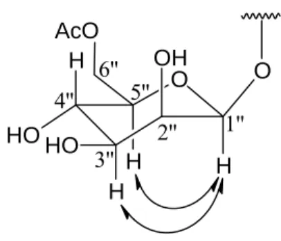
Bionectide A (1), a colorless powder, has a molecular formula of C43H76O16 as deduced from its HRFABMSand 13C-NMR spectral data. The IR spectrum of 1 indicated the presence of hydroxyl (3347 cm-1) and carbonyl (1705 cm-1) groups. The 13C-NMR and DEPT spectra of 1 exhibited a C43 skeleton closely compatible with that of TMC-151 A [3], except for two additional carbon signals at δC 21.2 and 172.7 ascribable to an acetyl functionality. In 1H NMR spectrum of 1, the signals corresponding to the aliphatic chain (H-3 ~ H3-29), mannitol moiety (H2-1′ ~ H2-6′), and glycopyranoside (H-1′′ ~ H2-6′′) were also closely consistent with those of TMC-151 A [3], except that its H-6′′a and H-6′′b shifted from δΗ 3.49 and 3.70 to lower fields of δΗ 4.25 and 4.42, respectively. The glycopyranoside was further deduced to be a 6′′-O-acetyl-β-mannopyranoside as evidenced from a large vicinal coupling constant between H-3′′ and H-4′′ (3J = 9.6 Hz) and a non-detectable coupling constant between H-1′′ and H-2′′, mutual nOe correlations between H-1′′, H-3′′ and H-5′′ indicating the axial orientations of H-1′′, -3′′ and -5′′ (Fig. 1), and the oxymethylene protons H2-6′′ at δ 4.25 and 4.42 exhibiting a long range correlation with an acetyl carbonyl carbon signal at δC172.7 in the HMBC spectrum. The residual key connectivities of other fragments were also accomplished by the complete assignments of HMBC spectrum where the anomeric proton H-1′′
signal at δΗ 4.45 correlated with oxygenated carbon C-13 resonance at δC 87.4, the mannitol H-3′
signal at δH 5.20 interacted with carbonyl carbon C-1 resonance at δC 169.9. Further analysis of all the 2D NMR data allowed the complete assignment of 1H and 13C NMR spectra of 1, as illustrated in Table 1. Accordingly, compound 1 was concluded to be 6′′-O-acetyl analogue of TMC-151 A, as shown in Fig. 2.
Compounds 1 and 2 were evaluated for their cytotoxicities against MT-2 (human leukemia), A498 (human renal carcinoma), NPC-tw01 (human nasopharyngeal carcinoma), H-226 and A549
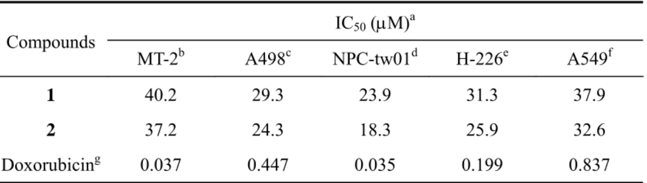
(non-small cell lung cancer) cells. The cell viabilities were assessed through MTT assay. As shown in Table 2, 1 and 2 with IC50 values ranging from 18.3 to 40.2 μM showed only minor toxicity toward five tested cancer cells when compared with the positive control, doxorubicin.
Experimental Section:
General
Optical rotations were measured on a JASCO P-1020 digital polarimeter (Kyoto, Japan). 1H- and 13C- NMR were acquired on a Bruker DMX-500 SB spectrometer (Ettlingen, Germany). Mass spectra were obtained using a Finnigan Thermo Quest MAT 95XL spectrometer (Bremen, Germany).
IR spectra were recorded on a Thermo Mattson IR 300 spectrometer (Califonia, USA). UV spectra were measured on a Hitachi U-2800 spectrophotometer (Tokyo, Japan). Column chromatography was carried out with Sephadex LH-20 gel (Amersham Biosciences, Uppsala, Sweden). Pre-coated Si gel plates (Si 60 F254, 0.2 mm, Merck, Darmstadt, Germany) were used for analytical TLC.
Fermentation of Bionectria ochroleuca
Bionectria ochroleuca (strain No. 91111210) was inoculated into 250 mL Erlenmeyer flasks containing 2 g malt extract (Bacto, Sparks, USA) and 100 mL deionized water. The fermentation was conducted under the static condition at 25 ºC for two weeks.
Extraction and Isolation
The mycelia together with the fermentation broth from the above fermentation were extracted three times with EtOAc with stirring at room temperature for 1 hour. Then, the organic layer was separated and concentrated in vacuum to dryness (6.5 g). This residue was re-dissolved in 20 mL MeOH and filtered, then applied onto a column of Sephadex LH-20 (3 × 65 cm) and eluted with MeOH (2.7 mL/min). Every 23 mL of eluent was collected as one fraction and each was analyzed by thin layer chromatography, and a solution of EtOAc/AcOH/H2O (85:10:10) for development.
UV 254 nm illumination and vanillin-sulfuric acid charring was used to group the compounds with similar skeleton. The bioactive fractions (#fr. 11 ~ 13), determined by anti-proliferation tests of human Colo205, HL60 and Hela cancer cells, were combined and evaporated under reduced pressure to yield 1.5 g of the bionectide mixture. The bionectide mixture was further purified by repetitive HPLC on a Hypersil ODS semi-preparative column (10 × 250 mm, Thermo Electron Corp., Bellefonte, USA) with MeCN/H2O (7:3) as eluent to afford 1 (7.5 mg) and 2 (10.0 mg), retention time: 16.2 and 18.0 min, respectively.
Bionectide A (1).–Colorless powder.–[α]20D +2.1° (c 0.1, MeOH).–UV (MeOH) λmax (log ε) = 208 nm (4.20).–IR νmax (KBr): ν = 3347, 2959, 2925, 2873, 1705, 1652, 1455, 1372, 1230, 1074, 1021 cm-1.–1H-NMR data: see Table 1.–13C NMR data: see Table 1.–MS (FAB): m/z 871 [M + Na]+.–MS (HRFAB): m/z 871.4998 (C43H76O16Na, calc. 871.5031).
Bionectide B (2).–Colorless powder.–[α]20D −0.85° (c 0.1, MeOH).–UV (MeOH) λmax (log ε) = 207 (4.25).–IR νmax (KBr): ν = 3353, 2960, 2873, 1705, 1651, 1455, 1373, 1270, 1228, 1072, 1023 cm-1.–MS (FAB): m/z 841 [M + Na]+.
Cell Culture
Human leukemia MT-2 cells, non-small cell lung cancer H-226 and A549 cells were maintained in RPMI-1640 medium supplied with 10% fetal bovine serum, 100 units/mL penicillin and 100 μg/mL streptomycin. Human renal carcinoma A498 cells and nasopharyngeal carcinoma
NPC-tw01 cells were maintained in MEM medium supplied with 10% fetal bovine serum, 100 units/mL penicillin and 100 μg/mL streptomycin.
Growth Inhibition Assay
Cell growth in the presence or absence of experimental agents was determined using the MTT-microculture tetrazolium assay [5]. Briefly, 100 μL of cell suspension in logarithmic growth phase were seeded into 96-well plate (MT-2, 1.5×10⁴/well; NPC-tw01, 2×10³/well; A498, H-226, and A549, 4×10³/well). After 24 hours, the cells were exposed to various concentrations of the test compound in a volume of 50 μL for 72 hours. Two hours prior to the end of incubation, 15 μL MTT solution (5 mg/mL) was added into the culture medium. Cells were lysised with 75 μL of MTT lysis buffer (20% SDS-50% DMF) and cell lysis solution were incubated at 37℃ for another 12 hours to dissolve the dark blue crystals. The absorption of formazan solution at 570 nm was measured using a microplate reader.
Acknowledgements: This research was supported by the grant from National Science Council, Taipei, Taiwan. We are grateful to Ms. Shwu-Huey Wang and Ms. Shou-Ling Huang for the NMR data acquisition in the Instrumentation Center of Taipei Medical University and Instrumentation Center of the College of Science, National Taiwan University, respectively.
References
[1] H. J. Schroers, G.. J. Samuels, K. A. Seifert, W. Gams, Mycologia 91, 365 (1999).
[2] H. J. Schroers, Studies in Mycology, Vol. 46, p. 74, Fungal Biodiversity Centre, Utrecht, The Netherlands (2001).
[3] J. Kohno, M. Nishio, M. Sakurai, K. Kawano, H. Hiramatsu, N. Kameda, N. Kishi, T. Yamashita, T. Okauda, S. Komatsubara, Tetrahedron 55, 7771 (1999).
[4] J. Kohno, Y. Asai, M. Nishio, M. Sakurai, K. Kawano, H. Hiramatsu, N. Kameda, N. Kishi, T.
Okuda, S. Komatsubara, J. Antibiotics 52, 1114. (1999).
[5] M. B. Hansen, S. E. Nielsen, K. Berg, J. Immunol. Method 119, 203 (1989).
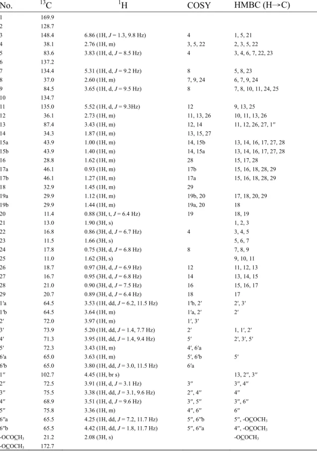
Table 1. 1H- and 13C-NMR data of 1 (CD3OD, 500 MHz for 1H and 125 MHz for 13C).
No. 13C 1H COSY HMBC (H→C)
1 169.9 2 128.7
3 148.4 6.86 (1H, J = 1.3, 9.8 Hz) 4 1, 5, 21
4 38.1 2.76 (1H, m) 3, 5, 22 2, 3, 5, 22
5 83.6 3.83 (1H, d, J = 8.5 Hz) 4 3, 4, 6, 7, 22, 23 6 137.2
7 134.4 5.31 (1H, d, J = 9.2 Hz) 8 5, 8, 23
8 37.0 2.60 (1H, m) 7, 9, 24 6, 7, 9, 24
9 84.5 3.65 (1H, d, J = 9.5 Hz) 8 7, 8, 10, 11, 24, 25 10 134.7
11 135.0 5.52 (1H, d, J = 9.3Hz) 12 9, 13, 25 12 36.1 2.73 (1H, m) 11, 13, 26 10, 11, 13, 26 13 87.4 3.43 (1H, m) 12, 14 11, 12, 26, 27, 1′′
14 34.3 1.87 (1H, m) 13, 15, 27
15a 43.9 1.00 (1H, m) 14, 15b 13, 14, 16, 17, 27, 28 15b 43.9 1.40 (1H, m) 14, 15a 13, 14, 16, 17, 27, 28
16 28.8 1.62 (1H, m) 28 15, 17, 28
17a 46.1 0.93 (1H, m) 17b 15, 16, 18, 28, 29
17b 46.1 1.27 (1H, m) 17a 15, 16, 18, 28, 29
18 32.9 1.45 (1H, m) 29
19a 29.9 1.12 (1H, m) 19b, 20 17, 18, 20, 29
19b 29.9 1.44 (1H, m) 19a, 20 18
20 11.4 0.88 (3H, t, J = 6.4 Hz) 19 18, 19
21 13.0 1.90 (3H, s) 1, 2, 3
22 16.8 0.86 (3H, d, J = 6.7 Hz) 4 3, 4, 5
23 11.5 1.66 (3H, s) 5, 6, 7
24 17.8 0.75 (3H, d, J = 6.8 Hz) 8 7, 8, 9
25 11.0 1.62 (3H, s) 9, 10, 11
26 18.7 0.97 (3H, d, J = 6.9 Hz) 12 11, 12, 13 27 16.7 0.95 (3H, d, J = 6.8 Hz) 14 13, 14, 15 28 21.0 0.90 (3H, d, J = 7.5 Hz) 16 15, 16, 17
29 20.7 0.89 (3H, d, J = 6.4 Hz) 18 17
1′a 64.5 3.53 (1H, dd, J = 6.2, 11.5 Hz) 1′b, 2′ 2′, 3′
1′b 64.5 3.64 (1H, m) 1′a, 2′ 2′
2′ 72.0 3.97 (1H, m) 1′, 3′
3′ 73.9 5.20 (1H, dd, J = 1.4, 7.7 Hz) 2′ 1, 1′, 2′
4′ 71.3 3.95 (1H, dd, J = 1.4, 9.4 Hz) 5′ 2′, 3′, 5′
5′ 72.3 3.43 (1H, m) 4′, 6′a
6′a 65.0 3.63 (1H, m) 5′, 6′b 5′
6′b 65.0 3.80 (1H, dd, J = 3.0, 11.5 Hz) 6′a
1′′ 102.7 4.45 (1H, br s) 13, 2′′, 3′′
2′′ 72.5 3.91 (1H, d, J = 3.1 Hz) 3′′ 3′′, 4′′
3′′ 75.5 3.38 (1H, dd, J = 3.1, 9.6 Hz) 2′′, 4′′ 4′′
4′′ 68.9 3.51 (1H, d, J = 9.6 Hz) 3′′, 5′′ 3′′, 6′′
5′′ 75.8 3.36 (1H, m) 4′′, 6′′ 6′′
6′′a 65.5 4.25 (1H, dd, J = 7.2, 11.7 Hz) 5′′, 6′′b 5′′, -OCOCH3
6′′b 65.5 4.42 (1H, dd, J = 1.8, 11.7 Hz) 5′′, 6′′a 4′′, -OCOCH3
-OCOCH3 21.2 2.08 (3H, s) -OCOCH3
-OCOCH3 172.7
Table 2. IC50 values of 1 and 2 against five human cancer cell lines.
IC50 (μM)a Compounds
MT-2b A498c NPC-tw01d H-226e A549f
1
40.2 29.3 23.9 31.3 37.92
37.2 24.3 18.3 25.9 32.6Doxorubicing 0.037 0.447 0.035 0.199 0.837
a Cells were treated with various concentrations of test compounds for 3 days. Cell growth was
determined by MTT assay. The IC50 value resulting form 50% inhibition of cell growth was calculated.
Each value represents the mean of three independent experiments.
b MT-2 as human leukemia cell line.
c A498 as human renal carcinoma cell line.
d NPC-tw01 as human nasopharyngeal carcinoma cell line.
e H-226 as human non-small cell lung cancer cell line.
f A549as human non-small cell lung cancer cell line.
g Doxorubicin, the chemotherarpeutic drug, as a reference compound in this study.
O O OH
HO HO AcO
H H
H
H 4'' 1''
6''
3'' 2'' 5''
Fig. 1. Key NOESY correlations of 6′′-O-acetyl-β-mannopyranoside in 1.
R
O OH OH
O O
OH
HO HO
OAc
1 R =
2 R = HO
OH OH
O
1 20
21 22 23 24 25 26 27 28 29
1'' 3''
6''
OH OH
1' 6' 5' 4' 3' 2'
5 9 13
HO
OH
OH
O OH
Fig.2. Structures of bionectide A (1) and B (2) identified in this report.
研究計畫成果自評:
本計畫成果與原計劃預定內容與進度完全相符,並達成了預定的目標,計自台灣株子囊菌 中分離、純化及鑑定出兩個極少見且結構特殊的多酮體化合物,其中化合物 1 為未曾發表的 新化合物,此外,化合物所具有的抗癌活性,或具有應用價值,仍有待評估。以上內容已投稿 於德國的化學期刊Zeitschrift für Naturforschung B,目前正審稿中。