行政院國家科學委員會專題研究計畫 成果報告
研究溫度對牙科修復材料誘發發炎反應的影響
計畫類別: 個別型計畫
計畫編號: NSC94-2314-B-040-020-
執行期間: 94 年 08 月 01 日至 95 年 07 月 31 日 執行單位: 中山醫學大學牙醫學系
計畫主持人: 黃富美 共同主持人: 張育超
報告類型: 精簡報告
處理方式: 本計畫可公開查詢
中 華 民 國 95 年 10 月 1 日
Induction of heme oxygenase-1 expression by dentin bonding agents in human pulp cells
Abstract
Aim The purpose of this study was to investigate the effects of dentine bonding
agents on the expression of HO-1 in human pulp cells.
Methodology Set specimens from Clearfil SE Bond (CB), Prime & Bond 2.1 (PB),
and Single Bond (SB) were eluted with culture medium. Cytotoxicity and Western
blot assays were used to investigate the effects of human primary pulp cells exposed
to dentine bonding agents.
Results Our data showed that CB, PB, and SB were cytotoxic to pulp cells in a
concentration-dependent manner (p<0.05). The exposure of quiescent pulp cells to CB,
PB, and SB resulted in the induction of HO-1 protein expression in a time-dependent
manner (p<0.05). The influence of the cytotoxicity and HO-1 expression depended on
the materials tested.
Conclusion Taken together, HO-1 expression might be one signal transduction
pathway linked to the induction of stress-inducible protein by dentine bonding agents.
Key words: dentin bonding agents; heme oxygenase-1; pulp cells
Introduction
Dentine bonding agents are designed to form strong bonding with dentine.
These agents are used to improve the bonding strength between resin and the tooth
structure, increase the retention of restoration, and reduce the microleakage across
dentine-resin interface and scatter the occlusal stress. They usually remain in close
contact with living dental tissue over a long period of time. Ideally, dentine bonding
agents should be biocompatible.
Monomers eluted from dentine bonding systems and bacterial microleakage
have been implicated as possible causes of pulpal irritation after composite resin
restoration (Akimoto et al. 1998, Cox et al. 1998). Investigators have measured cell
growth, cytotoxicity, and genotoxicity as indicators of cellular response to dentine
bonding agents using murine fibroblast cell line L929 (Schedle et al. 1998, Hashieh et
al. 1999, Kaga et al. 2001), mouse odontoblast-like cell line MDPC-23 (de Souza
Costa et al. 1999), human gingival fibroblasts (Szep et al. 2002, Huang et al. 2003)
and human pulp cells (Huang & Chang 2002, Huang et al. 2005a). However, the
intracellular mechanisms altered by dentine bonding agents are still not clear.
Heme oxygenase (HO) is originally identified as an enzyme that catalyzes the
initial reaction in heme catabolism the oxidative cleavage of the
-meso carbon bridge of b-type heme molecules to yield equimolar quantities of biliverdin IXa,cabon monoxide (Kageyama et al. 1992). HO has long been known to undergo
adaptive regulation in response to heme. There are two major isoforms: HO-1 and
HO-2. HO-2 is the major isoform that presents under physiological conditions,
localized in microsomes, and the stress-inducible isoform HO-1 is localized in
mitochondria. HO-1 expression is very sensitive to stress, and is induced by many
stimuli, including heme, heavy metal, heat shock, endotoxin, inflammatory cytokines,
prostaglandins, and oxidative stress (Keyse et al. 1990, Savdana et al. 1992, Lautier et
al. 1992).
Intra-orally, cells are exposed to many stressors from dental materials or
bacteria. HO-1 functions as an anti-oxidant enzyme because locally produced
bilirubin works as an efficient scavenger of reactive oxygen species (Vogt et al. 1995).
However, there is little information about the ability of dentine bonding agents to
modulate the expression of HO-1. We hypothesized that if dentin bonding agents
stress cells, this stress may be manifest by the induction of HO-1 expression. To
investigate this hypothesis, we exposed human pulp cells to dentin bonding agents by
using cytotoxicity and Western blot assays.
Materials and methods
Sample preparation
Three dentine bonding agents were evaluated: Clearfil SE Bond (CB), Prime &
Bond 2.1 (PB), and Single Bond (SB) (Table 1). In order to evaluate the effects of the
photo-polymerized dentine bonding agents, 60 mm x 10 mm cellulose strips were
exposed to UV light for 30 min in order to prevent bacterial contamination. On the
strips, 10 µl of each dentine bonding agent was applied and light-cured for 20 s. After
polymerization, each test specimen was eluted in 8 ml of culture medium at 37℃ for
2 days in a 5 % CO2 air atmosphere. The extraction media were then collected into
sterile syringes at the end of this period and passed through a 0.22 µm filter.
Subsequently, various dilutions (1:1, 1:2, and 1:4) of these extraction media were
prepared to be used in this study.
Cell culture
Cell culture
Human pulp cells were cultured using an explant technique as described
previously (Huang et al. 2004, Huang et al. 2005b). Briefly, impacted third molars
were obtained from healthy patients of the Oral Medicine Centre (Chung Shan
Medical University Hospital, Taichung, Taiwan) with the informed consent. Teeth
were sectioned horizontally below the cementoenamel junction with a number 330
high-speed bur with water spray. The pulp tissue was removed aseptically in lamina
flow,rinsed with Hanks’buffered salinesolution,and placed in a60 mm dish.Pulp
tissue was minced with a blade into small fragments and grown in Dulbecco’s
modified Eagle’smedium (DMEM)supplemented with 10 % foetal calf serum (FCS)
and antibiotics (100 U/ml penicillin, 100 g/ml streptomycin and 0.25 g/ml of fungizone). Cultures were maintained at 37C in a humidified atmosphere of 5 % CO2
and 95 % air. Confluent cells were detached with 0.25 % trypsin and 0.05 % EDTA
for 5 min, and aliquots of separated cells were subcultured. Cell cultures between the
third and eighth passages were used in this study.
Cytotoxicity assay
A 3-(4,5-dimethylthiazol-2-yl)-2,5-diphenyl tetrazolium bromide (MTT)
colorimetric assay was developed to monitor mammalian cell survival and
proliferation in vitro. Briefly, 2104 cells per well were seeded to 96 well plate and
left overnight to attach. Serial dilutions of various elute in 100 µl volumes were added,
and cells were treated for 24 h. After treatment, 50 µl of MTT (Sigma, St. Louis, MO)
solution (1mg/ml in PBS) was added to each well and incubated for another 4 hours at
37°C. To each well, 150 µl of dimethyl sulfoxide was added. Plates were then shaken
until crystals were dissolved. Reduced MTT was then measured
spectrophotometrically in a dual beam microtiter plate reader at 570 nm with a 650
nm reference. The percentage of the dehydrogenase activity at each material,
compared with that of the control, was calculated from the absorbance values.
HO-1 mRNA gene expression analysis
Cells arrested in G0by serum deprivation (0.5 % FCS for 48 h) were generally
used in these experiments. Prior to treatment, the cells were washed with serum-free
DMEM and immediately exposed for the indicated incubation times (1, 2, 4, 8, and 24
h) to various extraction media. The viability of cells exposed to these elutes were in
general cytoatatic according to the MTT assay.
Total RNA was prepared using TRIzol reagent (Gibco Laboratories, Grand
Island, NY, USA) following the manufacturer’s instructions.Single-stranded DNA
was synthesized from RNA in a 15 µl reaction mixture containing 100 mg random
hexamer and 200 units of Moloney murine leukemia virus reverse transcriptase
(Gibco Laboratories, Grand Island, NY, USA). The reaction mixture was diluted with
20 µl of water and 3 µl of the diluted reaction mixture was used for the polymerase
chain reaction (PCR). PCR reaction mixture contains 10 pmol of forward and reverse
primers and 2 units of Tag DNA polymerase. Amplification was performed at 25
cycles for GAPDH and 30 cycles for HO-1 in a thermal cycle. Each cycle consisted of
1 min of denaturation at 94°C, 1 min of annealing at 57°C, and 1 min of extension at
72°C. The sequences of primers used were as follows (Quan et al, 2004):
AGAPDH Forward: 5’-TCCTCTGACTTCAACAGCGACACC-3’ Reverse:5’-TCTCTCTTCCTCTTGTGCTCTTGG-3’
B) HO-1 Forward:5’-CAGGCAGAGAATGC TGAGTTC-3’
Reverse:5’-GATGTTGAGCAGGAACGCAGT-3’
The PCR products were analyzed by agarose gel electrophoresis and a 550 bp
band for HO-1 was noted. When the band densities were measured and compared
with the density of the band obtained for the housekeeping gene GAPDH, relative
proportions of mRNA synthesis could be determined within each experiment. The
intensity of each band after normalization with GAPDH mRNA was quantified by the
photographed gels with a densitometer (AlphaImager 2000; Alpha Innotech, San
Leandro, CA, USA).
Statistical analysis
Three replicates of each concentration were performed in each test. All assays
were repeated three times to ensure reproducibility. Statistical analysis was by
one-way analysis of variance (ANOVA). Tests of differences of the treatments were
analyzed by Duncan’stest.
Results
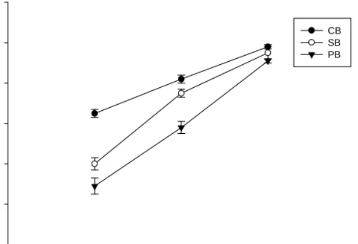
As shown in Fig. 1, the cytotoxicity of elutes of dentine bonding agents were
evaluated using MTT assay in pulp cells. The results showed that CB, PB, and SB
were cytotoxic to pulp cells in a concentration-dependent manner during 24-h
incubation period (p<0.05). The toxicity decreased in an order of PB>SB>CB. In vivo
acute toxicity is usually not associated with the clinical use of a dental material. Thus,
the sublethal doses of all dentine bonding agents (1:4 dilution) were selected in the
following experiments.
Investigations of the time dependence of HO-1 expression in CB-treated cells
revealed a rapid accumulation of the transcript, a significant signal first detectable
after 1 h of exposure and remained elevated throughout the 24-hour incubation period
(Fig. 2). Moreover, the peak of HO-1 level induced by CB was at 4 h (Fig. 2). The
quantitative measurement was made by the AlphaImager 2000. The levels of the
HO-1 increased about 1.7, 4.6, 5.9, 4.0, and 2.2 fold after exposure to CB for 1, 2, 4, 8,
and 24 h, respectively (Fig. 2).
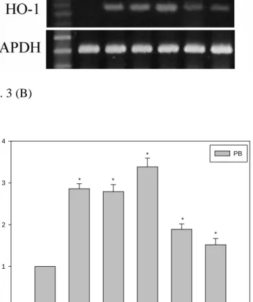
The induction of HO-1 expression by PB in pulp cells was similar to that of CB.
A significant signal first detectable after 1 h of exposure and remained elevated
throughout the 24-hour incubation period (Fig. 3). The peak of HO-1 level induced by
PB was at 4 h. The quantitative measurement was made by the AlphaImager 2000.
The levels of the HO-1 increased about 2.9, 2.8, 3.4, 1.9, and 1.5 fold after exposure
to PB for 1, 2, 4, 8, and 24 h, respectively (Fig. 3).
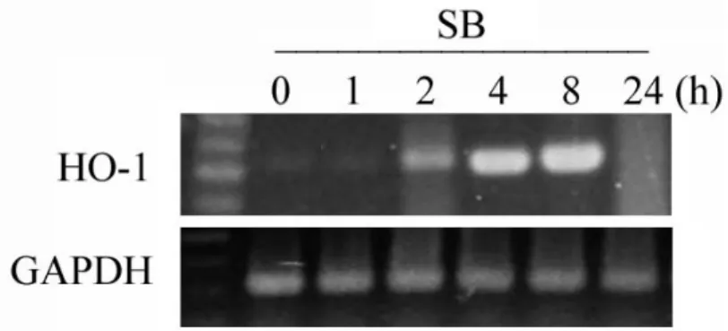
Investigations of the time dependence of HO-1 expression in SB-treated cells is
shown in figure 4. It demonstrated, a significant signal first detectable after 2 h of
exposure. The peak of HO-1 level induced by SB was at 8 h. The quantitative
measurement was made by the AlphaImager 2000. The levels of the HO-1 increased
about 1.1, 2.0, 5.4, 6.3, and 1.4 fold after exposure to SB for 1, 2, 4, 8, and 24 h,
respectively (Fig. 4).
Discussion
Many cell culture techniques have been applied to assess the biocompatibility of
dental materials. These methods are based on cell cultures with established or diploid
cell lines and tissue explant techniques. However, an increasingly number of authors
has stated that in vitro biocompatibility tests should be performed with the most
appropriate cells (i.e. cells homologous to the human tissues of ultimate concern)
(Feigal et al. 1985). Any material used for restorative will come into contact with, or
close proximity to the dental pulp. Thus, the effects of dentine bonding agents on this
cell type may have clinical significance.
In the present study, the cytotoxicity of elutes of three dentine bonding agents
was evaluated using MTT assay in pulp cells. It was found that all dentine bonding
agents were cytotoxic to pulp cells. Data suggest substantial differences in
cytotoxicity among three dentine bonding agents. Although the experimental
conditions in this study differed from these used in other studies, our results were in
agreement with previous studies (Schedle et al. 1998, Hashieh et al. 1999, de Souza
Costa et al. 1999, Kaga et al. 2001, Szep et al. 2002, Huang & Chang 2002, Huang et
al. 2003, Huang et al. 2005a) that CB, PB, and SB tested were cytotoxic.
HO-1 is known as a stress-inducible protein. HO-1 expression is very sensitive
to stress, and is induced by many stimuli. Detectable amounts of HO-1, however, can
be seen in oral cells, such as gingival fibroblasts (Huang & Chang in press) and pulp
cells (Min et al. 2006). In this study, HO-1 was first found to be upregulated in human
pulp cells stimulated with dentine bonding agents. Recently, similar results were
reported that HO-1 was found to be upregulated by resin based dental materials by
human gingival fibroblasts (Huang & Chang in press). Thus, HO-1 expression might
be one signal transduction pathway linked to the induction of stress response protein
by dentine bonding agents.
HO-1 may be induced by a variety of non-heme products, such as reactive
oxygen species, endotoxin, inflammatory cytokines, and nitric oxide (Choi et al.
1996). The functional significance of HO-1 induction is not well understood, as HO-1
activity can result in either cell protection or cell injury depending on the
experimental setting (Da Silva et al. 1996). Keyse & Tyrrell (1989) have
demonstrated that HO-1 is involved in the general cellular defense mechanisms
against oxidative injury. Thus, this enzyme is believed to play an important role in
maintaining cellular homeostasis in response to oxidant injury by dentine bonding
agents.
Conclusions
HO-1 is known as a stress-inducible protein and functions as an antioxidant
enzyme. Little is known about the induction of cellular signaling events after cell
exposure to dentine bonding agents. In summary, the current study has shown that
dentine bonding agents can significantly upregulated the expression of HO-1 at
sublethal concentrations in human pulp cells. The response can be upregulated,
depending on the material tested and time after stimulation.
ACKNOWLEDGMENTS
This study was supported by a research grant from the National Science Council,
Taiwan (NSC 94-2314-B-040-020).
References
Akimoto N, Momo Y, Kohno A, Suzuki S, Otsuki M, Cox CF (1998)
Biocompatibility of Clearfil Liner Bond 2 and Clearfil AP-X system on
non-exposed and exposed primate teeth. Quintance International 29, 177-88.
Choi AMK, Alam J (1996) Heme oxygenase-1: function, regulation, and implication
of a novel stress-inducible protein in oxidant-induced lung injury. American
Journal of Respiratory Cellular Molecular Biolology 15, 9-19.
Cox CF, Hafez AA, Akimoto N, Otsuki M, Suzuki S, Tarim B (1998)
Biocompatibility of primer, adhesive and resin composite systems on
non-exposed and exposed pulps of non-human primate teeth surface. American
Journal of Dentistry 10, 55-63.
Da Silva JL, Morishita T, Escalante D, Staudinger R, Drummond G, Goligorsky MS,
Lutton JD, Abraham NG (1996) Dual role of heme oxygenase in epithelial cell
injury: contrasting effects of short-term and long-term exposure to oxidant stress.
Journal of Laboratory & Clinical Medicine 128, 290-6.
de Souza Costa CA Vaeten MA, Edwards CA, Hanks T (1999) Cytotoxic effects of
current dental adhesive systems on immortalized odontoblast cell line MDPC-23.
Dental Materials 15, 434-41.
Feigal RJ, Yesilsoy C, Messer HH, Nelson J (1985) Differential sensitivity of normal
human pulp and transformed mouse fibroblasts to cytotoxic challenge. Archives
of Oral Biology 30, 609-13.
Hashieh IA, Cosset A, Franquin JC, Camps J (1999) In vitro cytotoxicity of one-step
dentin bonding system. Journal of Endodontics 25, 89-91.
Huang FM, Chang YC (2002) Cytotoxicity of dentine-bonding agents on human pulp
cells in vitro. International Endodontic Journal 35, 905-9.
Huang FM, Chou MY, Chang YC (2003) Dentin bonding agents induce c-fos and
c-jun protooncogenes expression in human gingival fibroblasts. Biomaterials 24,
157-63.
Huang FM, Yang SF, Hsieh YS, Liu CM, Yang LC, Chang YC (2004) Examination of
the signal transduction pathways involved in matrix metalloproteinases-2 in
human pulp cells. Oral Surgery Oral Medicine Oral Pathology Oral Radiology
and Endodontics 97, 398-403.
Huang FM, Tsai CH, Ding SJ, Chang YC (2005a) Induction of cyclooxygenase-2
expression in human pulp cells stimulated by dentin bonding agents. Oral
Surgery Oral Medicine Oral Pathology Oral Radiology and Endodontics 100,
501-6.
Huang FM, Chen YJ, Chou MY, Chang YC (2005b) Examination of the signal
transduction pathways leading to upregulation of tissue type plasminogen
activator by Porphyrmonas endodontalis in human pulp cells. International
Endodontic Journal 38, 860-5.
Huang FM, Chang YC (2006) Induction of heme oxygenase-1 expression by root
canal sealers in human gingival fibroblasts is augmented by oxidative stress.
Journal of Biomedical Materials Research (Part B: Applied Biomaterials) (In
press).
Kaga M, Noda M, Ferracane JL, Nakamura Q, Oguchi H, Sano H (2001) The in vitro
cytotoxicity of eluates from dentin bonding resins and their effect on tyrosine
phosphorylation of L929 cells. Dental Materials 17, 333-9.
Kageyama H, Hiwasa T, Tokunaga K, Sakiyama S (1992) Isolation and
characterization of a complementary DNA clone for a Mr.32,000 protein which
is induced with tumor promoters in BA2B/C 3T3 cells. Cancer Research 48:
4795-8.
Keyse SM, Tyrrell RM (1989) Induction of the heme oxygenase gene by hydrogen
peroxide and UVA: evidence for the involvement of the hydroxyl radical.
Carcinogenesis 11, 787-91.
Keyse SM, Applegate LA, Tromvoukis Y, Tyrrell RM (1990) Oxidant stress leads to
transcriptional activation of the human heme oxygenase gene in cultured skin
fibroblasts. Molecular Cell Biolology 10, 4967-9.
Lautier D, Luscher P, Tyrrell RM (1992) Endogenons glutathione levels modulate
both constitutive and UVA radiation/hydrogen peroxide inducible expression of
the human heme oxygenase gene. Carcinogenesis 13, 227-32.
Min KS, Kwon YY, Lee HJ, Kang KH, Kimura H, Lee SK, Kim EC (2006) Effects of
proinflammatory cytokines on the expression of mineralization markers and
heme oxygenase-1 in human pul cells.Journal of Endodontics32, 39-43.
Quan S, Yang L, Shnouda S, Schwartzman ML, Nasjletti A, Goodman AI, Abraham
NG (2004) Expression of human heme oxygenase-1 in the thick ascending limb
attenuates angiotensin II-mediated increase in oxidative injury. Kidney
International 65,1628-39.
Schedle A, Franz A, Rausch-Fan X, Spittler A, Lucas T, Samorapoompichit P, Sperr W,
Bolts-Nitulescu G (1998) Cytotoxic effects of dental composites, adhesive
substances, compomers and cements. Dental Materials 14, 429-40.
Szep S, Kumkel A, Ronge K, Heidemann D (2002) Cytotoxicity of mordern dentin
adhesives - in vitro testing on gingival fibroblasts. Journal of Biomedical
Materials Research (Part B: Applied Biomaterials) 63, 53-60.
Savdana MK, Sassa S, Kappas A (1982) Metal ion-mediated regulation of heme
oxygenase induction in cultured avian liver cells. The Journal of Biological
Chemistry 257: 4806-11.
Vogt BA, Alam J, Croatt AJ, Vercellotti GM, Nath KA (1995) Acquired resistance to
acute oxidative stress. Possible role of heme oxygenase and ferritin. Laboratory
Investigations 72, 474-83.
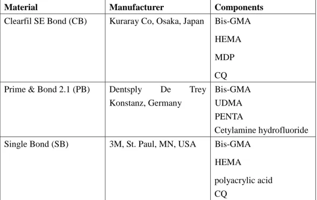
Table 1. Composition of the tested root canal sealers as given by the
manufacturers
Material Manufacturer Components
Clearfil SE Bond (CB) Kuraray Co, Osaka, Japan Bis-GMA HEMA MDP CQ Prime & Bond 2.1 (PB) Dentsply De Trey
Konstanz, Germany
Bis-GMA UDMA PENTA
Cetylamine hydrofluoride Single Bond (SB) 3M, St. Paul, MN, USA Bis-GMA
HEMA
polyacrylic acid CQ
Figures for legends
Fig. 1
(dilution)
cellviability(%ofcontrol)
0 20 40 60 80 100 120
CB SB PB
1:1 1:2 1:4
Fig. 1 Effect of elutes of CB, PB, and SB on human pulp cells by MTT assay for 24 h
incubation period. Percentage of cell viability compared with that of control
was calculated. Each bar represents a mean ± SD.
Fig. 2 (A)
Fig. 2 (B)
0 1 2 4 8 24
RelativeHO-1Level
0 1 2 3 4 5 6 7
CB
*
* *
*
*
(h)
Fig. 2 (A) Kinetics of HO-1 mRNA gene expression in human pulp cells exposed to
CB for 0, 1, 2, 4, 8, and 24 h, respectively. GAPDH was performed in order to
monitor equal protein loading. (B) Levels of HO-1 mRNA treated with CB
were measured by densitometer. The relative level of HO-1 mRNA expression
was normalized against GAPDH signal and the control was set as 1.0. Optical
density values represent the mean ± SD. * represents significant difference
from control values with p<0.05.
Fig. 3 (A)
Fig. 3 (B)
0 1 2 4 8 24
RelativeHO-1Level
0 1 2 3 4
PB
* *
*
*
*
(h)
Fig. 3 (A) Kinetics of HO-1 mRNA gene expression in human pulp cells exposed to
PB for 0, 1, 2, 4, 8, and 24 h, respectively. GAPDH was performed in order to
monitor equal protein loading. (B) Levels of HO-1 mRNA treated with PB
were measured by densitometer. The relative level of HO-1 mRNA expression
was normalized against GAPDH signal and the control was set as 1.0. Optical
density values represent the mean ± SD. * represents significant difference
from control values with p<0.05.
Fig. 4 (A)
Fig. 4 (B)
0 1 2 4 8 24
RelativeHO-1Level
0 1 2 3 4 5 6 7 8
SB
*
*
*
*
(h)
Fig. 4 (A) Kinetics of HO-1 mRNA gene expression in human pulp cells exposed to
SB for 0, 1, 2, 4, 8, and 24 h, respectively. GAPDH was performed in order to
monitor equal protein loading. (B) Levels of HO-1 mRNA treated with SB
were measured by densitometer. The relative level of HO-1 mRNA expression
was normalized against GAPDH signal and the control was set as 1.0. Optical
density values represent the mean ± SD. * represents significant difference
from control values with p<0.05.