Reconstruction of Defect Overlying the Achilles Tendon with Artificial Dermis (Terudermis
®) and Skin Graft
── A Case Report and Literature Review
Hsu-Tang Cheng, Chao-I Wu, Hsin-Han Chen, Erh-Kang Chou, Sophia Chia-Ning Chang, Yung-Chang Hsu
Division of Plastic and Reconstructive Surgery, Department of Surgery, China Medical University Hospital, Taichung, Taiwan
Background:
Soft tissue injuries over the Achilles tendon area often result in wound infection, delayed healing, adhesion, and limitation range of motion of the ankle joint. Soft tissue defects over the Achilles tendon has typically been treated with local and pedicled flaps or free flaps. Artificial dermis with skin grafts are rarely used on exposed Achilles tendons.
Terudermis®, an artificial dermis, which was previously used in the reconstruction after oral surgeries can be used in the reconstruction for the defects overlying the Achilles tendon.
Aim and Objectives:
The purpose of this article is to investigate the clinical feasibility of Terudermis® combined with skin grafts in defects overlying the Achilles tendon. We also review the management options for the defects around the Achilles tendon.
Materials and Methods:
We present a 73-year-old uremic woman with a full-thickness contact thermal burn over her right Achilles tendon. The wound was complicated with extended-spectrum β-lactamase Pseudomonas aeruginosa infection because of inadequate wound care. After the debridement, there is a 10*5 cm sized soft tissue defect overlying an exposed Achilles tendon.
Results:
Staged operation with the use of Terudermis® and then with skin grafting were performed in this patient. The skin grafts took well and there is minimal limitation in her ankle’s range of motion.
Conclusion:
Terudermis® may serve as an alternative way of management for defects overlying the Achilles tendon. It is suitable for those who are not suitable for flap reconstruction.
The operation time is shorter and there is no excessive tissue after the operation. It achieved good functional and aesthetic outcome. (J Taiwan Soc of Plast Surg 2010;19:244
∼249)
Key words: artificial dermis, achilles tendon injury, soft tissue defect Introduction
Reconstruction of the soft tissue defects overlying the Achilles tendon is a challenge to reconstructive surgeons. Regional or free tissue transferring have been provided, but none of these proposals have been universally adopted 1-15. There is limited literature about the use of artificial dermis with skin graft on exposed Achilles tendons 16. A bilayer artificial dermis (Terudermis®, Terumo Corporation, Japan), which was previously used as an alternative to split-thickness skin grafts on the defects in the oral mucosa after surgical treatment 17,18 and the donor site of free radial forearm flap 19 was applied to our presented case with a full thickness soft tissue defect on the Achilles tendon.
Materials and Methods
A 73-year-old uremic woman with a history of essential thrombocythemia under the control of anagrelide and hydroxyurea presented with a chronic wound at the posterior aspect of her right lower leg. She is not a diabetic patient and is a non-smoker.
Three weeks before admission, she had a contact thermal burn which resulted in a full thickness skin lesion at the posterior aspect of her right lower leg.
She received wound care at another hospital.
However, the wound deteriorated and was then complicated with extended-spectrum β-lactamase Pseudomonas aeruginosa infection. She was admitted to this hospital and received surgical debridement.
Postoperatively, there is a 12 * 3 cm sized lesion at the posterior aspect of her right lower leg. The wound was treated regularly with topical silver sulfadiazine.
Results
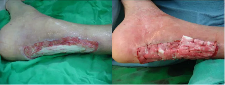
After 4 weeks of wound care at hospital and then at home, she received a staged reconstruction for the wound. After the initial debridement, there is a soft tissue defect on her right heel with a 9 * 3 cmsized exposure of Achilles tendon (Fig. 1). The total soft tissue defect was 10* 5 cm. After meticulous coagulation of the wound bed, an artificial dermis (Terudermis®, Terumo Inc, Japan) was applied to the defect of right heel and sutured with 4-0 catgut (Fig. 2). Saline-soaked gauze dressing was applied over the artificial dermis graft, followed by compressive dressing with elastic bandage. The right ankle joint was maintained in a neutral position by using a short leg splint.
The dressing was changed first on one day after operation, and followed by every two days until secondary split-thickness skin grafting. Three weeks after the artificial dermis grafting, the silicon sheet on the artificial dermis was removed. Split-thickness skin graft (STSG) about 10/1000 inch from the right lateral thigh, meshed 1.5:1, was grafted on the artificial dermis already taken on her right heel. Four days after the STSG, the skin graft took well (Fig. 3). Five months after the STSG, the skin is in good condition without any recurrence of ulcerations (Fig. 4). She can walk well without any assistance.
Discussion
Located at the low and distal part of the human body with scanty of subcutaneous tissue, well- vascularized soft tissue transferring for the reconstruction of defects overlying the Achilles tendon was considered.
Though they may achieve good coverage of the Achilles tendon, the excess tissue may make shoe- wearing discomfort.
Skin graft is thought not be taken on the freshly debrided recipient bed and too fragile to hold up the normal daily wear and tear that is exerted on Achilles tendon. Before Attinger CE et al. 16, there is limited literature about artificial dermis with skin graft on exposed Achilles tendons. According to their concepts, under good care with physiologically moist, granulation tissue will form over the tendon. They concluded that with a properly prepared wound bed, skin grafting can Fig. 1. Soft tissue defect about 10*5 cm in size with a 9*3 cm
sized exposure of the Achilles tendon.
Fig. 2. Two pieces of 5*5 cm sized artificial dermis was applied to the lesion suturing by 4-0 catguts.
Fig. 3. The split-thickness skin graft took well after the removal of the compressing dressings.
Fig. 4. Five months after the split-thickness skin grafting, the skin is stable without recurrence of ulceration.
be as effective as local or free flaps in successfully healing Achilles tendon wounds.
In the wound bed preparation of the presented case, it was commenced with aggressive debridement to remove the tissue infected by Pseudomonas aeruginosa.
It followed with maintenance of physiologically moist.
As in the studies of Attinger CE et al., silver sulfadiazine acts as an ideal topical agent in our patient. It not only prevents desication but also further infection. After achieving sterile wound bed, healthy granulation tissue may grow.
Higher risk of recurrent ulceration in skin-grafted Achilles tendon wounds has been reported16. The patient is elderly with less activity. An artificial dermis with overlying skin graft may prevent recurrent ulceration.
Terudermis® as an artificial dermis is a porous collagen of sponge type and composed of an atellocollagen complex that is extracted from bouvine tendon and chemically and hydrothermally treated 20. It consists of two layers (upper silicone and lower sponge). It has been known that neodermis will be made by fibroblast migration and capillary invasion from the wound.
Terudermis® was autolyzed and absorbed and substituted by neodermis 21.
Despite the disadvantage of the requirement of a secondary STSG, the use of artificial dermis in the defect overlying the Achilles tendon offers good soft-tissue augmentation and the graft-skin quality is similar to full-thickness skin graft. It achieves good functional and cosmetic outcome.
Reference
1. Upton J, Baker TM, Shoen SL et al. Fascial flap coverage of Achilles tendon defects. Plast Reconstr Surg. 1995;
95(6):1056-61.
2. Katsaros J, Tan E, Zoltie N. et al. Further experience with the lateral arm free flap.Plast Reconstr Surg. 1991;87(5):
902-10
3. Berthe JV, Toussaint D, Coessens BC. One-stage reconstruction of an infected skin and Achilles tendon defect with a composite distally planned lateral arm flap.
Plast Reconstr Surg. 1998;102(5):1618-22.
4. Lee HB, Lew DH, Oh SH et al. Simultaneous reconstruction of the Achilles tendon and soft-tissue defect using only a latissimus dorsi muscle free flap. Plast Reconstr Surg.
1999;104(1):111-9.
5. Wei FC, Chen HC, Chuang CC et al. Reconstruction of Achilles tendon and calcaneus defects with skin- aponeurosis-bone composite free tissue from the groin region. Plast Reconstr Surg. 1988;81(4):579-89.
6. Feibel RJ, Jackson RL, Lineaweaver WC et al.
Management of chronic achilles tendon infection with musculotendinous gracilis interposition free-flap coverage.
J Reconstr Microsurg. 1993;9(5):321-5
7. Papp C, Todoroff BP, Windhofer C.Partial and complete reconstruction of Achilles tendon defects with the fasciocutaneous infragluteal free flap. Plast Reconstr Surg.
2003;112(3):777-83.
8. Lee JW, Yu JC, Shieh SJ. Reconstruction of the Achilles tendon and overlying soft tissue using antero-lateral thigh free flap. Br J Plast Surg. 2000;53(7):574-7.
9. Kuo YR, Kuo MH, Chou WC et al. One-stage reconstruction of soft tissue and Achilles tendon defects using a composite free anterolateral thigh flap with vascularized fascia lata:
clinical experience and functional assessment. Ann Plast Surg. 2003;50(2):149-55.
10. Bullocks JM, Hickey RM, Basu CB. Single-stage reconstruction of Achilles tendon injuries and distal lower extremity soft tissue defects with the reverse sural fasciocutaneous flap. J Plast Reconstr Aesthet Surg. 2008;61(5):566-72.
11. Jepegnanam TS, Nithyananth M, Boopalan PR et al.
Reconstruction of open contaminated achilles tendon injuries with soft tissue loss. J Trauma. 2009;66(3):774-9.
12. Michlits W, Gruber S, Windhofer C. Reconstruction of soft tissue defects overlying the Achilles tendon using the super extended abductor hallucis muscle flap. J Trauma. 2008;
65(6):1459-62.
13. el-Khatib H.Island adipofascial flap for resurfacing of the Achilles tendon.Plast Reconstr Surg. 1996;98(6):1034-8.
14. Gruber S, Michlits W, Papp C.The new distal soleus adiposal pull-through composite flap for reconstruction of defects overlying the Achilles tendon: the anatomy and clinical experience.Surgery. 2008;143(3):441-6.
15. Hussein ElGhamry A. Peroneal tendofascial flap: a new fascial flap for Achilles tendon coverage, a preliminary report. Br J Plast Surg. 2003;56(3):284-8.
16. Attinger CE, Ducic I, Hess CL. Outcome of skin graft versus flap surgery in the salvage of the exposed achilles tendon in diabetics versus nondiabetics. Plast Reconstr Surg.
2006;117(7):2460-7.
17. Chen CM, Yang CF, Shen YS. The use of artificial dermis for surgical defects in the treatment of oral premalignant lesions. J Surg Oncol. 2008;97(3):291-3.
18. Ko EC, Shen YH, Yang CF. Artificial dermis as the substitute for split-thickness skin graft in the treatment of oral submucous fibrosis. J Craniofac Surg. 2009;20(2):443- 5.
19. Lee JW, Jang YC, Oh SJ. Use of the artificial dermis for free radial forearm flap donor site. Ann Plast Surg. 2005;
55(5):500-2.
20. Koide M, Osaki K, Konishi J. A new type of biomaterial for artificial skin: dehydrothermally cross-linked composites of fibrillar and denatured collagens.J Biomed Mater Res.
1993;27(1):79-87.
21. Nakamura T. A clinical evaluation of Terudermis, an artificial skin, for the repair of deep wound and soft tissue defect: results in fifty-five cases. Jpn J Plast Reconstr Surg.
1996;39:353
Reprints request from: Yung-Chang Hsu, M.D.
Department of Plastic Surgery, China Medical University Hospital Address: 2 Yuh Der Road, Taichung City, 404, Taiwan
Tel: (886)-4-22052121 ext. 1638 Fax: (886)-4-22030777
E-mail: [email protected]
人 工 真 皮 (Terudermis
®) 及植皮用於阿基里氏腱上 組 織 缺 損 之 重 建
── 病 例 報 告 及 文 獻 回 顧
鄭旭棠 吳肇毅 陳信翰 周爾康 張家寧 許永昌
背 景:
阿 基 里 氏 腱 區 域 為 一 特 殊 區 域 , 皮 膚 下 即 為 堅 強 的 腱 性 結 構 , 軟 組 織 條 件 差 , 因 此 阿 基 里 氏 腱 區 域 受 傷 後 常 會 有 感 染 、 延 遲 癒 合 、 粘 黏 以 及 踝 關 節 受 限 等 併 發 症 的 可 能 。 此 一 區 域 之 重 建 , 主 要 以 自 由 、 帶蒂 或 是 局 部皮 瓣 為 主 ,鮮 少 使 用 植皮 或 是 其 他皮 膚 替 代 物。 人 工 真 皮 (Terudermis®) 過去常使 用在口腔內之軟組織重建,可以配合植皮用在阿基里氏腱區域軟組織之重建。
目 的 及 目 標 :
探討阿基里氏腱區域損傷後導致組織缺損的重建方式以及使用人工真皮配合植皮手術的可行性。
材 料 及 方 法 :
我 們 提 出 一 位 七 十 三 歲 尿 毒 症 的 女 性 病 患 , 右 腳 阿 基 里 氏 腱 區 域 在 燙 傷 後 , 因 傷 口 照 顧 不 當 , 造
成具抗藥性的綠膿桿菌感染。在經過傷口清創手術後,留下10*5 公分大的組織缺損合併阿基里氏腱的
暴露。
結 果:
我們採階段手術的方式,先使用人工真皮 (Terudermis®),之後配合植皮手術,重建阿基里氏腱上
之皮膚及軟組織。病患術後傷口恢復良好,功能獲得好的改善。
結 論:
對 於 阿 基 里 氏 腱 上 之 軟 組 織 缺 損 , 人 工 真 皮 (Terudermis®) 配合植皮作重建是一種可行的替代方 式 。 此 一 方 法 可 以 使 用 在 下 肢 血 循 不 佳 不 適 合 作 皮 瓣 手 術 的 病 患 。 它 的 手 術 時 間 不 長 , 術 後 組 織 不 至 於過多,且兼顧了外觀以及功能上的考量。