1
105 學年度
休假研究報告書(封面)
休假研究計畫名稱:
The mechanism of how healthy epithelial cells evolve into
diseased cells
健康上皮細胞如何演化成病態細胞的作用機轉
單位:皮膚科姓名:王德華
中華民國 106 年 4 月 26 日
2
The mechanism of how healthy epithelial cells evolve into diseased cells Abstract
I spent my sabbatical and do research from 2016/9/1 to 2017/5/31 in Academician of Academia Sinica, Professor Tung-Tien Sun (孫同天院士)’s lab in the Department of Cell Biology, Medical School, New York University. Professor Sun is famous in teaching and research. He discovered a group of unique proteins on the urinary bladder cells which he named uroplakins. He discovered the eyes and skin stem cells. Now he is discovering a new class of stem cells in the bladder urothelium. It will become a breakthrough in stem cell research and the finding can have major impact in urology. His team also works with experts in urinary infectious diseases and bladder cancers.
In this report, I first describe the background of why I went to his lab and how I enjoy science research every day in his lab. In helping the search of a new class of stem cells in the bladder urothelium, I engage in molecular biology (RNA sequencing) which is totally new to me. We accidently found aggregation of keratin in the umbrella cells, the lining epithelial cells of urinary bladder after over-stretching by urine retention. The aggregation may relate to keratin degradation which may lead to dysfunction of the barrier of umbrella cells to toxic substances in urine. The studies are ongoing. After my return, I will collaborate with urologists in our hospital and Professor Sun to continue the studies in epithelial cells in urinary system. I sincerely encourage our young faculties to come his lab, the one of the top labs in the US to learn and to become an outstanding scientist and teacher.
3
1. Background
1.1 Why New York University the first chose?
New York University (NYU) is a private nonprofit research university founded in 1831, based in New York City. It is ranked amongst the top 32 universities in the world by Times Higher Education, U.S. News & World Report, and the Academic Ranking of World Universities. NYU is also operating university globally including NYU Abu Dhabi and NYU Shanghai, and centers in Accra, Berlin, Buenos Aires, Florence, London, Madrid, Paris, Prague, Sydney, Tel Aviv, and Washington, D.C.1
NYU alumni have profound influence in the country. There are 37 Nobel Laureates, more than 30 Pulitzer Prize winners, over 30 Academy Award winners, and hundreds of members of the National Academies of Sciences are from NYU faculty and alumni who include heads of state, royalty, eminent mathematicians, inventors, media figures, Olympic medalists, CEOs of Fortune 500 companies, and astronauts. NYU alumni are among the wealthiest in the world. According to The Princeton Review, NYU is consistently considered by students and parents as a "Top Dream College."1
1.2 NYU School of Medicine
The NYU School of Medicine was founded in 1841, ranking 11th in research according to U.S. News & World Report. As of 2016, it is one of the most selective medical schools in the United States, with an acceptance rate of 1.8%. In 2014, New York University School of Medicine attracted over $304.5 million in external research funding from the National Institutes of Health alone. The School of Medicine is part of NYU Langone Medical Center, locates at 550 First Avenue in New York City http://nyulangone.org/. The School of Medicine has 1,177 full-time faculties and 3,091 part-time faculties. Additionally, there are 104 endowed professorships, 1,078 residents/fellows, 68 M.D./Ph.D. candidates and 400 postdoctoral fellows as of 2011. The NYU Medical Center is home to the School of Medicine, the Sackler Institute of Graduate Biomedical Sciences, and the Charles C. Harris Skin & Cancer Pavilion.2
In 2016-17, NYU Langone Medical Center was also recognized on the U.S. News & World Report "Best Hospitals Honor Roll," ranking 10th among the top hospitals in the nation with 13 nationally ranked specialties including cancer, cardiology & heart surgery, neurology & neurosurgery, orthopedics, diabetes & endocrinology,
nephrology, geriatrics, gastroenterology, ear, nose & throat, rehabilitation, pulmonology, rheumatology, and urology.2
4 Fig. The NYU medical center at 550, 1 Ave, New York. The building in the left edge of the left photo is under construction, a new building for hosting labs for research purposes. Low, the night view of NYU medical center.
1.3 Academician of Academia Sinica, Professor Tung-Tien Sun (孫同天院士)’s lab Professor Sun is well known in the discovery of how acidic and basic keratin pairs in the epithelial cells http://sun-lab.med.nyu.edu/. He also found out where stem cells locate in corneal epithelium and hair follicle. He discovered a group of unique proteins on the urinary bladder cells which he named uroplakins.3,4 Now he is discovering a new class of stem cells in the bladder urothelium.5 It will become a breakthrough in stem cell research and the finding can have major impact in urology. His team also works with experts in urinary infectious diseases and bladder cancers. In addition, he also develops a training program in scientific methods for graduate and postdoctoral students.
1.4 Epithelial cell lining many organs of the body
Epithelial cells line internal and external surfaces of bodily organs to provide many important biological functions including protection, absorption and secretion. In human cancers, approximately 90% are derived from epithelial cells. It is critically
5 important to understand how a healthy epithelial cell transforms into diseased cell, especially into cancer.
The understanding basic science in epithelial cells is critical important to me as a dermatologist, a physician scientist, who is facing skin diseases every day. I am supervising 2 master and one PhD students, teaching under graduated students in Biochemistry, Microbiology and classes in general education. This is undoubtedly the study in one of the top labs in the US benefit me and my patients, students, the University, and ultimately our society. After finishing of this study, it will be the beginning of a new story. A long-term collaboration will be setup after my return to Taiwan.
2. Arrival
2.1 Do not hesitate to inquire
I started the application and contact Professor Sun in February, 2016 and planned to begin study in his lab in July. Professor Sun also affiliates with Department of Dermatology. He leads a research team including Mayumi Ito, associate professor of the department who is working on transgenic mice model on stem cells. Professor Sun assigned me to her lab at the beginning. My visa was applied through dermatology department. However, the secretary of the department was on vacation and my application was delayed. I was hesitated to ask because I felt impolite to push people working on my business. I asked only a few times and believe I could have the visa before July. Unfortunately, it turns out to be issued from the Cell Biology Department and I could go not until late August. It was delayed for almost 2 months. The lesson I learned before arrived to the US is you have to take care of yourself and do not hesitate on what it should be done aggressively. The final results are your own responsibility.
3. Basic research is the basic, fundamental of all research
3.1 Why study urothelial cells?
Science research is borderless. Professor Sun has discovered where skin stem cells6 and corneal stem cells7 locate. In recent years, his interest focus in an incredible epithelium, the outermost layer that lying on urinary bladder which protects us from the urine. He finds the urothelium stem cells may enrich in particular areas in the bladder and ureters. It is important to learn how to think, how to design, how to solve problems, how to do so called “thought experiments” before a hand on experiments, how to do risk assessments. It is of minimal importance of what organ, or what research area to study. It is needless to say how important to study epithelium as a
6 dermatologist. Urothelium is one of the models for me to learn. It is easy to apply what I have learned into skin epithelium studies.
3.2 Urothelium
3.2.1 Overview of urogenital organ development.
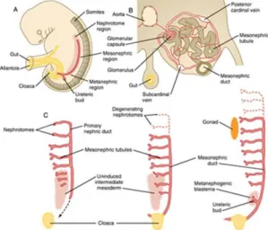
To study stem cells in urinary system, we need to understand the embryology first. The urogenital system arises from intermediate mesoderm which forms a urogenital ridge on either side of the aorta. The urogenital ridge develops into three sets of tubular nephric structures (from head to tail): the pronephros, the mesonephros, and the metanephros (Fig. 1).
Fig. 1. Overview of the development of the urogenital organ. Adopted from: Duke University Medical School Embryology Learning Resources
https://web.duke.edu/anatomy/embryology/urogenital/urogenital.html. Access date: April 22, 2017.
3.2.2 Development of the bladder.
Like many other tissue in our body, the bladder is composed of epithelium and fibromuscular tissue. The epithelium of the bladder is developed from endoderm derived from the embryonic cloaca (Fig. 2). The cloaca is subsequently divided by the urorectal septum into the primitive urogenital sinus and rectum. Epithelium of the upper urogenital sinus and the proximal portion of the allantois give rise to the lining of the bladder, a highly specialized urothelium.
7 Fig. 2. The development of the baldder. Adopted from: Duke University Medical School Embryology Learning Resources
https://web.duke.edu/anatomy/embryology/urogenital/urogenital.html. Access date: April 22, 2017.
3.2.3 Development of the ureters
The collecting tubules and ducts, minor and major calyces and ureters are derived from the ureteric bud whose epithelial lining is mesodermal in origin. Professor Sun’s lab is focusing on the urothelium involving below the ureteropelvic junction (Fig. 3).
Fig. 3. The development of the collecting tubules and ducts, minor and major calyces and ureters. Adopted from: Duke University Medical School Embryology Learning Resources https://web.duke.edu/anatomy/embryology/urogenital/urogenital.html. Access date: April 22, 2017.
3.2.4 The normal histology of urothelium
The human urothelium consists of 3-6 cell layers (Fig. 4, 5) including the basal layer, one or more intermediate layer(s) and the surface (or superficial) layer, arranged in a regular architecture.8,9 The smallest, condensed nuclei were situated in the basal layer, and the largest, least condensed, rounded nuclei were located in the superficial layer. Basal and intermediate urothelial cells were mononuclear, but superficial cells (umbrella cells) were mononuclear or binuclear. Prominent nucleoli could be
8 observed in all cell layers. Surface cell nuclei sometimes contained more than one nucleolus.
Fig. 4. Transitional epithelium of the urinary bladder. Note the rounded surface of the apical cells - a distinguishing characteristic of this type of epithelium. Adopted from Wikipedia, access date: April 22, 2017.
ureter near renal pelvis, mid-ureter, ureterovesical junction Fig. 5. The cross sections show urothelium of urinary bladder and different parts of the ureters of a 10 – 16-week-old Swiss Webster mouse.
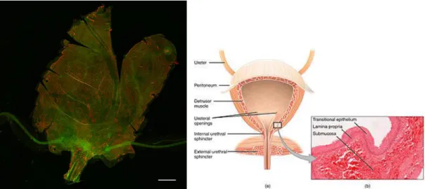
3.3 Whole mount immunofluorescence of the bladder and ureters
Immunofluorescence staining of different cell markers is an efficient way to explore where stem cells locate. However, the cross-section specimen only provides limited information because it demonstrates only few micrometer thickness of a fragment of a specimen. Whole mount methods offer a better whole picture of a tissue/organ
especially stem cell are expected to be less than 10% in the bladder.10 I learned how to dissect bladder and ureter from a mouse and how to prepare them and stain with
bladder
9 different antibodies of interest.
The first challenge is how to make a 3D bladder flat? High quality of photos only can be achieved by a good focusing on the images. We have to make the bladder flat in order to focus cells of interest under confocal microscope. The same problem challenges people who make a global map without distortion. The bladder is
stretchable. However, the cell-cell junction will break if it is stretched is too strong. The most challenge area is the trigone of the bladder. One way to overcome it is to make several incisions along the border of the bladder (Fig 7).
Fig. 6. How to make a global map from flat paper.
http://www.gma.org/surfing/imaging/globe.html access date: April 22, 2017.
Fig. 7. Whole mount of a 12-week-old Swiss Webster mouse bladder and ureters. The epithelium is stained with anti-keratin 5 (green) and anti-keratin 8 (red) antibodies. Noted the incisions around the edges of the border (bar = 1mm). Right figures show the relationship of ureters and the bladder (Wikipedia).
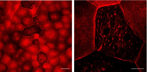
10 Unexpectedly, we found keratin aggregates on the umbrella cells of the bladder. Only few of them appeared in the bladder, especially cells in the bladder wall (Fig 8, 9). It was observed in mouse bladder with a relatively high volume of urine (around 300 uL). We suspect that the over-extension of bladder leads to a disruption of umbrella cells and the keratin started to degraded.11-13 Aggregation of keratin may be the first sign of keratin degradation. Interesting questions are: how large is the volume to induce keratin aggregation? What are the physiologic meanings of this aggregation? Does it affect the secretion of uroplakin which is supposed to be secreted to the cell surface to protect the cells from urine? Why only few cells are affected? Does it happen on the cells under the most extensive stretch? All these interesting questions are undergoing further studies.
Fig. 8. Aggregation of keratin on umbrella cells of urinary bladder. A. The mouse bladder was blocked with anti-Keratin 8 and examined under confocal microscope. Some cells are relatively dark. Speckles of aggregates were noted on the cells. B. Higher magnification to see the aggregates. A uniform keratin chicken wired network on cells around the cells with keratin aggregation. Bar: 100 um (left); 50 um (right).
11 Fig. 9. In some umbrella cells, the keratin 8 is aggregated into a cluster and locates around cell nucleus. Bar = 20 um.
3.4 RNA sequencing
To understand where the urothelial stem cells locate in bladder and ureters, we need to find out some specific biomarkers to differentiate bladder cells and ureter cells. The hypothesis is most of the stem cells are originated from the ureters. Some and few of the urothelial stem cells reside in the bladder. The reside stem cells in the bladder are sufficient to repair local minor damage in the bladder. Stem cells in the ureter activate and migrate to bladder to repair only in case of a severe damage in the bladder. Therefore, we need good biomarkers to differentiate urothelium of the bladder and ureters. To fulfill this purpose, RNA sequencing is one of the best solutions. We plan to isolate bladder urothelium and ureter urothelium respectively from 4 rats with same age, same sex and come from the same natural litter to reduce variations in animal. We expect that we can find out different biomarkers for stem cells from the bladder versus ureters. Then we can trace stem cells after we wipe off the bladder urothelium with cyclophosphamide to test our hypothesis. This is a project ongoing.
4. Social activity and cultural exposure
One of the achievement is invisible, the influence by the culture of New York City. When you go to work every day, you meet different people, different colors from different countries of the world. We have Asian, Americans, and French people working together in the same floor of the lab. We eat lunch together; we chat and
12 share our ideas on lives and on experiments. We go out to have fun together. I learn how their cultures are, how they handle problems, how they think differently from the East.
Professor Sun is very kind to us. He invited us to his place for dinner, to his house in Pennsylvania in holidays. It was a lot of fun and lot of memories. I saw how a great scientist, a great husband, and a great grandpa during my stay. He teaches patiently to my son how to play snooker. You can find out why he is one of the best teachers in the University by the way how he teaches. This is a really excellent role model to me and this is the purpose why I am here in his lab: to learn how to become a good teacher and how to become a better person. I believe I have them all in my mind and this will affect tremendously to my career and my life.
13 Fig. Upper, from the left to right: Professor TT Sun, Post-doctor Yi Liao, Professor Mayumi Ito, me. Middle, reunion party of Sun’s lab in his house. Low, good friends of lab members in Bhama Ramkhelawon’s lab.
5. Conclusion
I learned more than I expected. I will continue my works and collaborate with urologists in our hospital and Professor Sun after my return. The good news is Professor Sun opens new positions to our University to accept more young people to come to his lab and learn from him. I sincerely encourage our colleagues to come and they will find it is one of the best places to learn, to live and to enjoy.
6. Acknowledgements
I sincerely thank my university, my hospital, my department I am working, especially colleagues in my department for sharing my works. I also genuinely thank all
members in Professor Sun’s lab, administrations in NYUMC, and friends from
Professor Ramkhelawon’s lab for their wonderful help and friendship. Finally, I thank my wonderful family, Cindy and Brian to come with me to the US to share every moment I have.
14
References
1. Wikipedia. New York University.
https://enwikipediaorg/wiki/New_York_University - cite_note-13.access date: April 22, 2017.
2. Wikipedia. New York University School of Medicine.
https://enwikipediaorg/wiki/New_York_University_School_of_Medicine.
Access date: April 22, 2017.
3. Yu J, Manabe M, Wu XR, Xu C, Surya B, Sun TT. Uroplakin I: a 27-kD protein associated with the asymmetric unit membrane of mammalian urothelium. J Cell Biol. 1990;111(3):1207-1216.
4. Moll R, Laufer J, Wu XR, Sun TT. [Uroplakin III, a specific membrane protein of urothelial umbrella cells, as a histological markers for metastatic transitional cell carcinomas]. Verh Dtsch Ges Pathol. 1993;77:260-265. 5. Kong X-T, Deng F-M, Hu P, et al. Roles of uroplakins in plaque formation,
umbrella cell enlargement, and urinary tract diseases. The Journal of Cell
Biology. 2004;167(6):1195-1204.
6. Cotsarelis G, Sun TT, Lavker RM. Label-retaining cells reside in the bulge area of pilosebaceous unit: implications for follicular stem cells, hair cycle, and skin carcinogenesis. Cell. 1990;61(7):1329-1337.
7. Sun TT, Tseng SC, Lavker RM. Location of corneal epithelial stem cells.
Nature. 2010;463(7284):E10-11; discussion E11.
8. Jost SP, Gosling JA, Dixon J. Comparative morphology of normal and metaplastic human urothelium. Ann Urol (Paris). 1989;23(2):162-164. 9. Jost SP, Gosling JA, Dixon JS. The morphology of normal human bladder
urothelium. J Anat. 1989;167:103-115.
10. Hatina J, Schulz WA. Stem cells in the biology of normal urothelium and urothelial carcinoma. Neoplasma. 2012;59(6):728-736.
11. Shyy TT, Asch BB, Asch HL. Concurrent collapse of keratin filaments, aggregation of organelles, and inhibition of protein synthesis during the heat shock response in mammary epithelial cells. J Cell Biol.
1989;108(3):997-1008.
12. Russell D, Andrews PD, James J, Lane EB. Mechanical stress induces
profound remodelling of keratin filaments and cell junctions in epidermolysis bullosa simplex keratinocytes. J Cell Sci. 2004;117(Pt 22):5233-5243.
13. Chamcheu JC, Navsaria H, Pihl-Lundin I, Liovic M, Vahlquist A, Torma H. Chemical chaperones protect epidermolysis bullosa simplex keratinocytes from heat stress-induced keratin aggregation: involvement of heat shock proteins and MAP kinases. J Invest Dermatol. 2011;131(8):1684-1691.