Vol. 35: 175-185,1999 DISEASES OF AQUATIC ORGANISMS
Dis Aquat Org Published February 26
Specific genomic
DNA
fragment analysis of
different geographical clinical samples of shrimp
white spot syndrome virus
Chu-Fang Lol, Hui-Chen Hsul, Meng-Feng ~ s a i ' ,
Ching-Hui Hol, Shao-En peng1,
Guang-Hsiung
KOU',Donald V. ~ i ~ h t n e r ~ . *
'Department of Zoology, National Taiwan University. Taipei, Taiwan. ROC 'Department of Veterinary Science, University of Arizona, Tucson. Arizona 85721. USA
ABSTRACT. White spot syndrome (WSS) has been found in many species of shrimp and crabs, not just In Asia but globally. The causative agent is known as white spot syndrome virus (WSSV). In order to clarify the relatedness of WSSV from various geographic regions, we compared the viral DNA of a number of clinical samples of WSSV. (1) China96-116A from Penaeus chlnens~s, (2) India95-314 from Penaeus monodon, (3) grocery store95-204 and grocery store96-l15 from P. rnonodon possibly origi- nating from Thailand, (4) crayfish97-25 from Orconectespunctimanuscollected from the U.S. National Zoo, (5) Thailand95-46 from experimentally infected Penaeus vannamei. (6) South Carolina97-64 from P. vannamei, and (7) Texas95-242 and Texas96-7 from P. vannarnei. These specimens were first ex- amined by dot hybridization analysis with nucleic acid probes derived from a WSSV Taiwan isolate. Although the intensity of the hybridization signals varied, and although some specimens of India95- 314, crayfish97-25, Texas95-242 and Texas96-7 failed to give a detectable hybriduation signal with certain probes, the broad consistency of dot hybridization data suggests that these WSSV clinical samples from different geographical locations are closely related. Following this analysis, all the specimens were examined using 10 virus-specific polymerase chain reactions (PCR). The amplification products were subsequently digested with Cfo I, Hae 111, Hpa I1 and Rsa I restriction endonucleases to determine if there were any DNA fragment polymorphisms in the WSSV clinical samples. The results highlighted the genetic relatedness of all the WSSV clinical samples with the possible exception of a series of Texas viral samples which could be distinguished from the other geographic samples in some of the PCR-based tests.
KEY WORDS: Genomic DNA analysis. Restriction profiles of specific viral DNA fragments. White spot syndrome virus WSSV . WSBV . Geographical clinical samples
INTRODUCTION
The comn~ercial cultivation of marine shrimp is now a global industry, and one of the biggest problems facing this industry is disease. White spot syndrome (WSS) is a viral disease which affects many commer- cially cultivated marine shrimp species, not just in Asia but globally (Lightner 1996, Flegel 1997). The principal clinical sign of WSS is the presence of white spots on the proximal surface of the cuticle of the diseased 'Addressee for correspondence.
E-mail: [email protected]
shrimp. Affected individuals become lethargic and go off their feed. In shrimp ponds, they congregate in the shallows along the edges of the pond, and in culture tanks they sink inactively to the bottom, where they are frequently attacked and cannibalized by the healthier shrimp. WSS can cause up to l 0 0 % mortality (Chou et al. 1995, 1998), which leads to a correspond- ingly devastating economic impact. WSS has been formally recognized since 1992, but so far no signifi- cant resistance to this disease has been reported for any of the penaeid species (Lightner 1996).
The causative agent of WSS has, however, been identified: it is a non-occluded rod-shaped virus known O Inter-Research 1999
Lo et al.: DNA analysis of wh.ite spot syndrome virus 177
1996a) consisting of pms146F1 (5'-ACT ACT AAC TTC AGC CTA TCT AG-3') and pms146R1 (5'-TAA TGC GGG TGT AAT GTT CTT ACG A-3') was utilized for l-step WSSV diagnostic PCR (Lo et al. 1996a) in order to confirm the presence of the virus in the collected shrimp samples (pms: Penaeus monodon WSSV Sal 1 fragment). The l-step PCR was performed as follows. The DNA samples used for amplification totaled 0.1 yg in a 100 p1 reaction mixture containing 10 mM Tris- HC1, pH 8.8 at 25"C, 50 mM KC1, 1.5 mM MgC12, 0.1 % Triton X-100, 200 pM of each dNTP, 100 pm01 of each primer, and 2 units of TaqDNA Polymerase (Life Tech- nologies). The amplification was performed in a ther- mal cycler (Perkin-Elmer Corporation) for 1 cycle of 94°C for 4 min, 55°C for 1 min, 72°C for 2 min, then 39 cycles of 94°C for 1 min, 55°C for 1 rnin, 72°C for 2 rnin, plus a final 5 min extension at 72'C after 40 cycles. Control reactions containing no template DNA were run for all PCR reactions. A portion (10 pl) from each of the completed PCR reactions was mixed with 1 p1 loading buffer and subjected to electrophoresis on 1 % agarose gels containing ethidium bromide at a concentration of 0.5 pg ml-', and visualized by ultra- violet transillumination.
As described previously (Lo et al. 1996a), the quality of the DNA extracted from the collected shrimp sam- ples was checked with a primer set amplifying a deca- pod gene before the application of WSSV diagnostic PCR. For this purpose, 2 primers, 143F (5'- TGC CTT ATC AGC TNT CGA TTG TAG-S', where N represents G, A, T or C) and 145R (5'-TTC AGN TIT GCA ACC ATA CTT CCC-3'), derived from a highly conserved region of the 18s rRNA sequence of decapods (Kim & Abele 1990, Lo et al. 1996a) were used.
Dot blot hybridization analysis. DNA samples were boiled for 10 min and then quenched on ice. An aliquot
(1 y1) of each of the DNA samples was dotted onto a sheet of positively charged nylon paper (Boehringer Mannheim, Mannheim, Germany) that had been pre- soaked with 5 X SSC (1 X SSC = 150 mM NaCl, 15 mM Sodium Citrate, pH 7.0) for 5 min and air dried. After cross-linking the DNA with the membrane by UV light, the blot was used for hybridization with 11 WSSV probes which were non-radioactively labeled with digoxigenin (DIG)-dUTP (Boehringer Mannheim) by a random priming method. Following prehybridization at 65°C for 1 h in prehybridization solution (10 m1 5 X SSC with 100 mg of blocking reagent 11, 50 p1 of 20% sarkosyl, 20 p1 of 10 % SDS), the blot was hybridized at 65°C for 16 h with DIG-labeled probes. The detection of the DIG-labeled nucleotides in blots was accom- plished by an immunological method using anti-digox- igenin antibody conjugated to alkaline phosphatase (Boehringer Mannheirn) and CSPDB, (Boehringer Mann- heim) as a cherniluminescent substrate for alkaline
phosphatase. The blot was exposed to Kodak X-OMAT film at room temperature for 1 to 10 min to record the chemiluminescent signal. In this study, the blots were reused several times for hybridization analysis with different probes. For reprobing, the blots were rinsed in water for 1 min, treated with an alkaline solution (0.2 N NaOH, l % SDS) at 37°C for 30 min, and then rinsed in 2 X SSC. Following the prehybridization, the blot was then hybridized with a new probe.
The inserts of 11 plasmid clones were used as probes: pms473 (12 kbp), pms484 (8 kbp), pmslOO (6 kbp), pms321 (4.4 kbp), pms146 (1.5kbp), pmh2 ( > 9 kbp), pmhlO (>9 kbp), p m h l l ( > 9 kbp), pmhl3 ( > 9 kbp), pmh32 (>9 kbp), pmh34 (>9 kbp). Allthese clones were selected from Sal I and Hind 111 genomic libraries of a Taiwan WSSV clinical sample (Lo et al. 1996a). The figures in parentheses indicate the size of the insert of each clone. The first set of DNA samples (1 to 47, see Table 2), i.e. the Asian and the first 2 South Carolina specimens, were analyzed with all 11 probes. Only 3 probes were used, however, for the second batch of DNA samples (48 to 71, see Table 3), which included the new grocery store and South Carolina as well as the 2 Texas clinical samples.
Analysis of the DNA of WSSV geographical clinical samples by PCR with specific primer sets and restric- tion fragment length polymorphism. We used 10 primer pairs (Table l ) based on the DNA sequences (unpubl. data from Dr Guang-Hsiung Kou's laboratory) of 9 plasmid clones (pms54, pms94, pms98, pmsl20, pms146, pms321, pms473, p m h l l , pmhl3) selected from Sal I and Hind I11 genomic libraries of a Taiwan WSSV isolate from Penaeus monodon (Lo et al. 1996a). Ten amplicons, A p m d l F l / R l (0.96 kbp), Apms94Fl/ R1 (0.94 kbp), Apms98Fl/Rl (0.95 kbp), Apmsl20Fl/ R1 (0.95 kbp), Apmsl46Fl/Rl (1.5 kbp), Apms321F1/ R1 (0.9 kbp), Apms321F2/R2 (1.0 kbp), Apms473F3/R3 (1.1 kbp), ApmhllFl/Rl (1.0 kbp) and Apmh13Fl/Rl (1.2 kbp), were expected with l-step PCR and the primer sets. The size of each anticipated PCR product is indicated in parentheses. We used these 10 primer pairs and PCR to analyze the 71 DNA samples of vari- ous geographic WSSV clinical samples. To confirm that the amplified fragments were indeed virus specific, an additional internal primer set, pms98F2/R2 (Table l ) ,
was also used to perform 2-step PCR. For Texas speci- mens, we also used Southern hybridization with probes prepared from PCR products of Taiwan WSSV isolate using internal primer sets of pms98 and pms146 to de- tect WSSV-specific PCR products using a method de- scribed previously (Lo et al. 1996a,b). The Cfo I, Hae 111, Hpa 11, and Rsa I restriction profiles of some specific viral DNA fragments were also compared.
The thermal cycling program and reaction condi- tions for l-step PCR were the same as those described
178 Dis Aquat Org 35: 175-185, 1999
l'able 1. PCR pnmer sequences used i11 this study Primer set Primer sequence
1 143/145 143F: 5'-TGCCTTATCAGCTNTCGATTGTAG-3' 145R: 5'-TTCAGNT?TGCAACCATACTTCCC-3'd 2. prns54Fl/Rl pms54F1: 5'-CGTAACAGGCTCGGTGCC-3' pms54R1:5'-CAGCACGGATACGTTAAC-3' 3. pms94Fl/Rl pms94F1: 5'-CGGTCTCAGTAA'ITCGTC-3' pms94R1: 5'-CCTCCATITGCTGCAGTG-3' 4. pms98Fl/Rl pms98F1: 5'-GACAATGITGGTATCGGTAG-3' pms98R1: 5'-GAGCACGAGAAGCACGAC-3' 5, pmsl20Fl/Rl pmsl20Fl: 5'-GACATATACGCCACCAAGG-3' pmsl20R1: 5'-GGCAGCGTCCATACTGTTC-3' 6. pmsl46Fl/Rl pmsI46Fl. 5'-ACTACTAACTTCAGCCTATCTAG-3' pms146R1: 5'-TAATGCGGGTGTAATGTTCTTACGA-3' 7, pms321-lFl/Rl pns32;Fl. 5'-CGCCACCAAGGAATTCGAAC-3' pms321R1: 5'-GCAGACATGGCAGCGTCC-3' 8. pms321-2Fl/Rl pms321F2: 5'-GCGAGCGGCGTACTACGAC-3' pms32lR2 5'-GAGGCCACAGCCGAAGCTG-3' 9. pms473Fl/Rl pms473F3: 5'-AAGAGGAGGATTCTCCAGATCC-3' pms473R3: 5'-CCAACACGGTACACGTAATTC-3' 10. pmhl lFl/Rl pmhllF1: 5'-GGTGA'ITCTGCATCCAGC-3' pmhllR1: 5'-GCGGATTCTATGAGGCGAG-3' 11. pmhl3Fl/Rl pmhl3Fl: 5'-CAGGATGGTACAGAGGAC-3' pmhl3R1: 5'-GTCAATATAGCCATGGATGG-3' 12. pms98F2/R2 pms98F2: 5'-CTGGGCCGTAAAGTAGTG-3' pms98R2: 5'-CTGGACAATGCATGATGAG-3'
shown in Table 2, only the 3 grocery store95204 specimens consistently gave negative results in l-step WSSV diag- nostic PCR. As shown in Table 3, how- ever, 10 of the Texas specimens gave negative results, and only 1 Texas specimen was positive in l-step WSSV diagnostic PCR.
Dot blot hybridization analysis In Table 2, apart from grocery store95-204, only India95-314#3 a n d crayfish97-25#1 failed to hybridize with all 11 WSSV probes, although the intensity of the hybridization signals varied. The broad consistency of these data suggests that these WSSV clinical samples from different geographical locations are closely related.
In the subsequent analysis of South Carolina and Texas specimens, 4 of 10 Texas specimens failed to give a positive hybridization signal with the " N represents G , A , T or C
above for WSSV diagnostic PCR using the pms146F1/ R1 primer set. For l-step PCR, 10 p1 of the 1-step PCR reaction mixture was added to 90 p1 of PCR cocktail containing the inner primer pair, and this was then subjected to a second step of amplification in a thermal cycler (Perkin-Elmer Corporation) for 1 cycle of 94°C for 4 min, 55°C for 1 min, 72°C for 2 min, then 39 cycles of 94°C for 1 min, 55°C for 1 min, 72°C for 2 min, plus a final 5 min extension at 72°C after 40 cycles. Control reactions containing no template DNA were run for all PCR reactions. A portion (10 p1) from each of the com- pleted PCR reactions was mixed with 1 p1 loading buffer and subjected to electrophoresis on 1 % agarose gels containing ethidium bromide at a concentration of 0.5 pg ml-l, and visualized by ultraviolet trans- illumination.
RESULTS
Detection of WSSV in collected shrimp samples by WSSV diagnostic PCR with the use of pmsl46Fl/Rl
primer set
All the prepared templa.tes were amplifiable when assessed by the shrimp DNA-specific primer set 143/145 (Tables 2 & 3). Of the 47 tested DNA samples
pms146 and pms321 probes, while none of the Texas samples hybridized detectably with the p m h l l probe (Table 3). Examples of the dot hybridization results are shown in Fig. 1.
Analysis of the DNA of WSSV geographical clinical samples by PCR with specific primer sets and
restriction fragment length polymorphism As predicted, the sizes of the 10 amplicons were very close, ranging from 0.9 to 1.5 kbp (Fig. 2), but their restriction profiles varied (Fig. 3 ) . We used these primer pairs to amplify DNA fragments from 71 DNA samples from 7 geographical clinical samples as indi- cated in Tables 2 & 3. Using the 10 primer pairs and PCR, 10 specific DNA fragments were yielded from the 47 tested DNA samples of 6 WSSV geographical clinical samples (China96-116A, India95-314, cray- fish97-25, Thailand95-46, South Carolina97-64, grocery store 96-115). The sizes of major PCR products from 6 WSSV geographical clinical samples using the same primer pair were very similar and showed the ex- pected sizes (Fig. 4). Unlike the other 6 geographical clinical samples, however, only 1 of the 16 DNA sam- ples prepared from the 11 shrimp specimens of the 2 Texas clinical samples yielded PCR products with all 3.0 pnmer sets (Texas95-242-J60; Fig. 4 , Lane 56). The C f o I, Hae 111, Hpa 11, Rsa I RFLP profiles of the PCR
Lo et al.: DNA analysis of white spot syndrome virus 179
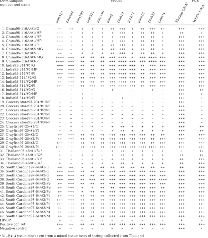
Table 2. Analysis of WSSV geographical strains by dot blot hybridization with 11 WSSV probes derived from Taiwan strain. Num- bers of plus symbols (i.e. between 1 and 4) corresponds to increasing levels of hybridization intensity on a n arbitrary scale (-) no hybridization. DNA sample names show collection areahndividual shrimp no./tissue source for DNA extraction (G: gill; HP: he- patopancreas; PI: pleopod; MG: midgut; M: muscle; H: hemolymph, Pe- pereiopod); HP/BP: homogenate of hepatopancreas of shrimp infected with Baculoviruspenaei. Positive control: WSSV Taiwan clinical sample. Negative control: DNA from WSSV-free Penaeus rnonodon. For probes pms: Penaeus monodon WSSV Sal I fragment, and pmh: P. monodon WSSV Hind 111 fragment
DNA samples number and name
Probes Q PCR C 4 Q
-F
A 1. China96-116A/#l/G 2. China96-116A/#l/HP 3. China96-1 16A/#l/HP 4. China96-1 16A/#l/HP 5. China96-116A/#l/Pl 6. China96-116A/#2/MG 7. China96-116A/#2/G 8. China96- 116A/#3/MG 9. China96-116A/#3/PI 10. India95-314/#1/G 11. India95-314/#1/HP 12. India95-314/#1/P1 13. India95-314/ #2/G 14. India95-314/#2/HP 15. India95-3 14/#2/P1 16. India95-3 14/#3/G 17. India95-3 14/#3/HP 18. India95-3 14/#3/P1 19. Grocery store95-204/#1/M 20. Grocery store95-204/#1/M 21. Grocery store95-204/#2/M 22. Grocery store95-204/#2/M 23. Grocery store95-204/#3/M 24. Grocery store95-204/#3/M 25. Crayfish97-25/#1/G 26. Crayfish97-25/#1/P1 27. Crayfish97-25/#2/G 28. Crayfish97-25/#2/P1 29. Crayfish97-25/#3/G 30. Crayfish97-25/#3/P1 31. Thailand95-46/#1/Bld 32. Thailand95-46/#l/BZa 33. Thailand95-46/#l/B3" 34 Thailand95-46/#1/B4a 35. South Carolina97-64/#1/H 36. South Carolina97-64/#2/G 37. South Carolina97-64/#2/G 38. South Carolina97-64/#2/G 39. South Carolina97-64/#2/Pe 40. South Carolina97-64/#2/Pe 41. South Carolina97-64/#2/Pe 42. South Carolina97-64/#2/Pl 43. South Carolina97-64/#2/P1 44. South Carolina97-64/#2/Pl 45. South Carolina97-64/#2/M 46. South Carolina97-64/#2/M 47. South Carolina97-64/#2/M HP/BP Positive control Negative control"Bl-B4: 4 tissue blocks cut from a mixed tissue mass of shrimp collected from Thailand 'Shr~mp DNA PCR using 143/145 primer set
180 Dis Aquat Org 35: 175-185, 1999
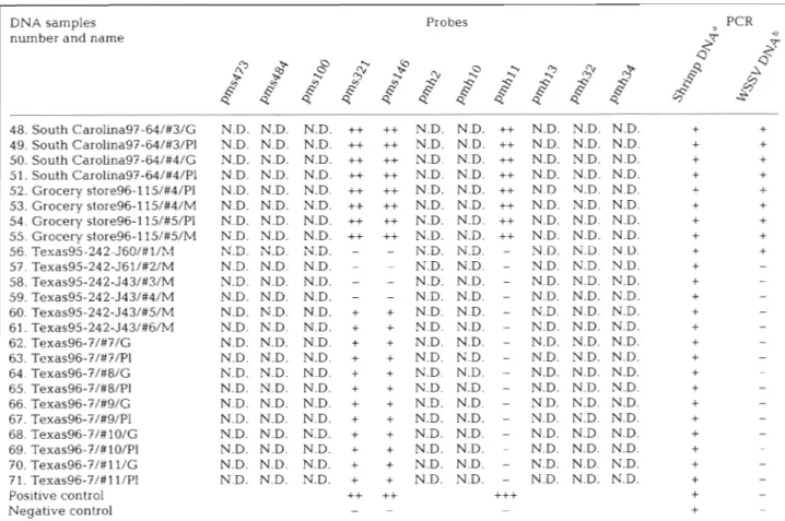
Table 3. Analysis of WSSV geographical strains by dot blot hybridization with 11 WSSV probes derived from Taiwan strain. Num- ber of plus symbols (i.e. between 1 and 4) corresponds to increasing level of hybridization intensity on an arbitrary scale. N.D.: test not done. (-) no hybridization. DNA sample names show collection area/individual shlimp no./tissue sources for DNA extrac- tion (G: g d ; PI: pleopod, M: muscle). Positive control: WSSV Taiwan clinical sample. Negative control: DNA from WSSV-free
Penaeus rnonodon
DNA samples Probes PCR
number and name
-$
0G3 Q
2
t
& " G ? & ' $ $0 5 , 3 4 " 4 =
A Q8
$
.
.
"
%
p
$
@
$
Q 48. South Carolina97-64/#3/G N.D. N.D. N.D.++
++
N.D. N.D. ++ N.D. N.D. N.D.+
+
49. South Carolina97-64/#3/P1 N.D. N.D. N.D.++
++
N.D. N D. ++ N.D. N.D. N.D.+
+
50. South Carolina97-64/#4/G N.D. N.D. N.D.++
++ N.D. N.D. ++ N.D. N.D. N.D.+
+
51. South Carolina97-64/#4/P1 N.D. N.D. N.D.++
++
N.D. N.D. ++ N.D. N.D. N.D.+
+
52. Grocery store96-115/#4/Pl N.D. N.D. N.D.++
++
N.D. N.D. ++ N.D N.D. N D. I+
53. Grocery store96-115/#4/M N.D. N.D. N.D.++
++
N.D. N.D. ++ N.D. N.D. N.D.+
+
54. Grocery store96-115/#5/Pl N.D. N.D. N.D.++
t + N.D. N.D.++
N.D. N.D. N.D.+
+
55. Grocery store96-115/#5/M N.D. N.D. N.D.++
++
N.D. N.D. ++ N.D. N.D. N.D.+
+
56. Texas95-242-J60/#l/hI: N.D. N.D. N.D. - - N.D. N.D. - N D. N.D N.D.+
+
57. Texas95-242-J61/#2/M N.D. N.D. N.D. - - N.D. N.D. - N.D. N.D. N.D.+
- 58. Texas95-242-J43/#3/M N.D. N.D. N.D. - - N.D. N.D. - N.D. N.D. N.D.+
- 59. Texas95-242-J43/#4/M N.D. N.D. N.D. - - N.D. N.D. - N.D. N.D. N.D.+
- 60. Texas95-242-J43/#5/M N.D. N.D. N.D. + + N.D. N.D. - N.D. N.D. N.D.+
- 61. Texas95-242-J43/#6/M N.D. N.D. N.D.+
+ N.D. N.D. - N.D. N.D. N.D.+
- 62. Texas96-7/#7/G N.D. N.D. N.D.+
+ N.D. N.D. - N.D. N.D. N.D.+
- 63. Texas96-7/#7/P1 N.D. N.D. N.D. + + N.D. N.D. - N.D. N.D. N.D.+
- 64. Texas96-?/#8/G N.D. N.D. N.D.+
+ N.D. N.D. - N.D. N.D. N.D.+
65. Texas96-7/#8/P1 N.D. N.D. N.D. + + N.D. N.D. - N.D. N.D. N.D.+
- 66. Texas96-7/#9/G N.D. N.D. N.D. + + N.D. N.D. - N.D. N.D. N.D.+
- 67. Texas96-?/#9/P1 N.D. N.D. N.D. + + N.D. N.D. - N.D. N.D. N.D.+
- 68. Texas96-7/#10/G N.D. N.D. N.D. + + N.D. N.D. - N.D. N.D. N.D.+
- 69. Texas96-7 /#lO/Pl N.D. N.D. N.D. + + N.D. N.D. - N.D. N.D. N.D.+
70. Texas96-?/#11/G N.D. N.D. N.D. ++
N.D. N.D. - N.D. N.D. N.D.+
- 71. Texas96-?/#l l/P1 N.D. N.D. N.D. + + N.D. N.D. - N.D. N.D. N.D.+
- Positive control++ ++
+++
+
- Negative control -+
-*Shrimp DNA PCR using 143/145 primer set
bl-step WSSV diagnostic PCR using pms146 primer set indicating the likely presence or absence of WSSV in samples products yielded by this Texas clinical sample suggest
that it is in fact very similar if not identical to the other 6 WSSV geographic clinical samples. One of these RFLP profile comparisons is shown in Fig. 5. The other Texas specimens were l-step PCR negative with
most of the primer sets (Fig. 4 , Lanes 57 to 71),
although some of them did yield PCR products in the tests with p m h l l F l / R l , p m h l 3 F l / R l , pms54Fl/Rl, pms94Fl/Rl, pms98Fl/Rl, pmsl2OFl/Rl, and pms321 F2/R2. Interestingly, the PCR products yielded by the Texas95-242 specimens were always of the antici- pated size (Fig. 4 , Lanes 56 to 61), while the faint PCR
products yielded by the Texas96-7 specimens in the
tests with primer sets p m h l l F R l / R l , pms94Fl/Rl, pms98Fl/Rl and pms321F2/R2 were all of unex- pected sizes (Fig. 4 , Lanes 62 to 71). When the inter- nal primer set 98F2/R2 was used to do 2-step PCR, however, all the Texas samples (Lanes 56 to 71) yielded bands of the expected size that were similar
to the other 6 geographic clinical samples (Fig. 4 ) . However, the intensity of 2-step PCR product bands yielded by Texas samples (Lanes 57 to 71) was much weaker than 1 Texas sample (Lane 56) and the other 6 geographic clinical samples (Fig. 4), this implies that the amount of virus in Texas samples (57 to 71) was much less than in the other clinical samples. Southern hybridization analysis also revealed that bands of ex- pected size were present in l-step PCR products of most of the Texas clinical samples (Fig. 6), thereby indicating the existence of WSSV in these specimens. Even so, there were some anomalies: some of the major PCR products of the Texas96-7 specimens (Fig. 6, Lanes 62 to 71) were visible in the Apms98F1/ R1 agarose gel but had a smaller than expected size and failed to hybridize with the Apms98F2/R2 probe, while those bands that successfully hybridized with the Apms98F2/R2 probe had the anticipated size, but were invisible in the Apms98Fl/Rl agarose gel.
Lo et al.: DNA analysis of white spot syndrome virus 181
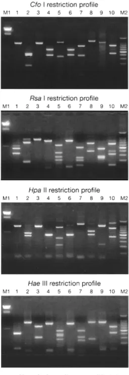
Cfo I restriction profile
M 1 1 2 3 4 5 6 7 8 9 1 0 M 2
Rsa I restriction profile
M 1 1 2 3 4 5 6 7 8 9 1 0 M 2
Hpa I I restrlctlon proftle
.afi
..a.
a.
M 1 1 2 , , 5 , 7 , 9 , 0 M 2 S 4 9 S-50 S-51 G52 G-53 C-54 G-55 Tx-56 Tx-S7 Tx-58me....
e
Tx-59 Tx-60 Tx-61 Tx-62 Tx-63 Tr-64 Tx-65 Tx-66 Tx-67 Tx-68 Tx-69 Tx-70 Tx-71 P Pmm .
Fiq 1 Dot hybridization of WSSV qeoqraphical chnical samples w t h (A) shrimp 1 8 s ~ R N A gene probe and (B)
%&'
probe derived from the ampli-Hae I l l restriction profile con Apms146Fl/Rl. The geographic origin of each DNA sample is indi-
cated above each dot (C: China; I: India; Cf: Crayfish; T: Thailand; S: South M 1 1 2 3 4 5 6 7 8 9 1 0 M 2 Carolina; G: grocery store; Tx: Texas), and the DNA sample numbers corre-
spond to those used in Tables 2 & 3. P: Taiwan clinical sample. Pm: healthy Penaeus monodon
M 1 2 3 4 5 6 7 8 9 1 0
Fig. 3. Restriction profiles of 10 amplicons amplified from A WSSV Taiwan clinical sample and cleared with cfo I, Rsa I,
Hpa 11, and Hae I11 nucleases. (Lane 1) Apms54Fl/Rl; (Lane 2) Apms94Fl/Rl; (Lane 3) Apms98Fl/Rl; (Lane 4) Apmsl2O Fl/Rl; (Lane 5) Apms146Fl/Rl; (Lane 6) Apms321Fl/Rl; (Lane 7) Apms321F2/R2; (Lane 8) Apms473F3/R3; (Lane 9) ApmhllFl/Rl; (Lane 10) Apmhl3Fl/Rl. M1: lambda phage
DNA Hind 111 fragments; M2: the 100 bp DNA ladder
Fiq. 2. Ten amplicons, Apmhl l F l / R l (Lane l), Apmhl3Fl/Rl When cleaved with Cfo I. Hae 111. Hwa I1 and Rsa I , A ( ~ a n e 21, A P ~ S ~ ~ F I / R ~ (Lane 3). ~ p m s g 4 ~ 1 / ~ 1 (Lane 41, restriction endonucleases, the RFLPs of China96-116A, Apms98Fl/Rl (Lane 5), Apmsl20Fl/Rl (Lane 6), Apms146
Fl/Rl (Lane 7). Apms321Fl/Rl (Lane 8), Apms321F2/R2 India95-314, crayfish97-25, Thailand95-46, South (Lane g), and Apms473F3/R3 (Lane 10), amplified from WSSV Car01ina97-64* grocery st0re96-1 l5 and Texas95-242 Taiwan clinicai sample. Lane M: pGEN
DNA
size marker were very similar, suggesting again that all 7 of thesel-step
PCR
M I 2 3 4 5 6 7 8 910 l1 1213 1415 161718192021 22232425262728293031 32333435363738394041 42434445464748495051 52535455505758596061 6263646566676860707lP 1431145 M I 2 3 4 5 6 7 8 91011 12131415I61718192021222324252627282930313233343536373839404142434445464748495051525354555657585960616263G;165GO676860707l P pms 146F 1 /R 1 000 bl I 2 3 4 5 h 7 8 91011 121314151617181920LI22232425262728293031323334353637383940414243444546474849505152535455%575859606162636465M6768607071l' pmh 11 FIIRl 4 M 1 2 3 4 5 6 7 8 91011 1213 1415 161718 192021 222324252627282930313233343536373839404142434445464748495051 52535455565758596061 6263h405(<)070869707IP.
pms 54FllRlI
pms 94FllRl(
M
1
2-step
PCR
,
M I 2m 5 6 7 8 91011 I213141516 718r ""~252627282930~33343536~39~42[ND~~40505152535155~6575859(~6I62b3646566676869707l1~ pms 98F2iR2Lo et al.: DNA analysis of white spot syndrome virus 183
Fig. 4. An~plification of amplicons 143/145, Apmsl46Fl/Rl. A p m h l l F l / R l , Apmhl3Fl/Rl, Apms54Fl/RI, Apms94Fl/Rl, Apms98 F l / R l , Apmsl20Fl/Rl, Apms321Fl/Rl, Apms321F2/R2, and Apms473F3/R3 by l-step PCR and Apms98F2/K2 by 2-step PCR from the WSSV geographical clinical samples indicated in Tables 2 & 3. (Lane number col-responds to DNA sample number.) M: PCR size marker. P: Taiwan WSSV. (*) Faint band with the expected size; (0) band with unexpected size. ND: not detected
Cfo I restriction profile
L. n 8 7 m n Y . .
..
Rsa I restriction profile M C I C f T S G T x P M
l
Hpa II restriction profileM C I C f T S G T x P M
Hae Ill restriction profile
M C I C f T S G T x P M
Fig. 5. Restriction profiles of Apmhl3Fl/Rl amplilied from China96-116A (Lane C), India95314 (Lane I), Crayfish97-25 (Lane Cf), Thailand9546 (Lane T), South CaroLina97-64 (Lane S), grocery store96-115 (Lane G ) ; Texas95-242-J60 (Lane Tx) and Taiwan WSSV (Lane P) cleaved with Cfo I, Rsa I, Hpa I1 and
Hae 111 endonucleases. M: pGEM DNA marker
Fig. 6. Southern blot hybridization analysis of PCR products. Ethibum bromide-stained agarose gel of PCR products of Apms98Fl/Rl (A) a n d Apmsl46Fl/Rl (C) are Southern blot hybridized with Apms98 F2/R2 probe (B) and Apms146F2/R2 probe (D), respectively. Lane numbers correspond to the DNA sample numbers used in Table 3
184 Dis Aquat Org 35: 175-185, 1999
WSSV geographical clinical samples belong to the results as a whole not only demonstrate the similarity same virus (group). Examples of RFLP analysis are of these clinical samples but also show that these clini-
shown in Fig. 5. cal samples can be easily detected using any of the 10
primer sets used for the present study.
The generally good yields seen in Fig. 4 suggest that
DISCUSSION all the primer sets annealed successfully with the DNA
from 48 (DNA samples 1 to 18 and 25 to 55) of WSSV
As in previous papers, the causal agent of the dis- clinical samples from 6 geographical areas (China ease or syndrome is referred to here as WSSV (white 96-116A, India95-314, crayfish97-25, Thailand95-46,
spot syndrome virus). However, as several viruses/ South Carolina97-64, grocery store96-115). This fur- baculoviruses have been described from the host ther suggests that there is very little sequence varia- species investigated here, Penaeus monodon (Lightner tion at least within the primer regions of these 6 WSSV 1996), it should be borne in mind that the possibility of geographical clinical samples. Furthermore, the 10
simultaneous infection by one or more viruses can not primer sets used in this study correspond to 10 entirely be completely excluded in the present studv. Accord- different DNA fragments of the entue WSSV genome.
ingly, since we did not purify the virus from the dis- These fragments, of approximately 1 kbp each, there- eased shrimp, the analyzed specimens were therefore fore constitute a reasonable sampling of the complete termed 'clinical samp!es1 ra!her than 'virus iso!atesl. In gcnome, so that, since the sizes of the mcijor PCR prod-
order that the impurity of the specimens might not lead ucts from all 6 WSSV geographical clinical samples
to difficulties in interpreting the results, the PCR using the same primer pair were very similar (Fig. 4), it
primers used here were derived from the genomic can be argued that substantial similarity exists in the
DNA extracted from purified virions of a WSSV Tai- genome structure as a whole.
wan isolate. These primer sequences (Table l ) , and For 2 Texas clinical samples, however, unexpected
even the fragment sequences from which they were reaction patterns were found: although the bands
derived, are unique. (unpubl. data from Dr Guang- shown by the Texas95-242 specimens were of the
Hsiung Kou's laboratory). These highly specific expected size, these bands were faint at best and, in
primers thus minimize the possibility of any interfer- many of the PCR tests, no PCR product was yielded at ence that might result from a mixed viral infection. all (Fig. 4 , Lanes 57 to 61). We therefore speculate that In the dot hybridization analysis, India95-314#3 and the virus in Texas95-242 specimens may well be dif-
crayfish97-25#1 (Table 2) failed to hybridize with some ferent from the Asian WSSV clinical samples. Further
probes. These anomalous results might have been sequence analysis will be required to confirm or refute
caused by the virus content in these specimens being this hypothesis.
very low. Alternatively, some mutants may have Texas96-7 specimens, on the other hand, yielded
existed in shrimp collected from the same geographi- faint PCR products in the tests with primer sets cal location or even from the same culture farm. The p m h l l F l / R l , pms94Fl/Rl, pms98Fl/R1 and pms321
PCR tests using 10 primer sets (Fig. 4, Lanes 16 to 18, F2/R2 but all of them had unexpected sizes (Fig. 4, 25, and 26), however, suggest that the virus in these Lanes 62 to 71). With 2-step PCR (Fig. 4) and Southern specimens is similar to other geographic clinical sam- hybridization (Fig. 6), however, we found at least some ples (e.g. China96-116, Thailand95-46, South Caro- bands with expected sizes in all of the Texas speci-
lina97-64), so that a low virus content in the samples is mens. Although this suggests that the Texas specimens very likely to have been the cause of the failure in the were all infected by WSSV or a WSSV-like virus, the
hybridization tests. evidence of the PCR reaction patterns argues that the
As shown in Fig. 4, the PCR tests using the 10 primer major virus population of Texas WSSV might not be sets derived from a WSSV Taiwan isolate provided identical to the other geographic clinical samples. In clear evidence that all the specimens of China96-116A, particular, the unexpectedly small size of the major
India95-314, crayfish97-25, Thailand95-46, South Apms98Fl/Rl product yielded by the Texas96-7 speci- Carolina97-64, and grocery store96-115 were infected mens (Fig. 6, Lanes 62 to 71) and its failure to hybridize
with WSSV or a WSSV-like virus. Almost all the ampli- with the Apms98F2/R2 probe suggest this amplicon
cons of the 10 primer sets from these DNA samples has little sequence homology to the expected Apms98
appeared as a single band with the expected sizes. Fl/Rl PCR product. Before further study can resolve
Even though a few primer sets gave rise to PCR prod- this question, however, the virus will need to be puri- ucts from WSSV negative DNA samples (e.g. grocery fied. PCR analysis could then check whether the PCR store95-204 which were somewhat degraded in stor- products of unexpected size still exist. If these anom- age) as either multiple bands (e.g. pms321Fl/Rl) or alous amplicons, were still found, we would expect single bands of unexpected size (e.g. pms98Fl/Rl), the DNA sequencing to reveal a low homology with the
Lo et a1 : DNA analysis of white spot syndrome virus 185
Asian isolates a n d that the genornic restriction profiles would b e distinct. This work would b e time consum- ing, but we believe it is important for the anomalies reported here to b e properly accounted for.
Acknowledgements. Thls work was supported by the National Sclence C o u n c ~ l Grant no. NSC86-2311-B-002-051 and Grant no NSC87-2311-B-002-070; Gulf Coast Research Laboratory Consortium M a n n e Shrimp Farmlng Program, CSREES, U. S. Dept. Agriculture Grant no. 95-38808-1424, National Sea Grant Program, U.S. Dept. of Commerce Grant no. NA56RG0617; and the National Fishery Institute. We are indebted to Mr Paul Barlow for his helpful criticism of the manuscript.
LITERATURE CITED
Chou HY, Huang CY, Wang CH, Chiang HC, Lo CF (1995) Pathogeniclty of a baculovirus infection causing white spot syndrome in cultured penaeid s h n m p in Taiwan. Dis Aquat Org 23:165-173
Chou HY, Huang CY, Lo CF, Kou GH (1998) Studies on trans-
mission of white spot syndrome associated baculovirus
(WSBV) in Penaeus monodon and P japonicus via water- borne contact and oral mgeshon Aquaculture 164:263-276 Flegel TW (1997) Major viral &seases of the black tiger prawn (Penaeus monodon) in Thailand. In. Inui Y (ed) New approaches to viral diseases of aquatic animals. NRIA International Workshop proceedings National Research Instltute of Aquaculture, Nansei, Wataral, Mle, J a p a n , p 167-187
Kim W, Abele LG (1990) Molecular phylogeny of selected decapod crustaceans based on 18s rRNA nucleotlde sequences. J Crust Biol 10: 1-13
Lightner DV ( e d ) (1996) A handbook of pathology and diag- nostic procedures for diseases of penaeid shnmp. World Aquaculture Society, Baton Rouge, LA, section 3.1 1 Lo CF, Leu J H , Ho CH, Chen CH, Peng SE, Chen YT, Chou Editorial responsibility. Larry Vaughan,
Arlington, Massachusetts, USA
CM, Yeh PY, Huang CJ, Chou HY, Wang CH, Kou G H (1996a) Detection of baculovirus associated with white spot syndrome (WSBV) in penaeld shrimps using poly- merase chain reaction Dis Aquat Org 25:133-141 Lo CF, Ho CH, Peng SE, Chen C H , Hsu HC, Chlu YL, Chang
CF, Liu KF, Su MS, Wang CH, Kou GH (1996b) Whlte spot syndrome baculov~rus (WSBV) detected in cultured and captured shrimp, crabs and other arthropods. DIS Aquat Org 27:212-225
Lo CF, Wang CH, Kou GH (1997) White spot syndrome (WSS): pathology, hosts and prevalence in captured shrimp a n d crabs in Taiwan. In: Inui Y ( e d ) New approaches to viral diseases of aquatic animals. NRIA International Workshop proceedings. National Research Instltute of Aquaculture, Nansei, Watarai, Mie, J a p a n , p 206-217
Wang CH, Lo CF, Leu J H , Chou CM, Yeh PY, Chou HY, Tung MC, Chang CF, Su MS, Kou G H (1995) Purification and genomic analysis of baculovirus associated with whlte spot syndrome (WSBV) of Penaeus rnonodon. Dis Aquat Org 23:239-242
Welsh J, McClelland M (1990) Fingerprinting genomes using PCR with arbitrary primers. Nucleic Acids Res 18(24):7213-7218
Wilharns CL, Goldson SL, Balrd DB, Bullock DW (1994) Geo- graphical origin of an introduced insect pest, Listronotus bonariensis (Kuschel), determined by RAPD analysis. Heredlty 72:412-419
W l h a m s J G , Kubelik AR, Livak KJ, Rafalski JA, Tingey SV (1990) DNA polymorph~sms amplified by arbitrary primers are useful as genetic markers. Nucleic Acids Res 18(22): 6531-6535
\Yilllams J G , Relter RS, Yong RM, Scolnik PA (1992) Genetic mapping of mutations uslng phenotyplc pools a n d mapped RAPD markers. Nucleic Acids Res 21(11):2697-2702 Wongteerasupaya C , Vlckers JE, Sriurairatana S, Nash GL,
Akarajamorn A, Boonsaeng V, P a n y m S, Tassanakajon A, Withyachumnarnkul B, Flegel TW (1995) A non-occluded, systemic baculovirus that occurs In cells of ectodermal a n d mesodermal ongin and causes high mortality in the black tiger prawn Penaeus rnonodon. Dls Aquat Org 21:69-77 Submitted. January 15, 1998; Accepted: November 10, 1998 Proofs received from author(s): February 22, 1999