Novel approach by nanobiomaterials in vascular tissue engineering
Huey-Shan Hung*¥, Hui-Chen Chen¥, Chang-Hai Tsai§,¶, Shinn-Zong Lin*,†,6
*
Center for Neuropsychiatry, China Medical University and Hospital, Taichung, Taiwan, R. O. C. ,
¥
Graduate Institute of Basic Medical Science, China Medical University, Taichung, Taiwan. R. O. C.
†
Graduate Institute of Immunology, China Medical University, Taichung, Taiwan, R. O. C.
§
Department of Pediatrics, China Medical University Hospital, Taichung, Taiwan, R. O. C.
¶
Department of Healthcare Administration, Asia University, Taichung, Taiwan
6
China Medical University Beigang Hospital, Yunlin, Taiwan, R. O. C.
Address correspondence to Shinn-Zong Lin, MD., Ph. D., Center for Neuropsychiatry, China Medical University and Hospital, Taichung, Taiwan. Tel: 886-4-22052121; Fax: 886-4-220806666; E-Mail: [email protected].
ABSTRACT
Interactions between vascular endothelial cells (EC) and biomaterials are important for engineered tissue substitute. The modification of biomaterial surfaces are designed to modulate EC adhesion and responses in order to improve implantation success rate. Specifically, it has now been well established that increased vascular tissue regeneration can be achieved on almost any surface by employing novel nano-fabricated surface features. To enhance EC adhesion and growth, material surface have been modified with physico-chemical and mechanical properties, such as bioactive molecules from the matrix, peptides and/or growth factors to control EC behavior. The advances in nanotechnology can bring additional functionality to vascular tissue engineering, optimize internal vascular graft surface, help to direct the differentiation of stem cells into the vascular cell phenotype, and most importantly, also provide a biomaterials-based cellularization process. Nanomaterials could promote in situ endothelialization by mobilizing endothelial progenitor cells (EPC) from the bone marrow, by encouraging cell-specific adhesion to the vascular graft and, once attached, by controlling the proliferation and differentiation of these cells. Interaction between different cell types and extracellular matrix continue to be a principal source of inspiration for material biological function and therefore, the understanding of the molecular mechanism trigger by the interaction is discussed.
A. Introduction
Synthetic materials used in vascular conduits applications has been indicated to be prone to clot and fail, and do not function well in a longer term. The urgent need in clinic has driven the search for a better and more biologically compatible blood vessel replacement. Numerous approaches have been used to fabricate the structure and the function of blood vessels [20,38,50]. The vascular tissue engineering approach is to use a good biocompatibility material that is seeded with cells. The advance in nanotechnology brings additional functionality to vascular conduits, it optimizes internal vascular grafts surface and even helps to direct the differentiation of stem cells into the vascular phenotype [1,26]. Nanotechnology also offers the key to understand the surface properties of materials on devices such as valves and stents. That promisingly improves biocompatibility of the device by regulating cell adhesion ability and inhibiting thrombosis and formation of blood clots [5]. There are plenty of evidences showing that development of nanotechnology-based methods in vascular tissue biofabrication represents one of the most important technological breakthroughs in vascular tissue engineering.
Nanostructuralized scaffolding can mimic the organization of a natural vascular extracellular matrix (ECM) and thus improve cell adhesion ability [38]. Several observations indicated that biomaterials seeded with differentiated endothelial cells (ECs) [15,30] or ECs in combination with other cell types such as smooth muscle cells (SMC) [56] promoted vascular tissue repairing after artificial grafts implantation. In order to improve the technology in vascular grafts, we need the knowledge and expertise in tissue engineering. Regarding previous findings, nanotechnology approaches seemed to exhibit an important role in the endothelialization processing, including endothelial cell migration, adhesion, proliferation and differentiation.
Therefore, this article will exploit nano-biomaterials in vascular tissue engineering that provide a mimic condition of the vascular tissue suitable for endothelial cell adhesion.
B. Nanoscale topography and vascular tissue
It is of interest to note that nano-structuralized biomaterials can modify the organization of a natural vascular ECM and thus improve cell attachment [2]. One explanation has provided by the evidence that nano-structured surface features can significantly improve vascular cell proliferation; such design criteria can be used in the synthesis of the more successful tissue-engineered vascular grafts [36]. For example, hydrogels have been modified with growth factors and ECM peptides on nano-structure biomaterials, which brought additional functionalities and improved the capacity of the hydrogel to direct cell differentiation and tissue regeneration [5,42]. Indeed, surface-aminated electrospun nanofibers that enhance adhesion and expansion of human umbilical cord blood hematopoietic stem progenitor cells has been observed [8]. Additionally, a study also shown that ECs, when cultured on gelatin-grafted electrospun poly(caprolactone) (PCL) nanofiber, were able to maintain the expression of three characteristic markers: platelet-endothelial cell adhesion molecule 1 (PECAM-1), intercellular adhesion molecule 1 (ICAM-1), and vascular cell adhesion molecule 1 (VCAM-1) [33]. The nano-bionic surface modified small intestinal submucosa (SIS) film possesses good and persistent antithrombogenicity, and also exhibits excellent blood compatibility [17]. Experiment was also performed to show that the collagen-blended poly(L-lactic acid)-co-poly(epsilon-caprolactone) nanofibers [P(LLA-CL) NFM] could enhance the viability, spreading and attachment of ECs, and moreover, preserve the ECs phenotype. The P(LLA-CL) NFM is a
potential material for tissue engineered vascular graft [19]. Indeed, the polyhedral oligomeric silsesquioxane attached by direct reaction onto a urethane segment (UCL-Nano) has been demonstrated to have similar viscoelastic properties to the walls of a natural artery which is resistant to degradation and is able to sustain endothelial cell seeding [42]. For example, endothelial progenitor cells respond to ridge-groove grating of 1200nmin period and 600nmin depth on nanotopography substrate with promoted alignment and elongation, reduced proliferation and enhanced migration [4]
Cells are known to be surrounded by nanoscale topography in their natural extracellular environment. It was observed that this surfaces comprise nanopatterned gratings with poly(methyl methacrylate) (PMMA) and PDMS can provide a valuable platform for the study of cell-substrate interactions and for the development of medical devices with nanoscale features of smooth muscle cells (SMC) [56]. It has been suggested that synthetic aligned poly(L-lactid-co-epsilon-caprolactone) [P(LLA-CL)] copolymer combines with the advantages of synthetic biodegradable polymers may represent an ideal tissue engineering scaffold, especially for blood vessel engineering [55]. Recently, synthetic poly-L-lactic acid (PLLA) nanofibers have been shown to not only improve biocompatible with outgrowth endothelial cells (OEC) but also promote and guide aligned OEC sustained proliferation [31]. Based on these findings, the aligned PLLA, that can serve as the scaffolds and promote cell growing during endothelialization of biomaterials, can provide a promising way to prevent intimal hyperplasia of small-diameter vascular grafts.
Engineered vascular grafts with polyhedral oligomeric silsesquioxane modified polycarbonate urea-urethane (POSS-PCU) that was crossed link with a bioactive RGD peptide and formed a biofunctionalised nanocomposite polymer-based small diameter
bypass graft, has been shown to have the potential for rapid endothelialization [12]. A similar approach using poly (carbonate) urethane-based nanocomposite polymer incorporating polyhedral oligomeric silsesquioxane (POSS)nanocages (UCL-NANO) showed anti-thrombogenicity and biostability as well as exhibited a good small calibre quadruple lamina vascular bypass grafts method [46].
C. Molecular and cellular interaction between nanobiomaterials and vascular microenvironment
Nanomaterials are known to be important in vascular tissue application. However, how the molecular signaling and its biological function is controlled are still unclear. It is extremely important to note that developing such biomaterials requires the modification of materials that mimic molecular mechanism of extracellular matrices [18,20]. These associated signal molecules could promote vascularization by the mobilization of ECs, induction of cell-specific adhesion to the vascular graft and, once attached, by controlling the proliferation and differentiation of these cells. The benefit from the nanobiomaterials is considered as the advantageous characteristics of the natural extracellular matrix for tissue repair processing [55]. It is likely that cells adhere to the ECM induces intracellular signaling cascades and migration of integrins into focal adhesion complexes [21]. For example, evidence indicated that manufactured aluminum oxide nanoparticles decrease the expression of tight junction proteins in brain vasculature. [6]. Indeed, cupper-nanoparticles is highly useful for regenerating of elastin matrices by adult vascular smooth muscle cells (SMCs). These results have immense utility in tissue-engineering vascular replacements [27]. There was further evidence suggesting that nanostructured hydroxyapatite (HA) crystals up-regulates FGF-2 expression and
activity in microvascular endothelium promoting angiogenesis in 3D matrices [41]. Current issues in nanotechnology and molecular self-assembly may provide novel solutions to cell transplantation deficiencies. An observation has shown that the delivery of vascular endothelial growth factor and fibroblast growth factor-2 from heparin-binding peptide amphiphiles (HBPAs) scaffolds significantly increases blood vessel density in the mouse omentum over control scaffolds without growth factors and significantly promotes islet engraftment [49]. The in vitro study demonstrated that cell adhesive proteins adsorbed (vitronectin and fibronectin) in different quantities and altered bioactivity on nanostructured which, at least in part, may account for the documented increased vascular cell adhesion on nanostructured PLGA [37].
A study from neovascuature has shown that nanosphere-aided targeting of the neovasculature with mutant Raf-1 causes regression of tumor progression [52]. Based on this finding, it seems reasonable to conclude that the nanoparticles coated with specific biological molecules may be useful for delivering signal molecules, which can cause the regression of tumor progression. The use of viscoelastic nanocomposites vascular conduit by exposing to shear stress preconditioning prior to physiological flow can increase endothelial growth factor receptor-2 (VEGFR-2) and transforming growth factor beta-1 (TGFbeta-1) on ECs [51]. Indeed, another approach also indicated that nanofibrous materials made from bioabsorbable and biocompatible polymers have promising applications as tissue-engineered scaffolds. Genetic analysis of human umbilical vein endothelial cells (HUVEC) that attached to Poly(glycolide) (PGA) nanofibrous materials prepared via electrospinning methods exhibited high expression of integrin v and VEGF receptor genes, which are known angiogenesis markers [24].
It is also of interest to note that conjunction with microvascular dysfunction, nanoparticle exposure also reduces nitric oxide (NO) activation through at least two functional distinct mechanisms that may mutually increase local reactive species [39]. A report also bring the attention that PU-Au nanocomposites activates FAK and PI3K/Akt signaling pathway in ECs which leads to proliferation and migration of ECs on a series of nanocomposites from polyurethane (PU) incorporated with various gold nanoparticles [23]. Other report also suggested that the nanometric micelles on PU surface may interact with ECs and accelerates their migration by increasing cytoplasmic Ca2+ and stimulating the PI3K/Akt/eNOS signaling pathway [22]. Indeed, carbon nanoparticles and microparticles have the ability to induce platelets activation and enhance vascular thrombosis, and this observation will be important for the pharmacological application [43]. These reports lead to the conclusion that biomaterials can successfully integrate into tissue's mechanical properties and material topography. The cellular molecular mechanism to a biomaterial may be enhanced in synthetic vascular conduits by mimicking the surface roughness that is created by the associated nano-structured extra-cellular matrix components of natural tissue [36].
D. Perspective on nanotechnology in manipulating vascular stem cells
Although stem cells hold great potential for the vascular tissue engineering application, the development of advanced and novel method to track and guide transplanted stem cells should be more detailed addressed [48]. For example, nanopatterns on the surface of a vascular graft with nanofilms can increase its adhesion for circulating endothelial progenitor cells [12]. Another way to influence stem cell differentiation and transplantation is engineered nanometer-scale scaffolds
[14]. For instance, functional nanofiber scaffolds with different spacers modulate adhesion and expansion of cryopreserved umbilical cord blood hematopoietic stem/progenitor cells [7]. This is especially noteworthy in the case of scaffold topography and cell-substrate interaction that regulate hematopoietic stem and progenitor cells (HSPC) proliferation and self-renewal in cytokine-supplemented expansion.
The formation of an endothelial cell layer on the luminal surface of vascular grafts is an important attribute to improve their potential for endothelialization. The formation of blood vessels is mainly due to the activity of endothelial progenitor cells (EPCs) [3]. It is of interest to note that nanocomposite-containing bioactive peptides could promote adhesion, spreading and formation of a confluent endothelial monolayer by circulated progenitor cells [1].
E. Application of nanoparticles for tracking stem cells
Many applications of new cellular magnetic labeling method to EPC exhibited therapeutic potential for revascularization. One report indicated that when magnetic nanoparticle-incorporated EPC was injected into a microfluidic channel by using a syringe pump can control the flow rate of cells [25]. Magnetic cell delivery and targeting offer a new opportunity for vascular tissue engineering and delivery of localized cell-based therapies. Using a magnetically tag device at a site of artery injury is able to enhance EPC localization [28]. This technology could be more adapted to trace cells in other organs and may provide a novel approach for the systemic injection of cell therapies in vascular tissue engineering application. When EPC is labeled with monocrystalline iron oxide nanoparticles (MIONs), it doesn’t adversely change their viability and its migration ability in vitro, and this allowed
successful detection of limited numbers of these cells in muscle [29]. MION labeled EPCs provides a non-invasive strategy to monitor the location of small signal molecules of these cells.
There is a technique using advanced ultra high-field 7-Tesla (7T) MRI of nanoparticle-labeled transplanted human EPCs in porcine ischemic hearts for clinical application [45]. Indeed, another report also point out that when magnetically labeled EPCs transplanted for therapeutic neovascularization in myocardial ischemia can be visualized by MRI [54]. It has been observed that self-synthesized superparamagnetic iron oxide particles (PLL-SPIO) can effectively improve EPC mobilization and proliferation [34]. Therefore, it can be assumed that PLL-SPIO can serve as a tracker for implanted EPCs.
F. Nanobiomaterials for the delivery of vascular genetic molecules
Strategies for vascular conduits application has been focused on the delivery of angiogenic growth factors such as vascular endothelial growth factor and fibroblast growth factor-2 to induce angiogenesis progress from the host vasculature [32,35]. Nanofibrous materials made from bioabsorbable and biocompatible polymers are promising applications as tissue-engineered scaffolds [9]. The nanocell comprising a nuclear nanoparticle within an extranuclear pegylated-lipid envelope is preferentially taken up by the tumor. The technology can be extended by using additional agents aimed to target multiple signaling pathways, such as hypoxia-inducible factor-1alpha (HIF1-alpha) as well as a temporal targeting of tumor cells [47]. Having observed in a NOD/SCID mouse hind limb vascular injury model, nanofiber-expanded cells exhibited more therapeutic potential in blood flow restoration after further applying VEGF (164), PDGF-BB and growth factor [10]. Additionally, overexpression of
proangiogenic growth factor can promote the cellular therapeutic efficiency in EPCs for ischemic disease [58]. In summary, nanofiber-based ex vivo expansion technology can provide a novel therapeutic strategy for stem cells in vascular tissue engineering. Moreover, the immobilization of CD34 antibodies on a lumenal vascular stent enhanced recruitment of circulated cells and accelerated post-implantational endothelialization in vitro [44].
G. Future prospects
The incorporation of vascular tissue engineering, stem cell application and nanotechnology research can improve regenerative medicine for current clinical application. Recently, the application of stem cell to vascular tissue engineering has undoubtedly become a novel technology. Having developed a fully biological vascular graft, functional concerns about its biological compatibility and mechanical property have become the focus of research effort. Although the main goal of nanotechnology is to enhance vascular tissue repairing ability, it is unclear how nano-biomaterials minics native properties of blood vessel grafts in a long-term implantation in vivo and this will be the important issue to be further pursued for a new therapy strategy. For example, development of genetic engineering methods [47,52] and the variety of cell types which are able to express signaling molecule [6,22,23,39], proteins [27,37], peptides [49] and growth factors [13,32,35,58] all show great promise for modification of vascular nano-biomaterials. However, a more advanced knowledge on the modulation of molecular mechanism will provide a medium closely mimicking the physiological environment within an engineered vascular graft. This may advance a better biocompatibility, ultimately provides a more durable vascular nano-biomaterials. Alternatively, advanced nano-biomaterials
fabrication and application will be essential for more functional vascular tissue in the regenerative medicine. For example, modification of nano-biomaterials has been shown to have a lower degradation rate in vivo, and this promotes the host immune response [57]. It remains to be a great concern for the trace amounts of potentially antigenic compounds of animal origins (lipids, DNA, glycosilation products) which have been reported to be present in various types of nano-biomaterial and may induce an inflammatory response at the implantation site [11,16].
A recent study has demonstrated that the tissue-specific matrix components cause significant different adhesion efficiency and phenotype [31,40] of vascular tissue, as well as endothelial cells. These observations suggest the need for more appropriate, tissue-specific nano-biomaterials from in vitro to in vivo study. Indeed, utilization of nano-biomaterials in clinical application may provide a likely successful vascular tissue regeneration for the patient. It is worthy of consideration that circulating hematopoietic blood stem cells from bone marrow [53], umbilical cord blood [7] or peripheral blood [12] have a high proliferative and regenerative potential, and they may be an optimal and ideal cell sources for mimicking in vivo endothelialization of synthetic nano-biomaterials. However, due to the heterogeneity of hematopoietic stem cells populations and the definition of stem cells surface molecules are still poorly identified, success of this approach depends on a better characterization and understanding of hematopoietic stem cell biology and property.
Conclusion
Nanotechnology has become an attractive method for the vascular tissue engineering from the view point of biology, i.e. stem cells, extracellular matrix, growth factors and signaling molecules. A range of nano-biomaterials have been
shown to provide an excellent biocompatibility for many processes that occur in the early stages of vessel repairing and allowing a long-term maintenance of neo-vascular tissue development. The endothelialization of synthetic nano-biomaterials is a promising therapeutic approach to create a non-thrombogenic barrier on surfaces, and could thereby improve the efficiency of vascular prosthesis of blood contacting devices. The design of athrombogenic nanosurfaces on vascular grafts and the development of nanopatterned biomimicking vascular grafts are already well established. The use of nanopatterned functionalized biomaterials for the expansion and directed differentiation of human stem cells into cells of the vascular lineage is a fast evolving and promising technology. The development of novel methods of vascular tissue assembly and rapid vascular biofabrication, such as magnetic-force-driven tissue engineering and simultaneous electrospinning of vascular scaffolds and living cells, represent probably the most important breakthroughs to date. Based on the above finding, evidencestrongly indicate the potential of nanotechnology in vascular tissue engineering for the development of successful and effective vascular-tissue engineered products. Immobilization of homing factor-mimetic molecules onto synthetic nano-biomaterials to increase the production of EPCs from circulating blood opens a new therapeutic strategy in regenerative medicine. This concept is based on the understanding that every patient will be able to seed his implants with his own progenitor cells after implantation, and this technology may bring new therapeutic strategy for vascular tissue engineering.
For reasons mentioned above, the application of signal molecules that can be incorporated into functional development of nano-biomaterials for regulatory of stem cell mobilization of the vascular biology. With the incorporation of the nanotechnology, biomaterial surface modification and stem cell biology for
developing a novel therapeutic strategy could be more useful advanced for the vascular tissue engineering.
REFERENCES
1. Alobaid, N.; Salacinski, H. J.; Sales, K. M.; Ramesh, B.; Kannan, R. Y.;
Hamilton, G.; Seifalian, A. M. Nanocomposite containing bioactive peptides
promote endothelialisation by circulating progenitor cells: an in vitro evaluation.
“Eur J Vasc Endovasc Surg.” 32:76-83; 2006.
2. Alsberg, E.; Feinstein, E.; Joy, M. P.; Prentiss, M.; Ingber, D. E.
Magnetically-guided self-assembly of fibrin matrices with ordered nano-scale
structure for tissue engineering. “Tissue Eng” 12:3247-3256; 2006.
3. Asahara, T.; Murohara, T.; Sullivan, A.; Silver, M.; van der Zee, R.; Li, T.;
Witzenbichler, B.; Schatteman, G.; Isner, J. M. Isolation of putative progenitor
endothelial cells for angiogenesis. “ Science” 275:964-967; 1997.
4. Bettinger, C. J.; Zhang, Z.; Gerecht, S.; Borenstein, J. T.; Langer, R.
Enhancement of In Vitro Capillary Tube Formation by Substrate
Nanotopography. “Adv Mater Deerfield” 20:99-103; 2008.
5. Chai, C.; Leong, K. W. Biomaterials approach to expand and direct
differentiation of stem cells. “Mol Ther” 15:467-480; 2007.
6. Chen, L.; Yokel, R. A.; Hennig, B.; Toborek, M. Manufactured aluminum oxide
nanoparticles decrease expression of tight junction proteins in brain vasculature.
“J Neuroimmune Pharmacol” 3:286-295; 2008.
Functional nanofiber scaffolds with different spacers modulate adhesion and
expansion of cryopreserved umbilical cord blood hematopoietic stem/progenitor
cells. “Exp Hematol” 35:771-781; 2007.
8. Chua, K. N.; Chai, C.; Lee, P. C.; Tang, Y. N.; Ramakrishna, S.; Leong, K. W.;
Mao, H. Q. Surface-aminated electrospun nanofibers enhance adhesion and
expansion of human umbilical cord blood hematopoietic stem/progenitor cells.
“Biomaterials” 27:6043-6051; 2006.
9. Chung, S.; Moghe, A. K.; Montero, G. A.; Kim, S. H.; King, M. W. Nanofibrous
scaffolds electrospun from elastomeric biodegradable
poly(L-lactide-co-epsilon-caprolactone) copolymer. “Biomed Mater” 4:15019;
2009.
10. Das, H.; Abdulhameed, N.; Joseph, M.; Sakthivel, R.; Mao, H. Q.; Pompili, V. J.
Ex vivo nanofiber expansion and genetic modification of human cord
blood-derived progenitor/stem cells enhances vasculogenesis. “Cell Transplant”
18:305-318; 2009.
11. Daly, K. A.; Stewart-Akers, A. M.; Hara, H.; Ezzelarab, M.; Long, C.; Cordero, K.;
Johnson, S. A.; Ayares, D.; Cooper, D. K.; Badylak, S. F. Effect of the alphaGal
epitope on the response to small intestinal submucosa extracellular matrix in a
12. de Mel, A.; Punshon, G.; Ramesh, B.; Sarkar, S.; Darbyshire, A.; Hamilton, G.;
Seifalian, A. M. In situ endothelialization potential of a biofunctionalised
nanocomposite biomaterial-based small diameter bypass graft. “Biomed Mater
Eng” 19:317-331; 2009.
13. Divya, P.; Sreerekha, P. R.; Krishnan, L. K. Growth factors upregulate deposition
and remodeling of ECM by endothelial cells cultured for tissue-engineering
applications. “Biomol Eng” 24:593-602; 2007.
14. Ferreira, L.; Karp, J. M.; Nobre, L.; Langer, R. New opportunities: the use of
nanotechnologies to manipulate and track stem cells. “Cell Stem Cell” 3:136-146;
2008.
15. Gafni, Y.; Zilberman, Y.; Ophir, Z.; Abramovitch, R.; Jaffe, M.; Gazit, Z.; Domb,
A.; Gazit, D. Design of a filamentous polymeric scaffold for in vivo guided
angiogenesis. “Tissue Eng” 12:3021-3034; 2006.
16. Gilbert, T. W.; Freund, J. M.; Badylak, S. F. Quantification of DNA in biologic
scaffold materials. “J Surg Res” 152:135-139; 2009.
17. Han, B. S.; Fan, C. Y.; Liu, S. H. Improvement of blood compatibility of small
intestinal submucosa used as engineering vascular scaffolds by nano-bionic
surface modification. “Zhonghua Yi Xue Za Zhi” 86:2065-2068; 2006.
extracellular matrix. “Nanomedicine” 2:37-41; 2006.
19. He, W.; Ma, Z.; Yong, T.; Teo, W. E.; Ramakrishna, S. Fabrication of
collagen-coated biodegradable polymer nanofiber mesh and its potential for
endothelial cells growth. “Biomaterials” 26:7606-7615; 2005.
20. He, W.; Yong, T.; Teo, W. E.; Ma, Z.; Ramakrishna, S. Fabrication and
endothelialization of collagen-blended biodegradable polymer nanofibers:
potential vascular graft for blood vessel tissue engineering. “Tissue Eng”
11:1574-1588; 2005.
21. Hodde, J.; Record, R.; Tullius, R.; Badylak, S. Fibronectin peptides mediate
HMEC adhesion to porcine-derived extracellular matrix. “Biomaterials”
23:1841-1848; 2002.
22. Hung, H. S.; Hsu, S. H. The response of endothelial cells to polymer surface
composed of nanometric micelles. “N Biotechnol” 25:235-243; 2009.
23. Hung, H. S.; Wu, C. C.; Chien, S.; Hsu, S. H. The behavior of endothelial cells on
polyurethane nanocomposites and the associated signaling pathways.
“Biomaterials” 30:1502-1511; 2009.
24. Igarashi, S.; Tanaka, J.; Kobayashi, H. Micro-patterned nanofibrous biomaterials.
“J Nanosci Nanotechnol” 7:814-817; 2007.
progenitor cells to a specific location within a microfluidic channel using
magnetic nanoparticles. “Biomed Microdevices” 11:287-296; 2009.
26. Kofidis, T.; Muller-Stahl, K.; Haverich, A. Myocardial restoration and tissue
engineering of heart structures. “Methods Mol Med” 140:273-290; 2007.
27. Kothapalli, C. R.; Ramamurthi, A. Copper nanoparticle cues for biomimetic
cellular assembly of crosslinked elastin fibers. “Acta Biomater” 5:541-553;
2009.
28. Kyrtatos, P. G.; Lehtolainen, P.; Junemann-Ramirez, M.; Garcia-Prieto, A.; Price,
A. N.; Martin, J. F.; Gadian, D. G.; Pankhurst, Q. A.; Lythgoe, M. F. Magnetic
tagging increases delivery of circulating progenitors in vascular injury. “JACC
Cardiovasc Interv” 2:794-802; 2009.
29. Li Calzi, S.; Kent, D. L.; Chang, K. H.; Padgett, K. R.; Afzal, A.; Chandra, S. B.;
Caballero, S.; English, D.; Garlington, W.; Hiscott, P. S. and others. Labeling of
stem cells with monocrystalline iron oxide for tracking and localization by
magnetic resonance imaging. “Microvasc Res” 78:132-139; 2009.
30. Li, J.; Ding, M.; Fu, Q.; Tan, H.; Xie, X.; Zhong, Y. A novel strategy to graft RGD
peptide on biomaterials surfaces for endothelization of small-diamater vascular
grafts and tissue engineering blood vessel. “J Mater Sci Mater Med”
31. Lu, H.; Feng, Z.; Gu, Z.; Liu, C. Growth of outgrowth endothelial cells on aligned
PLLA nanofibrous scaffolds. “J Mater Sci Mater Med” 20:1937-1944; 2009.
32. Luo, Z.; Asahara, T.; Tsurumi, Y.; Isner, J. M.; Symes, J. F. Reduction of vein graft
intimal hyperplasia and preservation of endothelium-dependent relaxation by
topical vascular endothelial growth factor. “J Vasc Surg” 27:167-173; 1998.
33. Ma, Z.; He, W.; Yong, T.; Ramakrishna, S. Grafting of gelatin on electrospun
poly(caprolactone) nanofibers to improve endothelial cell spreading and
proliferation and to control cell Orientation. “Tissue Eng” 11:1149-1158; 2005.
34. Mai, X. L.; Ma, Z. L.; Sun, J. H.; Ju, S. H.; Ma, M.; Teng, G. J. Assessments of
proliferation capacity and viability of New Zealand rabbit peripheral blood
endothelial progenitor cells labeled with superparamagnetic particles. “Cell
Transplant” 18:171-181; 2009.
35. Miki, A.; Rivas-Carrillo, J. D.; Navarro-Alvarez, N.; Soto-Gutierrez, A.; Chen, Y.;
Tanaka, K.; Narushima, M.; Tabata, Y.; Okitsu, T.; Noguchi, H. and others.
Maintenance of neovascularization at the implantation site of an artificial device
by bFGF and endothelial cell transplant. “Cell Transplant” 15:893-901; 2006.
36. Miller, D. C.; Thapa, A.; Haberstroh, K. M.; Webster, T. J. Endothelial and
vascular smooth muscle cell function on poly(lactic-co-glycolic acid) with
37. Miller, D. C.; Haberstroh, K. M.; Webster, T. J. Mechanism(s) of increased
vascular cell adhesion on nanostructured poly(lactic-co-glycolic acid) films. “J
Biomed Mater Res A” 73:476-484; 2005.
38. Nair, L. S.; Bhattacharyya, S.; Bender, J. D.; Greish, Y. E.; Brown, P. W.; Allcock,
H. R.; Laurencin, C. T. Fabrication and optimization of methylphenoxy
substituted polyphosphazene nanofibers for biomedical applications.
“Biomacromolecules” 5:2212-2220; 2004.
39. Nurkiewicz, T. R.; Porter, D. W.; Hubbs, A. F.; Stone, S.; Chen, B. T.; Frazer, D.
G.; Boegehold, M. A.; Castranova, V. Pulmonary nanoparticle exposure disrupts
systemic microvascular nitric oxide signaling. “Toxicol Sci” 110:191-203; 2009.
40. Park, J.; Bauer, S.; Schmuki, P.; von der Mark, K. Narrow window in nanoscale
dependent activation of endothelial cell growth and differentiation on TiO2
nanotube surfaces. “Nano Lett” 9:3157-3164; 2009.
41. Pezzatini, S.; Morbidelli, L.; Solito, R.; Paccagnini, E.; Boanini, E.; Bigi, A.;
Ziche, M. Nanostructured HA crystals up-regulate FGF-2 expression and activity
in microvascular endothelium promoting angiogenesis. “Bone” 41:523-534;
2007.
42. Punshon, G.; Sales, K. M.; Vara, D. S.; Hamilton, G.; Seifalian, A. M. Assessment
for seeding a novel vascular graft material. “Cell Prolif” 41:321-335; 2008.
43. Radomski, A.; Jurasz, P.; Alonso-Escolano, D.; Drews, M.; Morandi, M.; Malinski,
T.; Radomski, M. W. Nanoparticle-induced platelet aggregation and vascular
thrombosis. “Br J Pharmacol” 146:882-893; 2005.
44. Rotmans, J. I.; Heyligers, J. M.; Verhagen, H. J.; Velema, E.; Nagtegaal, M. M.; de
Kleijn, D. P.; de Groot, F. G.; Stroes, E. S.; Pasterkamp, G. In vivo cell seeding
with anti-CD34 antibodies successfully accelerates endothelialization but
stimulates intimal hyperplasia in porcine arteriovenous expanded
polytetrafluoroethylene grafts. “Circulation” 112:12-18; 2005.
45. Ruhparwar, A.; Ghodsizad, A.; Niehaus, M.; Bara, C.; Lotz, J.; Voelkel, T.;
Makoui, M.; Martin, U.; Wolf, F.; Gams, E. and others. Clinically applicable
7-Tesla magnetic resonance visualization of transplanted human adult stem cells
labeled with CliniMACS nanoparticles. “Thorac Cardiovasc Surg” 54:447-451;
2006. ulature with a nanoscale delivery system. “Nature” 436:568-572; 2005.
46. Sarkar, S.; Burriesci, G.; Wojcik, A.; Aresti, N.; Hamilton, G.; Seifalian, A. M.
Manufacture of small calibre quadruple lamina vascular bypass grafts using a
novel automated extrusion-phase-inversion method and nanocomposite polymer.
“J Biomech” 42:722-730; 2009.
Sasisekharan, R. Temporal targeting of tumour cells and neovasculature with a
nanoscale delivery system. “Nature” 436:568-572; 2005.
48. Solanki, A.; Kim, J. D.; Lee, K. B. Nanotechnology for regenerative medicine:
nanomaterials for stem cell imaging. “Nanomed” 3:567-578; 2008.
49. Stendahl, J. C.; Wang, L. J.; Chow, L. W.; Kaufman, D. B.; Stupp, S. I. Growth
factor delivery from self-assembling nanofibers to facilitate islet transplantation.
“Transplantation” 86:478-481; 2008.
50. Tsuda, Y.; Shimizu, T.; Yamato, M.; Kikuchi, A.; Sasagawa, T.; Sekiya, S.;
Kobayashi, J.; Chen, G.; Okano, T. Cellular control of tissue architectures using a
three-dimensional tissue fabrication technique. “Biomaterials” 28:4939-4946;
2007.
51. Vara, D. S.; Punshon, G.; Sales, K. M.; Sarkar, S.; Hamilton, G.; Seifalian, A. M.
Endothelial cell retention on a viscoelastic nanocomposite vascular conduit is
improved by exposure to shear stress preconditioning prior to physiological flow.
“Artif Organs” 32:977-981; 2008.
52. Wary, K. K. Signaling through Raf-1 in the neovasculature and target validation
by nanoparticles. “Mol Cancer” 2:27; 2003.
53. Webber, M. J.; Tongers, J.; Renault, M. A.; Roncalli, J. G.; Losordo, D. W.; Stupp,
“Acta Biomater” 6:3-11; 2010.
54. Weber, A.; Pedrosa, I.; Kawamoto, A.; Himes, N.; Munasinghe, J.; Asahara, T.;
Rofsky, N. M.; Losordo, D. W. Magnetic resonance mapping of transplanted
endothelial progenitor cells for therapeutic neovascularization in ischemic heart
disease. “Eur J Cardiothorac Surg” 26:137-143; 2004.
55. Xu, C. Y.; Inai, R.; Kotaki, M.; Ramakrishna, S. Aligned biodegradable
nanofibrous structure: a potential scaffold for blood vessel engineering.
“Biomaterials” 25:877-886; 2004.
56. Yim, E. K.; Reano, R. M.; Pang, S. W.; Yee, A. F.; Chen, C. S.; Leong, K. W.
Nanopattern-induced changes in morphology and motility of smooth muscle
cells. “Biomaterials” 26:5405-5413; 2005.
57. Zaveri, T. D.; Dolgova, N. V.; Chu, B. H.; Lee, J.; Wong, J.; Lele, T. P.; Ren, F.;
Keselowsky, B. G. Contributions of surface topography and cytotoxicity to the
macrophage response to zinc oxide nanorods. “Biomaterials” 31:2999-3007;
2010.
58. Zemani, F.; Silvestre, J. S.; Fauvel-Lafeve, F.; Bruel, A.; Vilar, J.; Bieche, I.;
Laurendeau, I.; Galy-Fauroux, I.; Fischer, A. M.; Boisson-Vidal, C. Ex vivo
priming of endothelial progenitor cells with SDF-1 before transplantation could
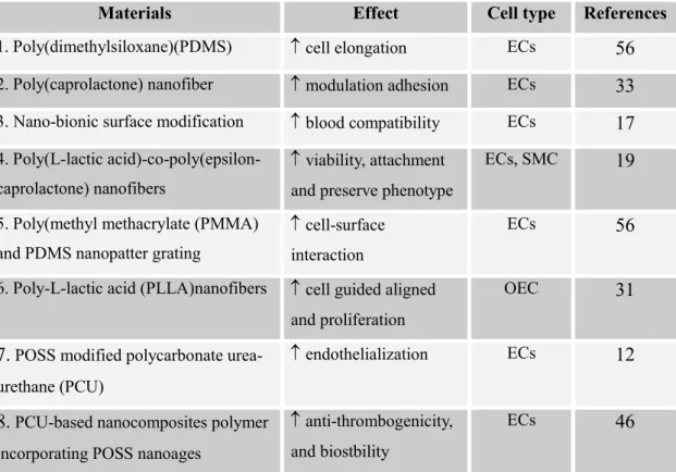
Table 1: Summary of key materials of vascular tissue engineering
Materials Effect Cell type References
1. Poly(dimethylsiloxane)(PDMS) ↑ cell elongation ECs 56
2. Poly(caprolactone) nanofiber ↑ modulation adhesion ECs 33
3. Nano-bionic surface modification ↑ blood compatibility ECs 17
4. Poly(L-lactic acid)-co-poly(epsilon- caprolactone) nanofibers
↑ viability, attachment and preserve phenotype
ECs, SMC 19
5. Poly(methyl methacrylate (PMMA) and PDMS nanopatter grating
↑ cell-surface interaction
ECs 56
6. Poly-L-lactic acid (PLLA)nanofibers ↑ cell guided aligned and proliferation
OEC 31
7. POSS modified polycarbonate urea- urethane (PCU)
↑ endothelialization ECs 12
8. PCU-based nanocomposites polymer incorporating POSS nanoages
↑ anti-thrombogenicity, and biostbility
ECs 46
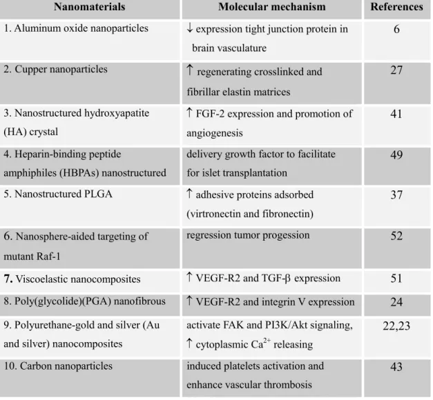
Table 2: Molecular and cellular interaction between nanobiomaterials and vascular microenvironment
Nanomaterials Molecular mechanism References
1. Aluminum oxide nanoparticles ↓ expression tight junction protein in brain vasculature
6
2.Cupper nanoparticles ↑ regenerating crosslinked and
fibrillar elastin matrices
27
3. Nanostructured hydroxyapatite (HA) crystal
↑ FGF-2 expression and promotion of angiogenesis
41
4. Heparin-binding peptide
amphiphiles (HBPAs) nanostructured
delivery growth factor to facilitate for islet transplantation
49
5. Nanostructured PLGA ↑ adhesive proteins adsorbed (virtronectin and fibronectin)
37
6. Nanosphere-aided targeting of mutant Raf-1
regression tumor progession 52
7. Viscoelastic nanocomposites ↑ VEGF-R2 and TGF-β expression 51
8. Poly(glycolide)(PGA) nanofibrous ↑ VEGF-R2 and integrin V expression 24
9. Polyurethane-gold and silver (Au and silver) nanocomposites
activate FAK and PI3K/Akt signaling, ↑ cytoplasmic Ca2+
releasing
22,23
10. Carbon nanoparticles induced platelets activation and enhance vascular thrombosis
43
PLGA: poly(lactic-co-glycolic acid); VEGF-R2: Vascular endothelial growth factor-receptor 2; TGF-ββββ: Transformer growth factor-beta; FAK: focal adhesion kinase; PI3K/Akt: Phosphatidylinositol
Figure legends
Figure 1: Schematic representation of the proposed mechanism by which nanomaterials contribute to the modulation of vascular tissue engineering. As a result of vascular tissue injuring, the nanomaterials (including nanoparticles, nanofibers, nanocarries, nanostructure pattern, nanosphere and nanofluore) trigger molecular mechanism by virtue of their expression of tight junction protein, adhesion molecules (e.g., FAK, integrin V), signaling molecules (e.g., PI3K/Akt, eNOS and Ca2+), growth factors (e.g., FGF-2, VEGF-R2 and TGF-β) and extracellular matrix (e.g., elastin, fibronectin and vitronectin). The interaction of endothelial cells with injure tissue may lead to activation of signal molecules that may contribute to the recovery of vascular injury (e.g., increase the adhesion, migration, proliferation, angiogenesis), promotion of drug delivery (e.g., regulatory of cancer) and tracing of stem cells stem cells differentiation. These molecular effects of nanomaterials contribute to restored vascular tissue regeneration of the injured area and result in the induction of endothelialization.
Figure 1
Tight junction protein
Adhesion molecules
(ex: FAK, Integrin V) Signaling molecules (ex: PI3K/Akt; eNOS; Ca2+; Raf-1)
Growth factors
(ex: FGF-2, VEGF-R2, TGF-β) Extracellular matrix
(ex:Elastin; Fibronectin; Vitronectin)
Vascular replacement
Endothelialization
Drug delivery Cell-surface interaction
(ex:Angiogenesis & Proliferation)
(ex: Regulatory of cancer) (ex: Adhesion & Migration)