Apigenin, 17α-estradiol 和flavone引起人類胃癌細胞株(SC-M1)生長的抑制和調節細胞週期相關基因的表現; Apigenin, 17α-estradiol and flavone induced growth inhibition and modulated cell cycle related gene expression in human stomach adenocarcinoma cell line (SC-M1)
107
0
0
全文
(2) 而成的,導致癌症形成的因子中飲食占 33%、抽煙占 30%、濾過性病毒感染占 10%、性行為占 7%以及酒精、工業化職業、遺傳、環境污染、放射線、紫外線、 食品添加物、不明原因等,而依流行病學統計及研究報告顯示,有超過 80%以上 的人類癌症與化學致癌物有關(4)。而暴露在職業和環境中的化學致癌物有芳香胺 類(arylamines)、多環芳香胺類、無機化合物、有機化合物、食品添加物及其他天 然性的致癌物,如黃麴毒素(aflatoxin)、紫外線… 等。在芳香胺類方面,已有研究 發現經由餵食芳香胺類化合物可能導致動物產生癌症,但將芳香胺類化合物直接 注射於動物器官卻不會產生癌症 (5-7)。經研究顯示許多化學致癌物須經過體內酵 素代謝活化後,才能形成活性更強的化學致癌物,再與細胞內 DNA 形成 DNA adducts , 最 後 造 成 基 因 上 的 突 變 而 引 發 癌 症 (8) 。 乙 醯 轉 移 酵 素 (N-acetyltransferase; NAT) 即是參與芳香胺類化合物代謝的其中一個重要酵素 (9-10) 。在多環芳香胺類方面,日本科學家 Yamagiwa 與 Ichikawa 將柏油塗抹於兔 子上導致皮膚癌而證實柏油中含有致癌物質 (11)。後來經科學家發現柏油中含有多 環芳香胺碳氫化物 benzo[α]-pyrene (12)是一種致癌物質。在無機化合物方面,如砷 化物(Arsenic)、鎘化物(Cadmium)、鉻(Chromium)等,已有報告指出與皮膚癌、 攝護腺癌、肺癌的產生有關。在有機化合物方面,如氯仿(Chloroform)、苯(Benzene) 和硝化物(nitrate)等則與肝癌、血癌和胃癌 (13)有關。而天然性致癌物,如發霉的 花生中的黃麴毒素,已被整時會引發人類肝癌 (14),而長期暴露在紫外線下則會造 成引發皮膚癌 (15)。 當長期暴露於職業、環境或食物中的芳香性胺類化合物(2-aminofluorene ; 2-AF) 後 , 2-aminofluorene (2-AF) 先 經 乙 醯 轉 移 酵 素 (NAT) 乙 醯 化 後 形 成 2-Acetylaminofluorene (2-AAF),再經細胞中的 Cytochrome P450 代謝後即形成活 性強的致癌物質(ultimate carcinogen),可以與 DNA 形成 DNA adducts 而導致癌症 (9,19) 。依乙醯轉移酵素(NATs) 的乙醯化能力,又分為快和慢乙醯轉移酵素(NATs)(9, 16-19) 。根據流行病學統計結果,快乙醯轉移酵素者(fast NAT1)易得大腸、直腸癌 (20-23) .,慢乙醯轉移酵素者(slow NAT2)易得膀胱癌 (21-24)。因此參與芳香胺類致癌 物乙醯化的酵素近年備受重視,若能發現影響此類酵素的藥物或化合物,使化學 致癌物不會被活化或許能預防癌症的產生。 我們都知道胃癌與飲食有極大相關性 (25),因此如果能由植物中搜尋出對胃癌 或其他癌症有效用的成份,如可以預防癌症的發生或治療癌症的療效,而將來有 機會應用於臨床上。 Apigenin、flavone、estradiol 屬於黃酮類(flavonoid)植物性賀爾蒙,在蔬菜及 水果含量相當豐富大多存在豆科植物、堅果類、茶葉及柑橘類水果中 (26-28)。 Flavonoids 成分主要多具有很強抗氧化作用 (29) ,有許多文獻報導指出此類成分對 於許多癌症都有很好抗癌作用,例如:大腸直腸癌、前列腺癌、血癌、乳癌及皮 膚癌等, (30-35) 目前已經有幾種此類藥物應用於臨床上 (36,37) ,因此本研究將檢測 apigenin、flavone 和 estradiol 對於人類胃癌細胞生長的影響、基因表現的影響及 對乙醯轉化酵素活性的影響。. 2.
(3) 第二章. 緒論. 第一節 化學致癌物質在體內之致癌流程 在我們生活與職業的環境中,有許多的致癌物質暴露在其中,例如:輻射、 香菸、食品添加物及有機與無機化合物等,其中以化學性的致癌物質占最多,有 研究報導已指出,人類罹患癌症,有 80%是由化學致癌物質所引起(4)。芳香胺類 如 2-aminofluorene (2-AF)、PAH、4-aminobiphenyl 是目前已知最強致癌物質 (38-40), 在自然界和食物中本身很安定活性低,在體內會經過乙醯轉移酵素 (N-acetyltransferases:NAT)或 cytochrome P450 代謝成 hydroxylamine 後,在經 NAT1、NAT2、OAT、GSTM1 或 CYP1A1 等酵素作用 (39-41) ,會形成具有親電性 的 nitrenium ions 與 DNA 結合產生 DNA adducts 而造成 DNA 突變最後使癌症產 生。(如 Figure 2.). NH2. NO2. Cytochrome P450 H N OH NAT1, NAT2, GSTM1, CYP1A1 N. H O-Ac. Carcinogen-DNA binding adducts Cancer Figure 2. The carcinogensis steps of chemical carcinogens. 3.
(4) 第二節 乙醯轉移酵素 乙醯轉移酵素(N-acetyltransferase)簡稱 NAT。在許多不同物種都已被證實有 乙醯轉移酵素的存在,例如:哺乳類中的人、兔子、老鼠和豬等各組織中 (45-46), 在兩棲類的青蛙,微生物中的白色念珠菌 (47)、沙門氏菌 (48)、大腸桿菌和胃幽門螺 旋桿菌 (49-50)均有。 在人類控制乙醯轉移酵素的基因位於 8p22 上,包含 NAT1、NAT2 和假基因 NATP Locus 的 genotypes(51, 52) ,此外,依乙醯轉移酵素對藥物(Dapsone 、 Isoniazid、 Sulfamethazine)或 Caffeine 乙醯化的速度又可分為快速乙醯者(Fast acetylators)及 慢速乙醯者(Slow acetylators) (42, 52)。目前有研究報導指出,當人類快速已醯者暴 露在化學致癌物時,較易罹患大腸直腸癌,慢速已醯者暴露在化學致癌物時,則 較易罹患膀胱癌 (38,53) 。NAT1 在許多組織中均有,包括肝臟、膀胱及大腸主要功 能是參與 N-acetyltranferasc 和 O-acetyltransferase 的活性,與快速乙醯有關。NAT2 的活性在肝臟及小腸組織中,與慢速乙醯有關(52)。 在人類體內,乙醯轉移酵素分子量為 34 Kda,基本作用是代謝乙醯化外來的 藥物及化合物,以及內生性的生化合成和去活性的功能(54),也是化學致癌物質代 謝的第一道關卡,接著再由體內其他酵素 (如 : Cytochrome P450 、 Glutathione S-transferase)進一步代謝成具有活性之致癌物而導致標器官組織癌症的產生。而 且目前已有許多研究報告指出,乙醯轉移酵素和芳香胺類化合物的致癌性有極大 相關性,如果乙醯轉移酵素活性提高,同時又接觸芳香胺類化物及抽煙,則會大 大增加罹患癌症產生的機率 (38, 40, 41, 42, 43, 52, 55)。. 4.
(5) 第三節 胃癌 民國 89 年衛生署統計結果,在台灣胃癌居癌症死因的第四位,以全世界而 言,日本胃癌的死亡率居第一位,其次為智利等中南美洲及東歐國家 (57, 58)。胃癌 好發年齡以 50-70 居多,但是已經有年輕化的趨向。一般而言,胃癌的發生和幽 門螺旋桿菌的感染、飲食習慣與社會經濟階層有關(25, 44,56)。根據統計資料顯示喜 歡吃煙燻及鹽漬物者,胃癌的發生率較高,除此之外食物中的硝酸鹽(nitrates), 經腸道細菌還原成亞硝酸鹽(nitrites),再形成亞硝酸胺(nitrosamine),經動物實驗 證實,亞硝酸胺為一致癌物,可引起胃癌,下列幾項因素與胃癌的形成也有關: 1. 遺傳,2. 萎縮性胃炎併腸上皮化生,3. 胃腺瘤性息肉,4. 曾經胃切除手術, 5. 惡性貧血。一般胃癌是指胃腺細胞癌,另有胃平滑肌癌、胃惡性淋巴瘤、胃神 經細胞瘤等,其中胃腺細胞癌佔 90%以上,胃惡性淋巴瘤佔 3-5%,胃平滑肌癌 佔 1-3%。世界衛生組織將胃癌分成 Signet-ring cell, papillary, tubular, mucinous 及 undifferentiated。目前胃癌的治療以手術治療為主,化學治療的效果較不佳,而 早期發現早期手術治療有 90%機率可以存活超過五年以上。 胃癌與飲食有極大關係,因此我們期望能從食物中找出對於胃癌有預防作用 的成分,藉由多攝取此類食物而達到預防癌症,以減少胃癌的發生率。但是減少 對含有致癌物的食品的攝取也是事先預防胃癌發生的根本方法。. 第四節 胃癌與乙醯轉移酵素之關係 根據流行病學研究統計指出乙醯轉移酵素基因的多型性與癌症發展有相關 包括尿道膀胱癌、大腸直腸癌、頭與頸部的癌症、乳癌、攝護腺癌及前列腺癌和 肺癌等 (18, 30, 31, 42)。但是對於胃癌與乙醯轉移酵素之間的關聯性卻是了解不多,在 1995 年 Bell 等科學家發現 NAT*10 基因型可能與胃腺細胞癌有極大關聯性,由 於帶有 NAT1*10 快乙醯化基因者具有較高 NAT 活性,而使罹患胃腺癌細胞危險 性增加 2.1 倍(59)。如果加上有胃幽門螺旋桿菌感染、吸煙或飲食不當則更增加罹 患胃腺癌細胞危險性 (44, 60)。. 5.
(6) 第五節 Apigenin、17α-estradiol 和 Flavone Apigenin 和 Flavone 均屬於類黃鹼素類(Flavonoids)主要存在蔬菜水果 (26, 30, 31, 32, 61) ,例如:芹菜、豆類、柑橘類水果及一些堅果類之中。此類成分毒性低且兼 具有抗氧化作用 (62),目前已經有許多研究報導指出此類成分也具有抗癌的效果, 例如:大腸直腸癌、血癌、皮膚癌、乳癌和前列腺癌 (30, 31, 32, 33, 34, 35, 61,),主要是引 起癌細胞的細胞週期停止而造成細胞的凋亡使癌細胞增生受到抑制 (35, 61, 63)。也具 有防癌的效果,因為 apigenin 可以抑制 TPA 調節的 Tumor promotion 而達到防止 癌症的產生(61)。目前已經有將此類藥物的結構加以改過,使其穩定性增加後,已 經應用在臨床上例如:Flavopiridol(36)。 Estradiol 即是雌激素,為哺乳動物體內重要的性荷爾蒙之一,它具有刺激細 胞 DNA 合成的作用是藉由刺激上皮生長因子(epidermal growth factor:EGF) (64)。 研究報導指出它可調節正常與癌症的乳房細胞增生與分化,主要活化調節細胞週 期的蛋白質例如 G1 cyclins 及刺激細胞增生所需要的 proto-oncogene c-myc 的基 因表現,因此它與乳癌形成有相關性。在抑制癌症方面,17β-estradiol 可以抑制 血癌(65)、皮膚癌(melanoma)(66)和乳癌,主要抑制細胞週期的進行及調節細胞週期 的蛋白質例如:cyclin A和 p27 或抑制 IL-8 的釋放,eatradiol 具有雙面作用。而 17α-estradiol 目前並無相關文獻指出它對胃癌細胞生長的影響及胃癌基因表現的 影響,且其結構式又與 flavonoids 相似,因此本實驗選取 apigenin、17α-estradiol 和 flavone 擬探討它們是否能對胃癌細胞乙醯轉移酵素的活性、癌細胞生長和對 胃癌細胞基因表現有影響,並進一步研究證明此三種藥物對胃癌細胞生長的機 轉。. A.. B.. C. OH. O. O. HO. Figure 3. The structure of A. apigenin, B. estradiol, C. flavone. 6.
(7) 第六節 細胞週期與其相關酵素 細胞週期是連串有規律的步驟,使細胞成長而後分裂為兩個子細胞之過程。 生物必須藉由此來完成繁衍或發育,細胞增殖受到生長因子的控制而涉及了生物 體生長、再生、修復、死亡及癌症的發生,同時它與細胞分化成組織、器官及系 統有關 (67)。細胞週期可分為四期:1. G1 期:G0 期是細胞週期的停止期,當細胞 受到適當刺激即可誘導 G0 期進入 G1 期,是產生新的子細胞所必須經過的生長 發育階段,在 G1 早期合成 RNA、結構蛋白和脢蛋白; G1 後期則合成胸甘激脢、 脫氧核甘酸激脢等與 DNA 複製有關的蛋白質。S 期為 DNA 進行複製使 DNA 含 量增加一倍的過程(2N 變 4N),此期需要 6-8 小時。G2 期為 DNA 合成後至有絲 分裂所必經的時期,因此必須合成與有絲分裂有關的蛋白以及組成紡錘體的微管 蛋白(Microtublin),另外組成細胞膜的蛋白也在此期合成,此期需要 2-5 小時, DNA 的量仍為 4N。M 期為進行有絲分裂即核裂及細胞質分裂的階段(68)。 細胞的分裂受極複雜的機制所控制,當其中重要調控因子發生突變而導致細 胞週期調節失控變混亂,使細胞失去原來正常功能與生長方式最後產生癌症。調 節細胞週期進行的蛋白主要分為兩種: Cyclin-dependent kinase (簡 稱 CDK)及 Cyclins,再推動細胞週期進行時, CDK 與 Cyclins 必須結合形成複合物才具有功 能,才能使細胞由 G1 期進入 S 期或由 G2 期進入 M 期。其中 Cyclin D (1, 2, 3) 結合 CDK4 及 CDK6 作用於 G1 中期至晚期之間,Cyclin E 與 CDK2 結合作用在 G1 晚期,Cyclin A與 CDK2 結合作用在 S 至 G2 期,Cyclin A及 cyclin B1 結合 CDK1 (cdc 2)作用在 G2 至 M 期,此外 Cyclin-CDK 複合物受兩組抑制劑調控, 一是專一性抑制 Cyclin D-CDK4/6 的 P16、P15、P18、P19,二是可抑制許多種 Cyclin-CDK 複合物的 P21、P27、P59 (69, 70, 71)。(Figure 4). Figure 4. The pathway of cell cycle. 7.
(8) 第七節 細胞死亡 外來化學致癌物質在細胞中代謝成活性更強的致癌物後,會與 DNA 結合而 可能造成細胞死亡。細胞死亡過程可分為兩種形式: 1. 細胞壞死(necrosis):又稱為意外性死亡(accidental cell death),因為細胞受 到某些外界因素,如物理、化學或生物性損傷,透過細胞內粒腺體的感應器執行 細胞死亡現象使細胞急速性死亡。細胞壞死的過程剛開始細胞膜通透性增加,外 形變的不規則,細胞內有內質網擴張及染色質不規則移位現象,接著細胞核腫 脹,溶小體破裂,最後細胞膜破裂,細胞內容物流出,細胞死亡。細胞壞死通常 會伴隨發炎反應的發生。 2. 細胞凋亡 (apoptosis):又稱細胞計畫性死亡 (normal or programmed cell death),它是一種極特殊的、自然的細胞自殺過程,主要由多種遺傳基因在導控, 如 pro-apoptotic gene:p53、Bax、Bad、Bak,anti-apoptotic gene:Bcl-2、Bcl-xL、 Bcl-w 等,細胞會按照自身設定的程序進行,直到細胞被吞噬,目的在保持細胞 或組織的恆定。細胞凋亡具有四個重要外在特徵:1. 細胞膜分割(shrinkage),2. 染 色體濃縮(condensation),3. DNA 切段(fragemenation),4. 凋亡小體(apoptotic body) 的產生。它的特性是細胞膜不會破裂,細胞內容物不會流出,因此不會有發炎反 應的產生。在細胞凋亡過程中,細胞內雙股 DNA 會被內切脢(Caspase)切斷,先 形成約 300 bp 大小,再進一步裂解為約 185 bp 的核小體,最後形成凋亡小體, 被吞噬細胞吞噬清除。而細胞凋亡在動物發育上有許多重要的功能,如形態改 變、去掉不需要構造、控制細胞數目、去除不正常或失去功能或是有害的細胞以 及產生分化細胞等。在偵測細胞死亡的實驗分析方法包括:1. 電泳分離技術:對 凋亡細胞 DNA 的提取後,利用電泳分離技術,可以觀察到 DNA Ladders 梯狀圖 譜,可了解 DNA 裂解程度。 2. Annexin V:細胞凋亡過程中,細胞膜內的 Phosphatidylserine (PS) 會移位到細胞膜外, Annexin V 是一種 Ca2+ dependent phospholipid-binding protein 對 PS 有高度親合力,因此可利用接有螢光的 Annexin V 偵測細胞凋亡存在與否。. 8.
(9) 第三章. 材料與方法. 第一節 實驗藥物 (一) 藥物 1. 17α-Estradiol,、Flavone、Apigenin、Propidium Iodide (PI) 、Ethidium bromide、Sodium azide、 Trypan blue、 Trition-X 100、Mineral oil、 TEMED、Sigmacoat、Ammonium persulfate、Tris-base、Ethylenediamine tetraaceticacid (EDTA) (Sigma Chemical Co) 2. Dimethyl Sulfoxide(DMSO)、BSA、Ethanol、Urea、Acetonitrile、Ethyl acetate、KH2PO4、KCl、Methanol、NaCl、Na2HPO4 (Merck Co) 3. 2-Aminofluorene (2-AF)、2-acetylaminoflurorene (2-AAF) (K and k Lab) 4. Agarose、Acryl/ Bis (19:1)、TBE (Amresco) 5. RNase A (CLONTECH) 6. RNeasy Mini KIT (Qiagen) 7. G-NOME DNA KIT (Bio101, Inc) 8. Complete Freund’s adjuvant and Incomplete Freund’s adjuvant PCR KIT (Gibco Laboratories) 9. Redivue[α35S]dATP AG1000 37MBq 1mCi (Amersham Pharmacia Biotech) 10. Anti-cyclin A、 B1、 E、 D1、 D2、 D3; anti-CDK1 、 CDK2 、 CDK6; FITC-conjugated goat anti-mouse IgG antibody (MDBio Inc.) 11. Trypsin (Gibco Laboratories) 12. PD 98059、SB 203580 (Promega) (二) 培養基 RPMI 1640、Fetal bovine serum、Penicilline /Streptomycine、L-glutamine (Gibco Laboratories) (三) 溶液配製 1. 1X PBS 8 g NaCl+0.2 g KCl+2.9 g Na2 HPO4-12H2O+0.2 g KH2PO4 加 D.D. Water 至 1000 ml 2. Medium RPMI 1640 440 ml+FBS 50 ml+10000 unit penicillin/ streptomycine 5 ml+L-glutamine 5 ml. 9.
(10) 3. Cell cycle stain reagent 1X PBS 550 µl+5 % Triton-X 100 200 µl+20 µM PI 200 µl+RNase A 50 µl 4. TE buffer 10 mM Tris pH7.5+1 mM EDTA pH7.5 5. HPLC solvent 17α-estradiol:20 mM KH2PO4+100 % Acetonitrile (60:40) Apigenin、flavone:20 mM KH2PO4+100 % Acetonitrile 45:55) (四) 儀器 1. High performance liquid chromatography (HPLC) (Beckman) 2. Flow cytometry (Becton Dickinson) 3. Gene Amp PCR 2400 (Perkin Elmer) 4. RP 18 column (Bischoff) 5. Cell culture incubator (Revco 3000 TVBA) 6. Water bath (TKS Zeta ZC-4000) 7. 高速離心機 (Hettich EBA 12R) 8. 冷凍乾燥離心機 (Labconco) 9. 倒立式位像差顯微鏡 (Nikon Ellipse TE300) 10. 無菌操作台 11. DNA 電泳槽 (Mupid-2) 12. BioMax Flim (Kodak) 13. 直立式 DNA 電泳槽 (Gibco/BRL) (五) 細胞株 SC-M1:Human stomach adenocarcinoma cell line 此細胞為貼附性之細胞株,來源為國家衛生研究院。. 10.
(11) 第二節 實驗方法 本研究將實驗分為三大部分,第一部分是分別檢測 apigenin、17α-estradiol 及 flavone 對人類胃癌細胞株(SC-M1)生長的影響,包括檢測此三種藥物對人類胃 癌細胞株的增生、細胞形態、 DNA、細胞週期、cyclins 及 cyclin-dependent kinases (CDKs)的影響。第二部分,利用 differential display RT-PCR (DD RT-PCR)的方法 檢測此三種藥物對胃癌細胞株基因表現影響的情形。第三部分主要檢測此三種藥 物對人類胃癌細胞株的乙醯轉化? (N-acetyltransferase:NAT)活性的影響,包括 藥物對胃癌細胞 SC-M1 將 2-AF 乙醯化形成 2-AAF 的影響及藥物對胃癌細胞株 細胞內 NAT mRNA 量的表現。本實驗所使用的藥物以 100% DMSO 當作溶劑, 配製 0.006、0.06、0.6、2、6、10、15、30 和 60 mM 等不同濃度。 第一部分 檢測 apigenin、17α-estradiol 及 flavone 對人類胃癌細胞株(SC-M1)生 長方面的影響 (一) 檢測 apigenin、17α-estradiol 和 flavone 對人類胃癌細胞株 SC-M1 細胞 增生的影響 取 5×10⁵ 個細胞放入 12 well 培養皿中的每一 well,接著每一 well 放入 2 ml 細胞培養液,再移入 37 ℃ 5% CO2 培養箱中培養 over night,加入 20 µl 不同濃度藥物(apigenin:DMSO、0.006、0.06、0.6、2、6、10 和 15 mM。 17α-estradiol:DMSO、0.6、3、6、9 和 15 mM。flavone:DMSO、0.06、0.6、 2、6、10、15 和 30 mM),分別培養 6、12、18、24、36 和 48 小時後,收集 離心,加 4 µg /µl PI solution 500 µl,接著利用流式細胞計數儀(Flow cytometor) 計數存活的細胞數。 (二) 檢測 apigenin、17α-estradiol 和 flavone 對人類胃癌細胞株(SC-M1)細胞 形態的影響 此法即是在檢測藥物對胃癌細胞株細胞膜的影響。取 5×10⁵ 個細胞放入 12 well 培養皿中的每一 well,接著每一 well 放入 2 ml 細胞培養液,再移入 37 ℃ 5% CO2 培養箱中培養 over night,加入 20 µl 不同濃度藥物(apigenin: 0.006、0.06、0.6、2、6、10 和 15 mM。17α-estradiol:DMSO、0.6、3、6、 9 和 15 mM。flavone:DMSO、0.06、0.6、2、6、10、15 和 30 mM),分別 培養 6、12、18、24、36 和 48 小時後,直接於光學顯微鏡下觀察細胞形態 上改變並照相。. 11.
(12) (三) 檢測 apigenin、17α-estradiol 和 flavone 對人類胃癌細胞株(SC-M1)細胞 DNA 的影響 利用電泳法來檢測藥物是否會使胃癌細胞株產生細胞凋亡的情形。取 5-6× 10⁶ 個細胞放入 6 well 培養皿中的每一 well,加 RPMI 1640 細胞培養液 使最終體積為 3 ml,移入 37 ℃ 5% CO2 培養箱中培養 over night,加入 30 µl 不同濃度藥物(apigenin: DMSO、0.6、6、10、15 和 30 mM。17α-estradiol: DMSO、0.6、3、6、10 和 15 mM。flavone:DMSO、0.6、6、10、15 和 30 mM), 分 別 培 養 24 和 48 小 時 後 收 集 離 心 並 移 除 培 養 液 , 利 用 DNA 抽 取 KIT(G-NOME BIO101 Inc.)將 DNA 提取出來。首先將細胞收集放在 15 ml 離心管中並小心將 PBS 吸乾淨,接著加入 1.85 ml Cell Suspension Solution, 左右搖晃使細胞完全均勻分散,加入 50 µl RNase Mix.搖晃均勻,再加入 100 µl Cell Lysis/ Denaturing Solution 搖晃均勻,放入 55℃水浴鍋中 15 分鐘, 取出加入 25 µl Protease Mix.搖晃均勻,再放入 55℃水浴鍋中 90 分鐘,加入 500 µl Salt-Out Mixture 小心混合均勻並將檢體分裝到 1.5 ml 離心管中,放入 4 ℃冰箱 10 分鐘,接著離心 12000 rpm 10 分鐘後小心吸取上清液放入 15 ml 離心管中,加入 2 ml TE buffer 混合均勻,接著緩慢加入 8 ml 100 % 酒精, 小心緩慢混合均勻,接著離心 1500 rpm 15 分鐘,將上清液倒掉並利用乾燥 方式把酒精去除,倒立放在室溫下並放置過夜,最後加入適量 TE bufer 將 DNA 溶解。取出固定量 DNA 以 6:1 比例與 DNA Loading dye 混合,接著 用電壓 100 mV, 1.5 % 瓊脂凝膠(Agarose)跑電泳,以 Eithidium bromide 染 色,最後放入 UV 燈下檢查並照相。 (四) 檢測 apigenin、estradiol 和 flavone 對人類胃癌細胞株(SC-M1)細胞週期 (Cell cycle)的影響 取 5×10⁵ 個細胞放入 6 well 培養皿中的每一 well,接著每一 well 放入 2 ml 細胞培養液,再移入 37 ℃ 5% CO2 培養箱中培養 over night,加入 20 µl 不同濃度藥物(apigenin:DMSO、0.006、0.06、0.6、6、15、30 和 60 mM。 17α-estradiol:DMSO、0.006、0.06、0.6、6、15、30 和 60 mM。flavone: DMSO、0.06、0.6、2、6、10 mM、15 和 30 mM),分別培養 6、12、18、 24、36 和 48 小時後收集離心,除去培養基,將細胞拍散並加入 3 ml PBS 再 次離心,除去 PBS 後,將細胞拍散緩慢加入冰浴過的 70%酒精置於-20℃冰 箱中隔夜,取出細胞離心後以 PBS 清洗去除酒精,加入 500 µl cell cycle stain reagent 並置於室溫下避光反應 30 分鐘,再以流式細胞儀(Flow cyctometor) 分析細胞週期 G0/G1、S、G2/M 期 DNA 的量。. 12.
(13) (五) 檢測 apigenin、17α-estradiol 和 flavone 對人類胃癌細胞株(SC-M1)細胞 內細胞週期素(Cyclin)的影響 取 1×10⁶ 個細胞放入 6 well 培養皿中的每一 well,接著每一 well 放入 2 ml 細胞培養液,再移入 37 ℃ 5% CO2 培養箱中培養 over night,加入 20 µl 不同濃度藥物(apigenin:DMSO、0.006、0.06、0.6、6、15、30 和 60 mM。 17α-estradiol:DMSO、0.006、0.06、0.6、6、15、30 和 60 mM。flavone: DMSO、0.06、0.6、2、6、10 mM、15 和 30 mM),培養 24 小時後收集離心, 除去培養基,將細胞拍散並加入 100 µl PBS,再把細胞移入 96 well 培養皿 中,離心 1500 rpm, 4℃ ,4 分鐘,小心倒掉上清液,加入 100 µl 1% formaldehyde 並置於冰上反應 5 分鐘後,再加入 100 µl 99% methanol 放置於 冰上反應 30 分鐘,離心去除 formaldehyde 及 methanol 並用含 0.1% BSA 的 PBS 離心於 1500 rpm 5 分鐘清洗 2 次後,加入 100 µl 含 0.1% Triton X-100 及 0.1% Sodium citrate 的 PBS 在冰上反應 45 分鐘,離心並以含 0.1% BSA 的 PBS 離心於 1500 rpm 5 分鐘清洗 2 次後,加入 50 µl anti-cyclin 及 anti-CDK 單株抗體並於室溫下反應 2.5 小時,離心並以含 0.1% BSA 的 PBS 清洗 2 次 後,加入 50 µl 二次抗體(FITC-conjugated goat anti-mouse IgG antibody)並於 室溫下避光反應 30 分鐘,離心並以含 0.1% BSA 的 PBS 離心於 1500 rpm 5 分鐘清洗 2 次後,加入 100 µl 含 0.1% BSA 的 PBS 接著用流式細胞儀(Flow Cytometor)分析細胞內 cyclins 及 CDKs 的變化。此實驗用抗體均用含 0.1% FBS 及 0.1% Sodium azide 的 PBS 稀釋。 (六) 檢測 apigenin、17α-estradiol 和 flavone 對人類胃癌細胞株(SC-M1)細胞 內細胞週期素(Cyclin)基因表現的影響 此實驗主要利用 RT-PCR 方法偵測 apigenin、17α-estradiol 和 flavone 對 人類胃癌細胞株細胞內調節細胞週期的相關酵素基因表現的影響。 (1) 反轉錄酵素(Reverse Transcriptase)反應 取 5×10⁶ 細胞培養在 6 well 培養皿中的每一 well,加入細胞培養液 使總體積為 3 ml,再移入 37 ℃ 5% CO2 培養箱中培養 over night,加入 30 µl 不同濃度藥物(apigenin:DMSO、0.06、0.6、6、15 和 30 mM。 17α-estradiol:DMSO、0.6、3、6、10 和 15 mM。flavone:DMSO、0.6 、 6、10、15 和 30 mM),經 24 小時培養後收集離心,以 RNA kit 抽出細 胞全部 RNA,接著於 260 nm 下測吸光值(OD 值)(假使 OD 值= 1 則 RNA= 40 ng/µl),每一個檢體均取 1500 ng RNA 置於 200 µl PCR 反應管中,加 入 RNA free water 使最終體積為 11.5 µl,再加入 1 µl oligo(dT)(0.5 µg/µl),接著置於 70 ℃下反應 10 分鐘後迅速置於冰上 2 分鐘並 spin. 13.
(14) down,再加入 7.5 µl reaction solution (2 µl 100 mM DTT+1 µl 10 mM dNTP+0.5µl Reverse transcriptase+4 µl 5X buffer),混合均勻並 spin down 後置於 37 ℃下反應 1 小時。反應後可得 75 ng/ µl cDNA,加入 40 µl 滅菌過的二次水使 cDNA 最終濃度為 25 µg/ µl,保存於 4 ℃冰箱。 (2) 聚合酵素連鎖反應(Polymerase Chain Reaction) 取 2 µl cDNA (50 ng cDNA)加入 23 µl PCR reaction solution (18.05 µl d.d.water+2.5 µl 10X buffer+0.75 µl 50 mM MgCl 2+0.5 µl 10 mM dNTP +0.5 µl forward primer+0.5 µl reward prmer+0.2 µl Taq DNA polymerase) 經混合後 spin down,進行聚合酵素連鎖反應(PCR)。先預熱 95 ℃ 5 分 鐘,再進行 35 個循環期:95 ℃ 1 分鐘,55 ℃ 1 分鐘,72 ℃ 1 分鐘, 結束後再於 72 ℃ 反應 10 分鐘,最後反應終止於 4 ℃。加入 5 µl DNA loading dye 混合且 spin down 後,取 10 µl sample 跑 1.5% agarose gel,然 後利用 Ethidium bromide 染色約 10 分鐘,於 UV 光下觀察並照相。 Table 1. The PCR primers were used in this study. Primers. Sequence 5’-3’. Act-b1 Act-b2 Cyclin D1. 5’-GCTCGTCGTCGACAACGGCTC-3’ 21 5’-CAAACATGATCTGGGTCATCTTCTC-3’ 25 5’-GAGACCATCCCCCTGACGGC-3’ 18 5’-TCTTCCTCCTCCTCGGCGGC-3’ 19 5’-CTGGCCATGAACTACCTGGA-3’ 20 5’-CCAGGAAATCATGTGCAATC-3’ 20 5’-AAGGCGAAGATCAACATGGC-3’ 20 5’-AGTCACCAATTTCTGGAGGG-3’ 20 5’-GTTGCACCAGTTTGCGTATGTG-3’ 22 5’-GGCCCTCCACAGCTTCAAGC-3’ 20 5’-GCTTTCTGCCATTCTCATCG-3’ 20 5’-GTCCCCAGAGTCCGAAAGAT-3’ 20 5’-AGCATGGAGCCTTCGGCTGACT-3’ 22 5’-CTGTAGGACCTTCGGTGACTGAT-3’ 23 5’-AGTGGACAGCGAGCAGCTGA-3’ 20 5’-TAGAAATCTGTCATGCTGGTCTG-3’ 23 5’-AAACGTGCGAGTGTCTAACGGGA-3’ 23 5’-CGCTTCCTTATTCCTGCGCATTG-3’ 23 5’-CAGCCAAGTCTGTGACTTGCACGTAC- 3’ 26 5’-CTATGTCGAAAAGTGTTTCTGTCAT C-3’ 26. Cyclin D3 Cyclin B1 Cyclin E CDK2 p16 p21 p27 p53. Size (bp). 14. References 72 72. 73. 74 74 74 75.
(15) 第二部分 檢測 apigenin、17α-estradiol 及 flavone 對人類胃癌細胞株(SC-M1)基 因表現的影響 此實驗主要利用 DD RT-PCR 方法來偵測 apigenin、17α-estradiol 及 flavone 這三種藥物對胃癌細胞株基因表現的影響並觀察未加藥物與加藥物培養 24 小時 之後基因表現是否有受到影響,藉由此實驗結果去推論藥物影響細胞死亡訊號傳 遞路徑(Signal transduction pathway)。 (1) 反轉錄酵素(Reverse Transcriptase)反應 取 5×10⁶ 細胞培養在 6 well 培養皿中的每一 well,加入細胞培養液使總 體積為 3 ml,再移入 37 ℃ 5% CO2 培養箱中培養 over night,加入 30 µl 不 同濃度藥物(apigenin:DMSO、0.6 mM、6 mM。17α-estradiol:DMSO、0.6 mM、 6 mM。flavone:DMSO、0.6 mM、6 mM),經 24 小時培養後收集離心,以 RNA kit 抽出細胞全部 RNA,接著於 260 nm 下測吸光值(OD 值)(假使 OD 值 = 1 則 RNA= 40 ng/ µl),將 RNA稀釋使為 0.1µg/ µl 並將 RNA分成三組(T11A、 T11C、 T11G)(Table 2.),每一組各取 3 µl 後分別再加入 14.85 µl RNA free water,接著第一組加入 3 µl﹐2 µM H-T11A,第二組加入 3 µl﹐2 µM H-T11C, 第三組加入 3 µl﹐2 µM H-T11G,混合均勻且 spin down 後在 70 ℃下反應 10 分鐘,取出立即放入冰上靜置 1 分鐘以上,接著取出 spin down 每一管均加 入 9.15 µl reaction solution (6 µl 5X buffer (M-MLV RT 5X buffer)+2.4 µl 250 µM dNTP +0.75 µl MMLV RT),混合均勻並 spin down,在 37 ℃下反應 1 小 時後 75℃反應 5 分鐘將酵素去活性,最後終止反應於 4 ℃。 Table2. The primers of H-T11A、H-T11C and H-T11G 1.. H-T11A. 5’-AAGCTTTTTTTTTTTA-3’. 2.. H-T11C. 5’-AAGCTTTTTTTTTTTC-3’. 3.. H-T11G. 5’-AAGCTTTTTTTTTTTG-3’. (2) 聚合酵素連鎖反應(Polymerase Chain Reaction) 配製 PCR reaction solution (d.d. H2O 5 µl+1 µl 10X buffer+0.8 µl 25 µM dNTP+1 µl 2 µM H-AP primer (Table 3.)+1 µl H-T11 A(H-T11C、H-T11G)+0.1 µl [α35S]dATP+0.1 µl Taq DNA polymerase)分裝成每一管為 9 µl,加入 1 µl 的 RT 產物的 cDNA 使最終體積為 10 µl,每一管均滴入 1 滴礦物油(Mineral oil),混合均勻並 spin down 後放入 PCR 機器中反應,進行 40 cycles:94 ℃ 30sec,40 ℃ 2 min,72 ℃ 30sec,結束後 72 ℃反應 5 分鐘,最後終止反 應於 4 ℃。PCR 反應結束後將產物取出加入 2 µl Stop solution (98% deionized formamide + 10mM EDTA (pH 8.0) + 0.025% Xylene cyanol + 0.025% 15.
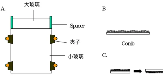
(16) Bromophenol blue),混合均勻並 spin down 後放入 0 ℃冰箱保存。 Table 3.. The DD RT-PCR primers were used in this study. Number of primers 1. 2. 3. 4. 5. 6. 7. 8. 9. 10. 11. 12. 13. 14. 15. 16.. Primers sequence. H-AP9 H-AP10 H-AP11 H-AP12 H-AP13 H-AP14 H-AP15 H-AP16 H-AP73 H-AP74 H-AP75 H-AP76 H-AP77 H-AP78 H-AP79 H-AP80. 5’-AAGCTTCATTCCG-3’ 5’-AAGCTTCCACGTA-3’ 5’-AAGCTTCGGGTAA-3’ 5’-AAGCTTGAGTGCT-3’ 5’-AAGCTTCGGCATA-3’ 5’-AAGCTTGGAGCTT-3’ 5’-AAGCTTACGCAAC-3’ 5’-AAGCTTTAGAGCG-3’ 5’-AAGCTTAGTTATC-3’ 5’-AAGCTTCAAGTTT-3’ 5’-AAGCTTTTATTCG-3’ 5’-AAGCTTGTTATAG-3’ 5’-AAGCTTTGAATTC-3’ 5’-AAGCTTAAATCGA-3’ 5’-AAGCTTGTCTAAA-3’ 5’-AAGCTTCTATTTC-3’. (3) 製備 6% Acrylamide/ urea 電泳膠 (I) 製備 6% Acrylamide/ urea solution: 取 40% Acrylamide solution (Tris:Bis=19:1) 60 ml 加入 168 g urea 及 80 ml 5X TBE buffer 最後加入二次水至總體積為 400 ml,放入磁石混合均 勻,以濾紙 (0.45- µ pore size) 過濾,最後放入冰箱保存備用。 (II) 電泳膠製備: 取 80 ml Acrylamide/ urea solution 加入 0.8 ml 10% Ammonium persulfate 混合均勻,再加入 20 µl TERMD 混合均勻,迅速倒入由兩片玻璃所組合而成 的灌膠槽中(如 Figure 5.所示),再插入鋸齒狀的 Cone 夾上夾子,蓋上保鮮膜, 放置隔夜等待膠凝固。切記在灌膠過程中膠不能有氣泡的產生,否則會影響 跑膠結果造成 Band 扭曲而不平整。. 16.
(17) A.. 大玻璃. B. Spacer 夾子 小玻璃. Comb C.. Figure 5. 電泳膠配置裝置示意圖. (4) 跑膠與壓片 將 Acrylamid/ urea Gel 拿 至 放 射 線 實 驗 室 並 撕 下 下 端 膠 帶 , 把 Acrylamide/ urea Gel 垂直立於電泳槽中且將玻璃夾緊,上層倒入 0.5X TBE buffer,下層倒入 1X TBE buffer,而 buffer 必須蓋過膠,接著將 Cone 拔出後, 用滴管沖一沖與 buffer 接觸的膠面,插上電極,用 1100 voltage 電壓預跑 30 分鐘,關掉電極,將 comb 倒插入槽中(如 Figure 5. (C)) ,取 3 µl sample loading 入孔中,將電壓調至 1900 voltage 插入電極,每隔 5 至 10 分鐘觀察跑膠的溫 度,不可使溫度超過 55 ℃,跑至第二條 dye 距下端 3 公分處即可將電極開 關關掉,取下玻璃並小心撬開,放上一張 Paper 將膠黏附於紙上後,放到乾 燥膠的機器上使膠乾燥,拿至暗房中以 X 光片進行壓片,放置-80 ℃冰箱保 存至隔夜沖片觀看結果,接著再壓一次片放置-80 ℃冰箱保存 3-5 天後再沖 片,而此張 X 光片作為結果保存用。 (5) Reamplification of cDNA probes 先將第一片 X 光片上有差異的 band 用針頭扎洞,接著將 Acrylamide/ urea Gel 與 X 光片對齊,依 X 光片上差異的 band 的位置用針頭在膠上扎洞以確 認 band 位置,用手術刀取下寬約 1 mm 的膠,而切下的膠即是 cDNA probe。 將取下的 cDNA probes 放入 200 µl PCR tube 中,加入 100 µl 二次水後 放置於室溫下 10 分鐘,在 95 ℃下反應 15 分鐘,離心 12000 rpm 2 分鐘,取 上清液加入 Salt-out solution (450 µl 100% iced ethanol+10 µl 3M Sodium acetate+5 µl 10 mM Glycogen) 混合均勻後,放置-80 ℃冰箱反應 30 分鐘, 離心 12000 rpm 10 分鐘,注意觀察有沒有 DNA pellet,小心去除上清液,加 入 200 µl 70% iced ethanol,離心 12000 rpm 約 2 分鐘,將上清液完全去除乾 淨,加入 10 µl 滅菌的二次水。. 17.
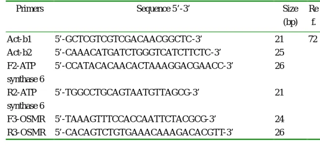
(18) 取 2.5 µl cDNA加入 47.5 µl PCR reaction solution(28 µl d.d. water+5 µl 10X Ex buffer+4 µl 250 µM dNTP+5 µl H-T11A (or H-T11C or H-T11G)+5 µl H-AP primer+0.5 µl Ex Taq DNA polymerase)經混合後 spin down,進行聚 合酵素連鎖反應(PCR)。進行 40 個循環期:首先於 94 ℃ 30 秒,接著於 50 ℃ 1 分鐘,在設於 72 ℃ 30 秒,結束後再於 72 ℃ 反應 10 分鐘,最後反應終 止於 4 ℃。 取出 5 µl Reamplificated cDNA 加入 1 µl DNA loading dye 混合且 spin down 後,跑 1.5% agarose gel,ethidium bromide 染色,於 UV 光下觀察並照 相。 取出 30 µl Reamplificated cDNA 放 入 1.5 ml 離心管中,標明清楚 Template 名稱,再取每一個檢體約 1-2 µl 20 µM primer 一起送至明欣生物科 技公司作 DNA 定序。 (6) 將已經被定序出的檢體利用 NCBI 網站進行 DNA 比對工作,比對是否有 無相同 DNA 序列,比對結果作為實驗設計參考用。 (7) 利用 RT-PCR 檢測不同濃度 17α-estradiol 作用 24 小時後,oncostatin M receptor 及 ATP synthase 6 mRNA 量是否隨著 17α-estradiol 濃度增加而表現 受到影響,再次證明 DD RT-PCR 實驗之結果。本實驗方法與檢測細胞內 Cyclins/ CDKs 基因表現的影響之實驗方法相同,所使用的 Primer 如 Table 4. 中所列。 Table 4. The PCR primers of β-actin、OSMR and ATP synthase Primers. Act-b1 Act-b2 F2-ATP synthase 6 R2-ATP synthase 6 F3-OSMR R3-OSMR. Sequence 5’-3’. Size (bp). 5’-GCTCGTCGTCGACAACGGCTC-3’ 5’-CAAACATGATCTGGGTCATCTTCTC-3’ 5’-CCATACACAACACTAAAGGACGAACC-3’. 21 25 26. 5’-TGGCCTGCAGTAATGTTAGCG-3’. 21. 5’-TAAAGTTTCCACCAATTCTACGCG-3’ 5’-CACAGTCTGTGAAACAAAGACACGTT-3’. 24 26. Re f. 72. (8) 將經由 DD RT-PCR 實驗所得到的結果,檢測 17α-estradiol 是否經由 Oncostatin M receptor 所影響的下游 MAPK pathway 而導致人類胃癌細胞死 亡。因此先加入 MAPK 抑制劑 pretreatment 3 小時後,再分成幾組實驗組: 1. Control (DMSO)、2. 60 µM 17α-estradiol、3. 100 µM 17α-estradiol、4. 10 µM 18.
(19) SB203580、5. 10 µM SB203580+60 µM 17α-estradiol、6. 10 µM SB203580+100 µM 17α-estradiol 、 7. 20 µM PD98059 、 8. 20 µM PD98059+60 µM 17α-estradiol、9. 20 µM PD98059+100 µM 17α-estradiol,培養 24 小時,將細 胞收集離心,加 4 µg /µl PI solution 500 µl,接著利用流式細胞計數儀(Flow cytometor)計數存活的細胞數。 (9) 加 入 MAPK inhibitors pretreatment 3 小 時 後 , 再 加 入 60 µM 17αestradiol,檢測 17α-estradiol 誘導人類胃癌細胞的細胞凋亡是否受到抑制, 進而證明 17α- estradiol 經由 MAPK 訊息傳導路徑而影響人類胃癌細胞的死 亡。本實驗方法與檢測 apigenin、17α-estradiol 和 flavone 對人類胃癌細胞株 (SC-M1)細胞 DNA 的影響方法相同,主要實驗分成 1. Control (DMSO)、2. 60 µM 17α-estradiol 、 3. 10 µM SB203580 、 4. 10 µM SB203580+60 µM 17α-estradiol、5. 20 µM PD98059、6. 20 µM PD98059+60 µM 17α-estradiol 等六組。 第三部分 檢測 apigenin、estradiol 及 flavone 對人類胃癌細胞株的乙醯轉化酵素 (N-acetyltransferase, NAT)活性及基因表現的影響 (1) 檢測 apigenin、estradiol 及 flavone 對人類胃癌細胞株將 2-AF 乙醯化成 2-AAF 的影響 取 5×10⁵ 細胞,將細胞培養於 24 well 培養皿中的每一 well 並加入 1 ml 培養液培養至隔夜,每一 well 先加入 10 µl 6.75 mM 2-AF 後接著加入 10 µl 不同濃度的藥物(apigenin:DMSO、0.006、0.06、0.6、6、15、30、60 mM。 17α-estradiol:DMSO、0.006、0.06、0.6、6、15、30、60mM。flavone:DMSO、 0.006、0.06、0.6、6、15、30、60mM),再移入 37 ℃,5% CO2 培養箱中, 分別培養 6、12、18、24、36 和 48 小時後收集上層的細胞培養液,分別加 入 2 ml 萃取液(Ethyl acetate:Methanol=95:5)混合均勻後靜置,離心收集 有機溶媒層並以冷凍乾燥機乾燥,加入 50 µl Methanol,取出 20 µl 利用高效 液相層析儀(High performance liquid chromatography;HPLC) 分析 2-AF 代謝 產物的量,然後利用已知標準濃度的 2-AF 和 2-AAF 而換算出實驗組的 2-AF 及 2-AAF 的量。 (2) 檢測 apigenin、17α-estradiol 及 flavone 對人類胃癌細胞株中乙醯轉化酵素 (NAT)基因的表現 此實驗主要利用 RT-PCR 方法偵測 apigenin、17α-estradiol 和 flavone 對 人類胃癌細胞株細胞內乙醯轉化酵素基因表現的影響。 本實驗方法與檢測細胞內 Cyclins/ CDKs 基因表現的影響之實驗方法相 同,所使用的 Primer 如 Table 4.中所列。. 19.
(20) Table 4. The PCR primers of β-actin and NAT1 Primers. Act-b1 Act-b2 B-MDIEANAT1 VPKHGDX-NAT1. Sequence 5’-3’. Size (bp). 5’-GCTCGTCGTCGACAACGGCTC-3’ 21 5’-CAAACATGATCTGGGTCATCTTCTC-3’ 25 5’-CACCCGGATCCCGGGATCATGGACATTGAAGC-3’ 31 5’-GGT CCT CGAGTCAATCACCATGTTTGGGCAC-3’. 20. 31. Re f. 72 75.
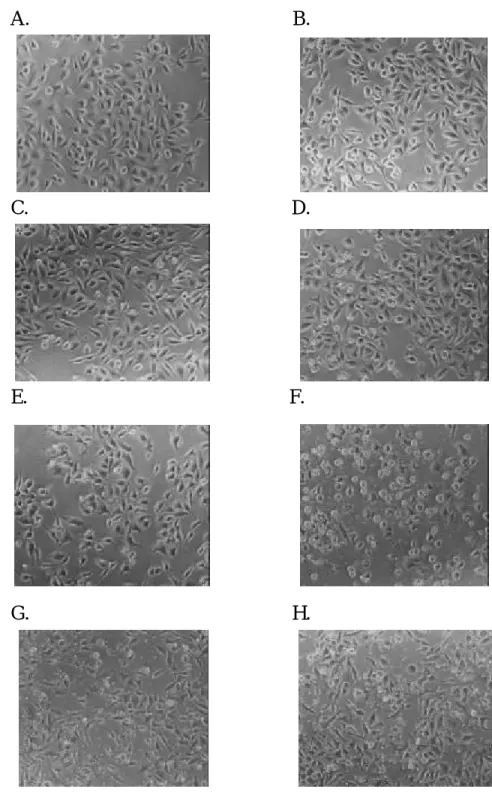
(21) 第四章 結果 第一節 對人類胃癌細胞株(SC-M1)細胞生長方面的影響 (一) 檢測 apigenin 對胃癌細胞株(SC-M1)細胞生長方面的影響 (1) 檢測 apigenin 對胃癌細胞株(SC-M1)細胞增生的影響 我們發現給予 apigenin 經不同時間培養之後,在 6 小時藥物對胃癌細胞 作用並不明顯,於 150 µM apigenin 細胞存活率仍然達 83%左右,在 12 小時 開始細胞生長的抑制,隨著藥物濃度的增加而抑制越明顯且當時間增長則細 胞存活率越來越低,因此我們從這個結果可以發現 apigenin 確實對胃癌細胞 的增生方面有很明顯的抑制。如 Figure 6.。 (2) 檢測 apigenin 對胃癌細胞株(SC-M1)細胞形態的影響 由實驗結果 Figure 7.至 Figure 12.發現當 apigenin (60、100、150 µM)濃度 增加,SC-M1 細胞的細胞膜漸漸漲大而且懸浮在細胞培養液的細胞也增加, 經 24、36 和 48 小時之後更可清楚看見細胞碎片,從照片中可以觀察到經高 濃度的 apigenin (100、150 µM)作用的 SC-M1 細胞都已經平平貼在培養皿,細 胞膜不像控制組細胞膜平滑有立體感。 (3) 檢測 apigenin 對胃癌細胞株(SC-M1) DNA 的影響 由細胞存活率及形態學上我們發現 apigenin 對胃癌細胞確實都有很明顯 的影響,因此我們利用電泳法去觀察 apigenin 對胃癌細胞 DNA 造成何種影 響,由實驗結果 Figure 13.發現將細胞加 apigenin 分別培養 24 和 48 小時後, 在 24 小時,6 µM 已觀察到細胞 DNA 斷裂,在 48 小時則沒有觀察到有 DNA Ladders 的產生,因此 apigenin 對 SC-M1 細胞造成凋亡(apoptosis)在 24 小時 較明顯,所以可以推論胃癌細胞在 apigenin 24 和 48 小時作用之後分別造成的 細胞死亡是使 SC-M1 細胞 DNA 產生斷裂造成細胞的凋亡及可能造成細胞壞 死。 (4) 檢測 apigenin 對胃癌細胞株(SC-M1)細胞週期的影響 我們由實驗結果 Figure 14. 觀察到 apigenin (60、150、300、600 µM)可使 SC-M1 細胞週期停止在 G2/M 期,隨著藥物濃度的增加停止於 G2/M 期細胞 漸增。而且隨著藥物作用時間增長 apigenin 在 36、48 小時作用後, SC-M1 細 胞停止在 G2/M 期更明顯增加,相對 G0/G1 期的細胞則是減少。而 G2/ M 停 止現象從 18 和 24 小時作用已初步觀察到,因為在 18 和 24 小時已經可以發. 21.
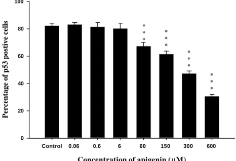
(22) 現隨著 apigenin 藥物濃度增加,停在 G0/G1 期的 SC-M1 細胞已經漸漸減少, 相對的 S 和 G2/M 期則是漸漸上升,最後在 36 及 48 小時,細胞週期停留在 G2/ M 期。 (5) 檢測 apigenin 對胃癌細胞株(SC-M1)細胞週期素(Cyclins)的影響 由細胞週期的結果已經發現在 36 和 48 小時 apigenin 可使 SC-M1 細胞的 細胞週期停止於 G2/M 期,而在 24 小時 apigenin 作用,SC-M1 細胞週期同時 發現 S 期 G2/M 期都有增加。利用 Flow cytometry 分析偵測 SC-M1 細胞內 Cyclins 和 CDKs 的結果 Figure 15.,經 24 小時 apigenin 作用後發現調控 G2/M 期的 Cyclin B1 和 CDK1 從 60 µM 開始隨著 apigenin 濃度增加很明顯受到抑 制。對於調節細胞週期由 S 期進入 G2/M 期的 cyclin E 沒有很大影響,但是 CDK2 則是隨著 apigenin 濃度增加一樣明顯受到抑制。 經 24 小時 apigenin 作用後,SC-M1 細胞內 p53 也隨著濃度增加而被抑制, 控制組 82.11±2.08,實驗組分別為 0.06 µM:82.88± 1.40、0.6 µM:81.18±3.17、 6 µM:80.03±3.92、60 µM:66.77±2.75、150 µM:61.02±2.75、300 µM:47.00 ±1.96、600 µM:30.45±1.50,此結果可由統計 Figure 16.顯示。 (6) 檢測 apigenin 對人類胃癌細胞株(SC-M1)細胞內細胞週期素(Cyclins)基因表現 的影響 經 24 小時作用後發現 SC-M1 細胞內調節細胞週期 G2/M 期的 cyclin B1 的 mRNA 的量隨著濃度增加而漸漸被抑制(Figure 17.),調節細胞進入 G0/G1 期的 cyclin D1 mRNA 的量也受到抑制使細胞無法進入 G0/G1 期(Figure 18.), 而 CDK2 mRNA 的量則是沒有很大的影響(Figure 19.)。對於 cyclin-dependent kinase inhibitor 包括 p21 (Figure 20.)和 p53 (Figure 21.)方面,p53 mRNA 的量 很明顯受到 apigenin 的抑制而 p21 mRNA 的量則是漸漸增加。. 22.
(23) Control 6 µM. 100. * ** ** *. 20 µM. * * *. 80. % viabile cells. * * * * * * * ** * *. * * *. 60 µM 100 µM. * * * * * ** * *. 60. * * *. 150 µM. * * * * * *. 40. 20. 0 6h. 12 h. 24 h. 36 h. 48 h. Incubation time (hr). Figure 6. Percentage of viable SC-M1 cells after cotreatment with different doses of apigenein for 6, 12, 24, 36 and 48 hrs incubation. After the treatment of various concentrations of apigenin, cells were harvested PI stain solution and assayed for % viable cells by FACS analysis. Data were analyzed by one-way ANOVA. Value= mean± SD, n=3 *P<0.05, **P<0.005, ***P<0.001. 23.
(24) A.. B.. C.. D.. E.. F.. G.. H.. Figure 7. The morphology of human stomach cancer (SC-M1) cells after exposure to the different doses of apigenin for 6 hr then were examined and photographed by phase microscope (200x). A:Control, B:0.06 µM, C:0.6 µM, D:6 µM, E:20 µM, F: 60 µM, G:100 µM, H:150 µM.. 24.
(25) A.. B.. C.. D.. E.. F.. G.. H.. Figure 8. The morphology of human stomach cancer (SC-M1) cells after exposure to thr different doses of apigenin for 12 hr then were examined and photographed by phase microscope (200x). A:Control, B:0.06 µM, C:0.6 µM, D:6 µM, E:20 µM, F: 60 µM, G:100 µM, H:150 µM.. 25.
(26) A.. B.. C.. D.. E.. F.. G.. H.. Figure 9. The morphology of human stomach cancer (SC-M1) cells after exposure to the different doses of apigenin for 18 hr then were examined and photographed by phase microscope (200x). A:Control, B:0.06 µM, C:0.6 µM, D:6 µM, E:20 µM, F: 60 µM, G:100 µM, H:150 µM.. 26.
(27) A.. B.. C.. D.. E.. F.. G.. H.. Figure 10. The morphology of human stomach cancer (SC-M1) cells after exposure to the different doses of apigenin for 24 hr then were examined and photographed by phase microscope (200x). A:Control, B:0.06 µM, C:0.6 µM, D:6 µM, E:20 µM, F: 60 µM, G:100 µM, H:150 µM.. 27.
(28) A.. B.. C.. D.. E.. F.. G.. H.. Figure 11. The morphology of human stomach cancer (SC-M1) cells after exposure to the different doses of apigenin for 36 hr then were examined and photographed by phase microscope (200x). A:Control, B:0.06 µM, C:0.6 µM, D:6 µM, E:20 µM, F: 60 µM, G:100 µM, H:150 µM.. 28.
(29) A.. B.. C.. D.. E.. F.. G.. H.. Figure 12. The morphology of human stomach cancer (SC-M1) cells after exposure to the different doses of apigenin for 48 hr then were examined and photographed by phase microscope (200x). A:Control, B:0.06 µM, C:0.6 µM, D:6 µM, E:20 µM, F: 60 µM, G:100 µM, H:150 µM.. 29.
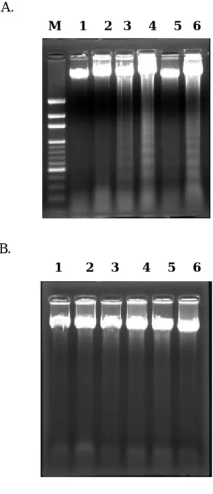
(30) A. M. 1. 2. 3. 4. 5. 6. B. 1. 2. 3. 4. 5. 6. Figure 13. An agarose electrophresis of DNA from human stomach cancer (SC-M1) cells after exposure to various concentrations of apigenin for 24 (A) and 48 hr (B). M: DNA Maker;Lane 1:DMSO;Lane 2:0.6 µM;Lane 3:6 µM;Lane 4:60 µM; Lane 5:150 µM;Lane 6:300 µM.. 30.
(31) A.. B. 100 G0/G1 S G2/M. 80. percentage of cell number. percentage of cell number. 100. 60. 40. 20. G0/G1 S G2/M. 80. 60. 40. 20. 0. 0 control 0.06. 0.6. 6. 60. 150. 300. 600. control 0.06. Concentration of apigenin (µM). C.. 150. 300. 600. 300. 600. 300. 600. G0/G1 S. 80. G2/M. percentage of cell number. percentage of cell number. 60. 100 G0/G1 S G2/M. 60. 40. 20. 80. 60. 40. 20. 0. 0 control. 0.06. 0.6. 6. 60. 150. 300. control. 600. 0.06. 0.6. 6. 60. 150. Concentration of apigenin ( µM). Concentration of apigenin ( µM). E.. F. G0/G1 S G2/M. 100. 80. percentage of cell number. percentage of cell number. 6. D.. 100. 100. 0.6. Concentration of apigenin (µM). 60. 40. 20. G0/G1 S G2/M. 80. 60. 40. 20. 0. 0 control. 0.06. 0.6. 6. 60. 150. 300. control. 600. 0.06. 0.6. 6. 60. 150. Concentration of apigenin ( µM). Concentration of apigenin ( µM). Figure 14. The cell cycle distribution of SC-M1 cells growing in the percentage of different doses of apigenin for (A) 6hr, (B) 12 hr, (C) 18 hr, (D) 24 hr, (E) 36 hr and (F) 48 hr incubated. After the treatments of cells were harvested and analyzed by FACS for cell cycle. Values= means± SD n=3. 31.
(32) 100. percentage of cell number. * *. 80 *. ** *. 60. control 0.06 µ M 0.6 µM 6.0 µM. * *. 6 0 µM 150 µ M 300 µ M 600 µ M. 40 *. *. 20. 0 cyclin B1. CDK1. CDK2. cyclin A. cyclin E. Figure 15. The data analysis for intracellular cyclins distribution of human stomach cancer (SC-M1) cells repressing in the presence of different doses of apigenin for 24 hr. The cell cyclins were analyzed by FACS. Data were analyzed by one-way ANOVA. Value= mean± SD, n=3 *P<0.05, **P<0.005, ***P<0.001. Percentage of p53 postive cells. 100. * * *. 80. * * * * * *. 60. * * *. 40. 20. 0 Control. 0.06. 0.6. 6. 60. 150. 300. 600. Concentration of apigenin ( µM). Figure 16. The data analysis for intracellular p53 distribution of human stomach cancer (SC-M1) cells repressing in the presence of different doses of apigenin for 24 hr. The p53 were analyzed by FACS. Data were analyzed by one-way ANOVA. Value= mean± SD, n=3 *P<0.05, **P<0.005, ***P<0.001. 32.
(33) A. Cyclin B1 (720 bp). β-actin (353 bp) 1. 2. 3. 4. 5. 6. B.. Ratio of cyclin B1 mRNA. 1.5. 1.0. 0.5. 0.0 Control. 0.6. 6. 60. 150. 300. Concentration of apigenin (mM). Figure 17. RT-PCR analysis of cyclin B1and beta-actin in human stomach cancer (SC-M1) cells for treatment with different doses of apigenin. (A) Gel electrophoresis. Lane 1: DMSO;Lane 2:0.6 µM;Lane 3:6 µM;Lane 4:60 µM;Lane 5:150 µM;Lane 6:300 µM. (B) The statistic figure of cyclin B1 mRNA levels. Values are mean± SD n= 2. 33.
(34) A. Cyclin D1 (495 bp). β-actin (353 bp) 1. 2. 3. 4. 5. 6. B.. Ratio of cyclin D1 mRNA. 1.0. 0.5. 0.0 Control. 0.6. 6. 60. 150. 300. Concentration of apigenin (mM). Figure 18. RT-PCR analysis of cyclin D1and beta-actin in human stomach cancer (SC-M1) cells for treatment with different doses of apigenin. (A) Gel electrophoresis. Lane 1:DMSO;Lane 2:0.6 µM;Lane 3:6 µM;Lane 4:60 µM;Lane 5:150 µM; Lane 6:300 µM. (B) The statistic figure of cyclin D1 mRNA levels. Values are mean± SD n= 2. 34.
(35) A. CDK2 (330 bp) β-actin (353 bp). 1. 2. 3. 4. 5. 6. B.. Ratio of CDK2 mRNA. 1.0. 0.5. 0.0 Control. 0.6. 6. 60. 150. 300. Concentration of apigenin (mM). Figure 19. RT-PCR analysis of CDK2 and beta-actin in human stomach cancer (SC-M1) cells for treatment with different doses of apigenin. (A) Gel electrophoresis. Lane 1:DMSO;Lane 2:0.6 µM;Lane 3:6 µM;Lane 4:60 µM;Lane 5:150 µM; Lane 6:300 µM. (B) The statistic figure of CDK2 mRNA levels. Values are mean± SD n= 2. 35.
(36) A. p21 (380 bp). β-actin (353 bp). 1. 2. 3. 4. 5. 6. B.. Ratio of p21 mRNA. 1.0. 0.5. 0.0 Control. 0.6. 6. 60. 150. 300. Concentration of apigenin (mM). Figure 20. RT-PCR analysis of p21 and beta-actin in human stomach cancer (SC-M1) cells for treatment with different doses of apigenin. (A) Gel electrophoresis. Lane 1: DMSO;Lane 2:0.6 µM;Lane 3:6 µM;Lane 4:60 µM;Lane 5:150 µM;Lane 6:300 µM. (B) The statistic figure of p21 mRNA leve ls. Values are mean± SD n= 2. 36.
(37) A. p53 (293 bp). β-actin (353 bp). 1. 2. 3. 4. 5. 6. B.. Ratio of p53 mRNA. 1.0. 0.5. 0.0 Control. 0.6. 6. 60. 150. 300. Concentration of apigenin (mM). Figure 21. RT-PCR analysis of p21 and beta-actin in human stomach cancer (SC-M1) cells for treatment with different doses of apigenin. (A) Gel electrophoresis. Lane 1: DMSO;Lane 2:0.6 µM;Lane 3:6 µM;Lane 4:60 µM;Lane 5:150 µM;Lane 6:300 µM. (B) The statistic figure of p53 mRNA levels. Values are mean± SD n= 2. 37.
(38) (二)檢測 17α-estradiol 對胃癌細胞株(SC-M1)細胞生長方面的影響 (1) 檢測 17α-estradiol 對胃癌細胞株(SC-M1)細胞增生的影響 Figure 12. 結果發現給予 17α-estradiol 經不同時間培養之後,在 6 小時對 胃癌細胞已有抑制細胞生長的作用,於 150 µM 17α-estradiol,細胞存活率為 66.12± 3.21%,在 12、18、24、36 和 48 小時,細胞生長抑制隨著藥物濃度的 增加及時間的增長而抑制作用越明顯,因此從這個實驗結果可以發現 17α-estradiol 確實對胃癌細胞的增生方面有很明顯的抑制。 (2) 檢測 17α-estradiol 對胃癌細胞株(SC-M1)細胞形態的影響 由實驗結果 Figure 23.至 Figure 28.發現當 17α-estradiol (60、100、150 µM) 濃度增加,SC-M1 細胞的細胞膜漸漸產生皺折而且懸浮在細胞培養液的細胞 也增加,經 24、36 和 48 小時之後更可清楚看見細胞碎片而且細胞也很明顯 減少,從照片中可以觀察到經高濃度的 17α-estradiol (100、150 µM)作用的 SC-M1 細胞都已經平平貼在培養皿,細胞膜不像控制組細胞膜平滑而具有立 體感。 (3) 檢測 17α-estradiol 對胃癌細胞株(SC-M1) DNA 的影響 由細胞存活率及形態學上發現 17α-estradiol 對胃癌細胞確實都有很明顯 的影響,因此利用電泳法觀察 17α-estradiol 對胃癌細胞 DNA 造成何種影響, 由實驗結果 Figure 29.發現將細胞加藥分別培養 24 和 48 小時後,在 30 µM 就 觀察到細胞 DNA 斷裂現象,而發現在 48 小時 17α-estradiol 對 SC-M1 細胞造 成凋亡(apoptosis)更明顯,因此我們可以推論胃癌細胞在 17α-estradiol 24 和 48 小時作用之後所造成的細胞死亡是使 SC-M1 細胞 DNA 產生斷裂造成細胞的 凋亡。 (4) 檢測 17α-estradiol 對胃癌細胞株(SC-M1)細胞週期的影響 由實驗結果 Figure 30.觀察到 17α-estradiol (60、150、300、600 µM)可使 SC-M1 細胞週期停止在 G2/M 期,隨著藥物濃度的增加停止於 G2/M 期細胞 漸增,且在 24 小時作用後,SC-M1 細胞停止在 G2/M 期的最明顯,而在 6 和 12 小時細胞週期並沒有受到影響。從 18 小時作用已經可以初步看到,因為在 18 小時已經可以發現隨著17α-estradiol 藥物濃度增加,停在 G0/G1 期的 SC-M1 細胞已經漸漸減少,相對的 S 和 G2/M 期則是漸漸上升。 (5) 檢測 17α-estradiol 對胃癌細胞株(SC-M1)細胞週期素(Cyclins)的影響 由細胞週期的結果已經發現在 24 小時 17α-estradiol 可使 SC-M1 細胞的細 胞週期停止於 G2/M 期最明顯。經 24 小時 17α-estradiol 作用後,偵測 SC-M1 細胞內 Cyclins 和 CDKs 的結果 Figure 31.發現調控 G2/M 期的 Cyclin B1 和. 38.
(39) CDK1 隨著濃度增加很明顯受到抑制,而調節細胞週期由 S 期進入 G2/M 期的 cyclin E 和 CDK2 及調節 G0/G1 期的 cyclin D1、cyclin D2、cyclin D3 和 CDK6 則是沒有很大影響。 SC-M1 細胞內 p53 經 24 小時 17α-estradiol 作用後,隨著濃度增加並沒有 很明顯受到抑制,控制組 88.40± 1.08,實驗組分別為 0.6 µM:85.87±0.24、6 µM:84.43±1.00、30 µM:81.23± 1.19、60 µM:80.03± 1.00、100 µM:78.47± 0.59、150 µM:77.88±0.41 及 300 µM:71.81± 0.75,此結果可由統計 Figure 32 顯示。 (6) 檢測 17α-estradiol 對人類胃癌細胞株(SC-M1)細胞內細胞週期素(Cyclins)基因 表現的影響 經 24 小時作用後發現 SC-M1 細胞內調節細胞週期 G2/M 期的 cyclin B1 mRNA 的量並沒有隨著 17α-estradiol 濃度增加而受到抑制(Figure 33.),調節細 胞進入 G0/G1 期的 cyclin D1 及 CDK2 mRNA 的量也沒有受到抑制(Figure 34、 35),但是 cyclin E 隨著濃度增加漸漸受到抑制 (Figure 36.) 。對於 cyclindependent kinase inhibitor 包括 p53 和 p21 方面,p53 mRNA 的量(Figure 37.) 沒有明顯受到 17α-estradiol 的抑制,而 p21 mRNA 的量(Figure 38.)則是漸漸增 加。. 39.
(40) 100. % viable cells. 80. * ** ** *. * * * * ** * *. 60. * * * * * *. * ** ** ** * * ** ** **. Control 6 µM. * ** ** * * *. * ** ** *. 20 µM. * * ** * **. 60 µM 100 µM 150 µM. ** ** *. 40. 20. 0 6h. 12 h. 18 h. 24 h. 36 h. 48 h. Incubation time (hr). Figure 22. Percentage of viable SC-M1 cells after cotreatment with different doses of 17α-estradiol for 6, 12, 18, 24, 36 and 48 hrs incubated. After the treatment of various concentrations of apigenin, cells were harvested PI stain solution and assayed for % viable cells by FACS analysis. Data were analyzed by one-way ANOVA. Value= mean± SD, n=3 *P<0.05, **P<0.005, ***P<0.001. 40.
(41) A.. B.. C.. D.. E.. F.. Figure 23. The morphology of human stomach cancer (SC-M1) cells after exposure to 17α-estradiol for 6 hr then were examined and photographed by phase microscope (200x). A:Control, B:6 µM, C:30 µM, D:60 µM, E:100 µM, F:150 µM.. 41.
(42) A.. B.. C.. D.. E.. F.. Figure 24. The morphology of human stomach cancer (SC-M1) cells after exposure to 17α-estradiol for 12 hr then were examined and photographed by phase microscope (200x). A:Control, B:6 µM, C:30 µM, D:60 µM, E:100 µM, F:150 µM.. 42.
(43) A.. B.. C.. D.. E.. F.. Figure 25. The morphology of human stomach cancer (SC-M1) cells after exposure to 17α-estradiol for 18 hr then were examined and photographed by phase microscope (200x). A:Control, B:6 µM, C:30 µM, D:60 µM, E:100 µM, F:150 µM.. 43.
(44) A.. B.. C.. D.. E.. F.. Figure 26. The morphology of human stomach cancer (SC-M1) cells after exposure to 17α-estradiol for 24 hr then were examined and photographed by phase microscope (200x). A:Control, B:6 µM, C:30 µM, D:60 µM, E:100 µM, F:150 µM.. 44.
(45) A.. B.. C.. D.. E.. F.. Figure 27. The morphology of human stomach cancer (SC-M1) cells after exposure to 17α-estradiol for 36 hr then were examined and photographed by phase microscope (200x). A:Control, B:6 µM, C:30 µM, D:60 µM, E:100 µM, F:150 µM... 45.
(46) A.. B.. C.. D.. E.. F.. Figure 28. The morphology of human stomach cancer (SC-M1) cells after exposure to 17α-estradiol for 48 hr then were examined and photographed by phase microscope (200x). A:Control, B:6 µM, C:30 µM, D:60 µM, E:100 µM, F:150 µM.. 46.
(47) A.. 1. 2. 3. 4. 5. 6. 7. 1. 2. 3. 4. 5. 6. 7. B.. Figure 29. An agarose electrophresis of DNA from human stomach cancer (SC-M1) cells after exposure to various concentrations of 17α-estradiol for A:24 hr;B:48 hr. Lane 1:Maker;Lane 2:DMSO;Lane 3:6 µM;Lane 4:30 µM;Lane 5:60 µM; Lane 6:150 µM;Lane 7:300 µM.. 47.
(48) A.. B. 100. G0/G1 phase S phase G2/M phase. 80. percentage of cell number. percentage of cell number. 100. 60. 40. 20. G0/G1 phase S phase G2/M phase. 80. 60. 40. 20. 0. 0 control. 0.06. 0.6. 6. 60. 150. 300. control. 600. 0.06. 0.6. 6. 60. 150. 300. 600. Concentration of 17α-estradiol ( µM). Concentration of 17α-estradiol ( µM). C.. D. 100. G0/G1 phase S phase G2/M phase. 80. percentage of cell number. percentage of cell number. 100. 60. 40. 20. G0/G1 phase S phase G2/M phase. 80. 60. 40. 20. 0. 0 control. 0.06. 0.6. 6. 60. 150. 300. control. 600. 0.06. E.. 6. 60. 150. 300. 600. F.. 100. 100 G0/G1 phase S phase G2/M phase. 80. percentage of cell number. percentage of cell number. 0.6. Concentration of 17α-estradiol ( µM). Concentration of 17α-estradiol ( µM). 60. 40. 20. 0. G0/G1 phase S phase G2/M phase. 80. 60. 40. 20. 0 control. 0.06. 0.6. 6. 60. 150. 300. 600. control. Concentration of 17α-estradiol ( µM). 0.06. 0.6. 6. 60. 150. 300. 600. Concentration of 17α-estradiol ( µM). Figure 30. The cell cycle distribution of SC-M1 cells growing in the percentage of different doses of 17α-estradiol for 6hr (A), 12 hr (B), 18 hr (C), 24 hr (D), 36 hr (E) or 48 hr (F) incubated. After the treatments of cells which were harvested and analyzed by FACS for cell cycle.. 48.
(49) A.. 100 Control 0.06 µM. 80. 0.6 µM 6 µM. % postive cells. 60 µM 150 µM 300 µM. 60. 600 µM. 40. 20. 0 Cyclin D1 Cyclin D2 Cyclin D3. Cyclin E. CDK2. CDK6. B.. 100. % postive cells. 80. 60. Control. *. * * * ** *. 0.6 µM. * *. * *. * *. 6µM. * *. 20 µM 60 µM. * *. 100 µM 150 µM 300 µM. * *. * *. 40. 20. 0 Cyclin B1. CDK1. Figure 31. The data analysis for intracellular cyclins distribution of human stomach cancer (SC-M1) cells repressing in the presence of different doses of 17α-estradiol for 24 hr. The cell cyclins were analyzed by FACS. Data were analyzed by one-way ANOVA. Value= mean± SD, n=3 *P<0.05, **P<0.005, ***P<0.001. 49.
(50) Percentage of p53 postive cells. 100. * *. * * *. * * *. * * *. * * *. * * *. 0.6. 6. 30. 60. 100. 150. 80. * * *. 60. 40. 20. 0 Control. 300. Concentration of 17α-estradiol (µM). Figure 32. The data analysis for intracellular p53 distribution of human stomach cancer (SC-M1) cells less repressing in the presence of different doses of 17α-estradiol for 24 hr. The cell cyclins were analyzed by FACS. Data were analyzed by one-way ANOVA. Value= mean± SD, n=3 *P<0.05, **P<0.005, ***P<0.001. 50.
(51) A. Cyclin B1 (720 bp) β-actin (353 bp) 1. 2. 3. 4. 5. 6. B.. Ratio of cyclin B1 mRNA. 1.0. 0.5. 0.0 Control. 6. 30. 60. 100. 150. Concentration of 17α-estradiol (µM). Figure 33. RT-PCR analysis of cyclin B1 and beta-actin in human stomach cancer (SC-M1) cells for treatment with different doses of 17α-estradiol. (A) Gel electrophoresis. Lane 1:DMSO;Lane 2:6 µM;Lane 3:30 µM;Lane 4:60 µM;Lane 5:100 µM; Lane 6:150 µM. (B) The statistic figure of cyclin B1 mRNA levels. Values are mean± SD n= 2. 51.
(52) A. Cyclin D1 (495 bp) β-actin (353 bp). 1. 2. 3. 4. 5. 6. B.. Ratio of cyclin D1 mRNA. 1.00. 0.50. 0.00 Control. 6. 30. 60. 100. 150. Concentration of 17α-estradiol (µM). Figure 34. RT-PCR analysis of cyclin D1 and beta-actin in human stomach cancer (SC-M1) cells for treatment with different doses of 17α-estradiol. (A) Gel electrophoresis. Lane 1:DMSO;Lane 2:6 µM;Lane 3:30 µM;Lane 4:60 µM;Lane 5:100 µM; Lane 6:150 µM. (B) The statistic figure of cyclin D1 mRNA levels. Values are mean± SD n= 2. 52.
(53) A. CDK2 (330 bp) β-actin (353 bp) 1. 2. 3. 4. 5. 6. 100. 150. B.. Ratio of CDK2 mRNA. 1.00. 0.50. 0.00 Control. 6. 30. 60. Concentration of 17α-estradiol (µM). Figure 35. RT-PCR analysis of CDK2 and beta-actin in human stomach cancer (SC-M1) cells for treatment with different doses of 17α-estradiol. (A) Gel electrophoresis. Lane 1:DMSO;Lane 2:6 µM;Lane 3:30 µM;Lane 4:60 µM;Lane 5:100 µM; Lane 6:150 µM. (B) The statistic figure of CDK2 mRNA levels. Values are mean± SD n= 2. 53.
(54) A. Cyclin E (375 bp) β-actin (353 bp) 1. 2. 3. 4. 5. 6. 100. 150. B.. Ratio of cyclin E mRNA. 1.00. 0.50. 0.00 Control. 6. 30. 60. Concentration of 17α-estradiol (µM). Figure 36. RT-PCR analysis of cyclin E and beta-actin in human stomach cancer (SC-M1) cells for treatment with different doses of 17α-estradiol. (A) Gel electrophoresis. Lane 1:DMSO;Lane 2:6 µM;Lane 3:30 µM;Lane 4:60 µM;Lane 5:100 µM; Lane 6:150 µM. (B) The statistic figure of cyclin E mRNA levels. Values are mean± SD n= 2. 54.
(55) A. p53 (293 bp) β-actin (353 bp) 1. 2. 3. 4. 5. 6. B.. Ratio of p53 mRNA. 1.00. 0.50. 0.00 Control. 6. 30. 60. 100. 150. Concentration of 17α-estradiol (µM). Figure 37. RT-PCR analysis of p53 and beta-actin in human stomach cancer (SC-M1) cells for treatment with different doses of 17α-estradiol. (A) Gel electrophoresis. Lane 1: DMSO;Lane 2:6 µM;Lane 3:30 µM;Lane 4:60 µM;Lane 5:100 µM;Lane 6:150 µM. (B) The statistic figure of p53 mRNA levels. Values are mean± SD n= 2. 55.
(56) A. p21 (380 bp) β-actin (353 bp) 1. 2. 3. 4. 5. 6. B. 0.7. Ratio of p21 mRNA. 0.6. 0.5. 0.4. 0.3. 0.2. 0.1. 0.0 Control. 6. 30. 60. 100. 150. Concentration of 17α-estradiol (µM). Figure 38. RT-PCR analysis ofp21 and beta-actin in human stomach cancer (SC-M1) cells for treatment with different doses of 17α-estradiol. (A) Gel electrophoresis. Lane 1: DMSO;Lane 2:6 µM;Lane 3:30 µM;Lane 4:60 µM;Lane 5:100 µM;Lane 6:150 µM. (B) The statistic figure of p21 mRNA levels. Values are mean± SD n= 2. (三) 檢測 flavone 對胃癌細胞株(SC-M1)細胞生長的方面. 56.
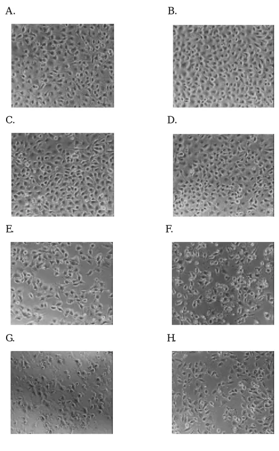
(57) (1) 檢測 flavone 對胃癌細胞株(SC-M1)細胞增生的影響 Figure 39 結果發現給予 flavone 經不同時間培養之後,在 6 小時 flavone 對胃癌細胞已經有抑制細胞的生長的作用,於 150 µM flavone 細胞存活率為 40%左右,於 300 µM flavone 細胞存活率只為 1.19%,在 12、24、36、48 小 時胃癌細胞生長抑制,從 60 µM 至 300 µM 隨 flavone 濃度的增加而抑制越明 顯且當時間增長則細胞存活率越來越低,因此從這個結果可以發現 flavone 確 實對胃癌細胞的增生方面有很明顯的抑制。 (2) 檢測 flavone 對胃癌細胞株(SC-M1)細胞形態的影響 由實驗結果 Figure 40 至 Figure 45 發現當 flavone (60、100、150、300 µM) 濃度增加,SC-M1 細胞的細胞形態越來越不規則且細胞膜有漲大的現象,懸 浮在細胞培養液的細胞也增加,經 12、24、36 和 48 小時之後,於高濃度 flavone 作用後更可清楚看見細胞碎片而且細胞也明顯減少,從照片中也可以觀察到 經高濃度的 flavone (150、300 µM)作用的 SC-M1 細胞的細胞膜不像控制組細 胞膜平滑有立體感。 (3) 檢測 flavone 對胃癌細胞株(SC-M1) DNA 的影響 由細胞存活率和形態學上發現 flavone 對胃癌細胞確實都有很明顯的影 響,為了觀察 flavone 對胃癌細胞 DNA 造成何種影響,因此利用電泳法分析 檢測,由實驗結果 Figure 46 發現將細胞加藥分別培養 24 和 48 小時後,在 24 小時,在 60 µM 可以清楚觀察到細胞 DNA 斷裂,在 48 小時即使是高濃度都 沒有觀察到有 DNA Ladders 的產生,因此 flavone 對 SC-M1 細胞造成凋亡 (apoptosis)在 24 小時較明顯,所以我們可以推論胃癌細胞在 flavone 24 和 48 小時作用之後分別造成的細胞死亡是使 SC-M1 細胞 DNA 產生斷裂造成細胞 的凋亡及可能造成細胞壞死。 (4) 檢測 flavone 對胃癌細胞株(SC-M1)細胞週期的影響 由實驗結果 Figure 47 觀察到 flavone (60、150、300、600 µM)可使 SC-M1 細胞週期停止在 G2/M 期,隨著藥物濃度的增加停止於 G2/M 期細胞漸增而且 在 48 小時作用後,SC-M1 細胞停止在 G2/M 期的最明顯,在 6 和 12 小時並 沒有影響。從 18 小時作用已經可以初步看到,因為在 18 小時已經可以發現 隨著 flavone 藥物濃度增加,停在 G0/G1 期的 SC-M1 細胞已經漸漸減少,相 對的 S 和 G2/M 期則是漸漸上升。 (5) 檢測 flavone 對胃癌細胞株(SC-M1)細胞週期素(Cyclins)的影響 由細胞週期的結果已經發現在 24 小時 flavone 可使 SC-M1 細胞的細胞週 期停止於 G2/M 期明顯。經 24 小時 flavone 作用後,偵測 SC-M1 細胞內 Cyclins 和 CDKs 的結果 Figure 48,發現調控 G2/M 期的 Cyclin B1、CDK1 及 Cyclin A. 57.
(58) 隨著濃度增加很明顯受到抑制,而對於調節細胞週期由 S 期進入 G2/M 期的 cyclin E 和 CDK2 則是沒有很大影響。 (6) 檢測 flavone 對人類胃癌細胞株(SC-M1)細胞內細胞週期素(Cyclins)基因表現 的影響 經 24 小時作用後發現 SC-M1 細胞內調節細胞週期 G2/M 期的 cyclin B1 的 mRNA 的量隨著濃度增加有明顯抑制(Figure 49),而 CDK2 mRNA 的量同 樣沒有很大的影響(Figure 50),但是 cyclin E 隨著濃度增加漸漸受到抑制 (Figure 51)。對於 cyclin- dependent kinase inhibitor 包括 p53 和 p21 方面,p53 mRNA (Figure 52)的量沒有明顯受到 flavone 的抑制而 p21 mRNA (Figure 53) 的量則是漸漸抑制。. 58.
(59) 100 Control. *. % Viable cells. 80. *. * * * *. 60. * * * *. * * *. * * *. 40. 0 6h. 12 h. * * * 18 h. 100 µM. * * *. * * *. 20 * * *. 60 µM 150 µM 300 µM. * * * * * *. * * *. * * *. 20 µM. *. * * *. * * *. * * * 24 h. * * *. 36 h. * * * 48 h. Incubation time (hr). Figure 39. Percentage of viable SC-M1 cells after cotreatment with different doses of flavone for 6, 12, 18, 24, 36 and 48 hrs incubated. After the treatment of various concentrations of flavone, cells were harvested PI stain and assayed for % viable cells by FACS analysis. Data were analyzed by one-way ANOVA. Value= mean± SD, n=3 *P<0.05, **P<0.005, ***P<0.001. 59.
(60) A.. B.. C.. D.. E.. F.. G.. H.. Figure 40. The morphology of human stomach cancer (SC-M1) cells after exposure to flavone for 6 hr then were examined and photographed by phase microscope (200x). A:Control, B:0.6 µM, C:6 µM, D:20 µM, E:60 µM, F:100 µM, G:150 µM, H:300 µM.. 60.
(61) A.. B.. C.. D.. E.. F.. G.. H.. Figure 41. The morphology of human stomach cancer (SC-M1) cells after exposure to flavone for 12 hr then were examined and photographed by phase microscope (200x). A:Control, B:0.6 µM, C:6 µM, D:20 µM, E:60 µM, F:100 µM, G:150 µM, H:300 µM.. 61.
(62) A.. B.. C.. D.. E.. F.. G.. H.. Figure 42. The morphology of human stomach cancer (SC-M1) cells after exposure to flavone for 18 hr then were examined and photographed by phase microscope (200x). A:Control, B:0.6 µM, C:6 µM, D:20 µM, E:60 µM, F:100 µM, G:150 µM, H:300 µM.. 62.
(63) A.. B.. C.. D.. E.. F.. G.. H.. Figure 43. The morphology of human stomach cancer (SC-M1) cells after exposure to flavone for 24 hr then were examined and photographed by phase microscope (200x). A:Control, B:0.6 µM, C:6 µM, D:20 µM, E:60 µM, F:100 µM, G:150 µM, H:300 µM.. 63.
(64) A.. B.. C.. D.. E.. F.. G.. H.. Figure 44. The morphology of human stomach cancer (SC-M1) cells after exposure to flavone for 36 hr then were examined and photographed by phase microscope (200x). A:Control, B:0.6 µM, C:6 µM, D:20 µM, E:60 µM, F:100 µM, G:150 µM, H:300 µM.. 64.
(65) A.. B.. C.. D.. E.. F.. G.. H.. Figure 45. The morphology of human stomach cancer (SC-M1) cells after exposure to flavone for 48 hr then were examined and photographed by phase microscope (200x). A:Control, B:0.6 µM, C:6 µM, D:20 µM, E:60 µM, F:100 µM, G:150 µM, H:300 µM.. 65.
(66) A. M. 1. 2. 3. 4. 5. 6. B. 1. 2. 3. 4. 5. 6. Figure 46. An agarose electrophresis of DNA from human stomach cancer cell line (SC-M1) after exposure to various concentrations of flavone for 24 and 48 hr. M:DNA makers;Lane 1:DMSO;Lane 2: 6 µM;Lane 3:20 µM;Lane 4:60 µM;Lane 5:150 µM;Lane 6:300 µM.. 66.
(67) A.. B.. 100. 100 G0/G1 phase G0/G1 phase S phase. G2/M phase. percentage of cell numbers. percentage of cell numbers. S phase. 80. 60. 40. 20. 80. G2/M phase. 60. 40. 20. 0. 0 control. 0.6. 6. 20. 60. 100. 150. control. 300. 0.6. 6. 20. 60. 100. 150. 300. Concentration of flavone (µM). Concentration of flavone (µM). C.. D.. 100 G0/G1 phase. 100. S phase. 80. percentage of cell numbers. percentage of cell numbers. G2/M phase. 60. 40. 20. 0. 80 G0/G1 phase. 60. S phase G2/M phase. 40. 20. 0. control. 0.6. 6. 20. 60. 100. 150. 300. control. 0.6. Concentration of flavone (µM). 6. 20. 60. 100. 150. 300. 150. 300. Concentration of flavone (µM ). E.. F.. 100. 80. G0/G1 phase G0/G1 phase. S phase G2/M phase. percentage of cell numbers. percentage of cell numbers. 100. 60. 40. 20. 0. 80. S phase G2/M phase. 60. 40. 20. 0. control. 0.6. 6. 20. 60. 100. 150. 300. control. Concentration of flavone (µM). 0.6. 6. 20. 60. 100. Concentration of flavone (µM ). Figure 47. The cell cycle distribution of SC-M1 cells growing in the percentage of different doses of flavone for (A) 6hr, (B) 12 hr, (C) 18 hr, (D) 24 hr, (E) 36 hr or (F) 48 hr incubated. After the treatments of cells which were analyzed by FACS for cells.. 67.
數據

+7


相關文件
• DNA analysis: cell cycle analysis, tumor monitoring, detection of ploidy,. •
Xianggang zaji (miscellaneous notes on Hong Kong) was written by an English and translated into Chinese by a local Chinese literati.. Doubts can therefore be cast as to whether
Central granular cell odontogenic tumour, report of the first malignant case and review of the literature. Human leucocyte antigen typing in
Persons with a phenotype mediated by one of these MC1R genetic variants are at greater risk of UV-induced skin cancers, because pheomelanin not only provides less effective
One of these enlargements is peripheral giant cell granuloma (PGCG), a lesion unique to the oral cavity, occurring only on the gingiva.. It is distinguishable from similar lesion
To evaluate the clinicopathologic features, prognostic factors, and management of patients in the North Chinese population with head and neck squamous cell carcinoma (HNSCC)
The predicted expression profiles of 24 chemokines and immunosuppressive biomarkers for SCC4, SCC15, and SCC25 were used in a decision tree format to sort cell lines into those
Specific deleterious gene mutation profiles were converted into a computational format and annotated into the HNSCC cancer network, simulated to induce the cell line - specific
