The Variations of Brain Activities of Acupuncture to TE5 of
Left Hand in Normal Subjects
Sheng-Feng Hsu2, Chien-Yue Chen1* , Ming-Da Ke1 , Chien-Hsun Huang 3, Yuan-Ting Sun4 1
Institute of Electronic Engineering, National Yunlin University of Science & Technology 2
Acupuncture Research Center, China Medical University, Taichung, Taiwan 3
Department of Community and Family Medicine, National Taiwan University Hospital Yun-Lin Branch 3
Graduate Institute of Chinese Medical Science, China Medical University 4
Department of Internal Medicine, National Cheng Kung University Hospital Dou-Liou Branch
*
chencyue@yuntech.edu.tw
Abstract
This paper presents the evident effects of acupoint stimulation, using EEG measurements. With acupuncture stimulation and the EEG measurement on the same meridian, EEG is able to accurately detect the effects of acupunctural point stimulation on brain waves.
In our study, 24 subjects without heart or nervous diseases were randomly separated into two groups of 12. In the first group, the subjects laid on a bed with eyes closed for 10 minutes. They then received acupuncture at their waiguan points(TE5)on their left hands for 20 minutes. After plucked the fine point, they were observed after a five-minute pause.
The other 12 subjects belonged to the control group. They followed the same procedures as the acupuncture group, but the needle was instead inserted at non-vital points in their left hands. The acupoints located on EEG waves were presented as: T3, T4, 01, 02. The study did not adopt needle twirling to develop our experiments.
According to our adopted continuous wavelet transform analysis, the brain waves are identified as: δ(0.5~4HZ), θ(4~8HZ), α(8~13HZ) and β(13~30HZ). During acupuncture stimulation, the θ wave’s energy increased more at all statistical points than before. Upon removing the needle, T3 and T4 points slowly declined and revealed the obvious differences in energy levels between rest and exposure to acupuncture. During acupuncture, only T3 on the α wave showed small statistical energy variations, but levels began decreasing after the first five minutes.
Using EEG readouts gathered during our research, we prove that acupuncture affects brain waves and that the stimulation changes based on the potential of the cranium and scalp tissue.
Introduction
Acupuncture, a form of traditional Chinese medicine, belongs to the field of physical therapy. Since the Old Stone Age, it has been well known in China [1]. In recent years, the method’s popularity has carried it across oceans to Western countries, where it is generally regarded as a cure for pain associated with chronic disease [2]. In 1980, the WHO (the World Health Organization) announced that 43 separate health conditions are curable by acupuncture therapy [3-7]. To date, no evidence of side effects exists.
Recently, a great number of observations about stimulated acupoints and their related brain wave changes have been made. Litscher (2004) wrote about the use of acupoints massage, as well as manual and laser acupuncture to observe the effect of brain waves’ bispectral index and to measure the Spectral Edge Frequency (SEF 95%) of his study participants [10]. Additionally, he adopted manual and laser acupunctural methods, and during two experiments in 2006, used Response entropy (RE) and Static entropy (SE) to observe and analyze the nine participants’ brain waves by inserting the acupoints of Shenting (GV 24), Yintang (Ex.1), Sedative Point 1 (Ex.8, Anmian I), Sedative Point 2 (Ex.9, Anmian II) and Shenmen (He.7). Litscher discovered that acupuncture treatment can be used as a sedative and that laser treatment may be used as a form of painless therapy [11].
Chen at al. (2006) discovered that after the θ wave energy was exposed to high frequency electric stimulation at the HeGu point, it drew transcutaneous energy from the intra-cranium to the surface of the scalp [12].
Wang, Li, Chen et al. (2006) tested 10 male volunteers with electrode-acupuncture at the Hegu point. After stimulation, both the topmost level (331ms) of maximum oscillation amplitude and the region strength had statistically increased. However, the counterpart in 45msn dropped [13].
Takamatsu et al. (2006) tested 40 female patients with electrode-acupuncture. All brain wave activity increased, according to the simulation strength [14].
Acupuncture therapy is a worldwide concern for researchers. Many have presented reports on acupuncture principles and mechanisms [8-9].
Rosted et al. (2001) is of the opinion, however, that acupuncture has no affect on the potential change of the scalp. The paper made no mention of an acupuncture method, an acupoint, or a measuring region for a brain wave.
To prove the relation between acupoints and brain waves’ reactive regions, we propose inserting a needle at the waiguan point in the left hand. This will allow us to observe a reaction in a specific region of the subject’s head. According to Chinese theory, we should be able to change the brain wave by using wavelet based time-frequency analysis.
Methods
According to the meridians theory of Chinese medicine, we adopted the research of the sanjiao channel of the hand shaoyang Meridian to be a mark for probing the circulation of Qi and the brain wave activity (Shown in Fig. 1). We chose this meridian because its waiguan point is in the wrist’s radius region, which is uneasily moved, thereby reducing the redundant
effect. Also, the meridian extends into the head and over the Jiao-Sun acupoint (TE20), which is measured as point T3. One of two meridians on the left side passes over T3; the other passes on the right side through T4. The two of meridians do not converge in the head. Therefore, the design of the experiment was to pierce the waiguan point (TE5) on the left hand with a fine needle at observe the measured point, T3. Members of the control group were lightly pierced at a point (not TE5) without de qi. We adopted a time-frequency analysis with the continual wavelet transform to calculate the data of energy change. We measured these changes before piercing and throughout the experiment on brainwave scanners. We searched the data for exceptional change.
Fig. 1 The sanjiao primary channel[29]
Subjects
The participants were 24 healthy males. A month before treatment, they stopped taking any therapeutic medicine. The day before the experiment, they slept over eight hours. We also asked them not drink tea or alcoholic beverages eight hours before the treatment.
The 24 participants were randomly assigned to either the test or control group, and were not told to which group they belonged, to prevent a
placebo effect. There were 12 people in the test group (at the average age 23.6±1.7). The remaining 12 were placed in the control group (at the average age of 22.3±1.3).
EEG recording
Brain wave detection helmets, made by the BIOPAC company, in accordance with the International 10-20 electrode placement standard, were used to adjust the measurement positions (e.g. Fig. 2 [15] [16]). BIOPAC MP-150 physiological data acquisition system recorded the four signals: T3, T4, 01 and 02. Exceptional change was especially noted for T4, 01and 02, after being compared to the EEG energy on various meridians.
The laboratory was kept silent, the indoor temperature was controlled at 26±1degrees and the light was natural. at the experiment time was from 14.00 to 17.00, during which, participants continuously relaxed. They remained silent, conscious, and were inactive with eyes closed.
Fig.2 The international 10-20 EEGelectorde placement system.[16]
Procedure
Members of the test group laid on their beds with eyes closed for 10 minutes, while their
brain activity was measured by EEG. During the eleventh minute, they were pierced with fine needles at the waiguan points(TE5)on their left hands for 20 minutes. after plucked the fine points, their brain waves were continuously recorded for 5 minutes (results shown in Fig. 3). When piercing with a fine needle, a acupunturist should feel a point like a fishhook swallowed by a fish [17]. Disinfected acupunctural needles were inserted at least 1.5 cm in depth, to ensure de qi. A non-twirling method was used to avoid technical variations.
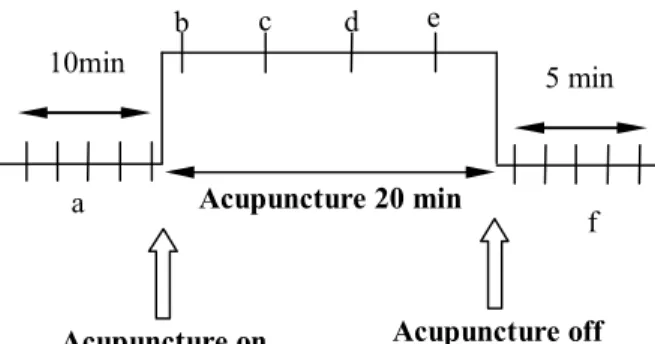
Fig.3 Experimental process. The (a) is rest state before acupuncture; (b-k) is during the acupuncture; (f) is the rest state after acupuncture. a: average of 2-10minute. b-e: average of every 5minute. f: average of final 5minute.
The control group underwent the same procedure and EEG measurements as the test group, but the needle was inserted at a non-acupoint, as shown in Fig. 4.
Fig. 4 The control point
The data analysis
Wavelet based time-frequency analysis identifies the time at which various signal frequencies are present, known as localization analysis. The Fourier analysis of the traditional method may be replaced by the wavelet analysis. A dual function exists in the transformation of a random function
f
L R
2( )
of the continuous wavelet transform to a random signal [31].dt a b t t f a b a Wf
1 () ( ) ) , ((1)
The signal of the wavelet transform Wf ratio (a, b) is the original signal f(t) nearby t = b, according the weighted average (a,b)(t). The “b” parameter represents the central point of the analytic time. The “a” parameter represents the extendable criterion, t = b is as the center of the scope. Its reciprocal is the frequency criterion.
The energy of an EEG signal can be calculated using the following equation:
2
E
c
t f
(2),
E is the brain wave energy, c is a calculated wavelet by (1), t is the unit of the signal translation and f is the frequency.
Finally, the EEG signals were recorded by the continual wavelet transformation method as (among the transform method of a & f was a = 142.86/f ): δ wave ( 0.5-4 Hz, a = 285.72~35.72 )、 θ wave ( 4-8 Hz, a = 35.72~17.86 )、 α wave ( 8-13 Hz, a = 17.86~8.93 ) and β wave ( 13-30 Hz, a = 8.93~4.76. Then, each wave of brain energy was
Control point
f a b c d e Acupuncture 20 min 10min 5 mindivided by the total brain wave energy. The average energy in the second to the tenth minute was used as the baseline. The “a” is the unit of during each five-minute interval of the procedure. The “c” is the energy averages in the eleventh to fifteenth minute. The “d” is the energy in the sixteenth to twentieth minute. The “e” represents the twenty-sixth to thirtieth minute. The “f” is the average energy, five minutes after removing the needles. The complete information was indicated from (mean) ± the tandard difference (SD), and through the ANOVA statistics examination.
Results
We performed acupuncture stimulation at the acupoint, and detected influence through the related meridian. The duration of the stimulation influence is separated into the beginning, the middle and the end.
Test Group
Acupuncture group 0 10 20 30 40 50 δ θ α β N o rm al iz ed (% ) T3 T4 O1 O4Fig.5 EEG average energy of nine minutes before acupuncture, while the test group lay on the beds with eyes closed.
θ wave 14 15 16 17 18 19 20 T3 T4 O1 O2 N o rm a li ze d Pθ /Pt (% ) a b c d e f
Fig.6 12 participants at the waiguan point of average power on θ wave, * means the statistical difference. α wave 24 26 28 30 32 T3 T4 O1 O2 N o rm al iz ed Pα /Pt (% ) a b c d e f
Fig.7 12 participants at the waiguan point of average power on α wave * indicates the statistical difference
Fig. 5, the average energy of nine minutes was, before acupuncture, divided into the positions δ、θ、α、β.
Fig. 6 presents the acupuncture stimulation from the sixteenth to twentieth minute. T3 is rising (p<0.034), as well as T4 (p<0.034). After removing the needle at minute 30 to 35, T3 abates (p<0.034), as well as T4 (p<0.042). Also, in acupuncture during the sixteenth to the twentieth minute, at O1 rose (p<0.034), as well as O2 (p<0.034). Upon removing the needle, the energy was abated, but no significant difference was noted.
According to Fig. 7, only at T3 was rising during the twenty-first to twenty-fifth minute. The others (T4, O1, O2) showed no statistical difference. Additionally, T3, T4, O1 and O2 on the δ, β waves did not present major differences
* * * * * * *
before or after acupuncture.
Control group
The control point is showed in Fig. 4, so that it can be compared to the acupoint. Fig. 8 shows the average energy nine minutes before needle insertion, divided into the positions δ、θ、 α、β. Sham group 0 10 20 30 40 50 δ θ α β N o rm al iz ed (% ) T3 T4 O1 O2
Fig.8 the average EEG energy nine minutes before needle insertion, measured while the control group lay on the beds with eyes closed.
In Fig. 9 during needle insertion, the energy grew at T3, T4, O1 and O2. Statistical increases were noted at the twenty-first to twenty-fifth minute at T3 (p<0.008), T4 (p<0.01), 01 (p<0.007), 02 (p<0.015) and in the twenty-sixth to thirtieth minute at θ wave 15 16 17 18 T3 T4 O1 O2 N o rm al iz ed Pθ /Pt (% ) a b c d e f
Fig.9 The control point of average power on θ wave, for 12 participants.
α wave 28 28.5 29 29.5 30 T3 T4 O1 O2 N o rm a li z ed Pα /Pt (% ) a b c d e f
Fig.10 The control point of average power on α wave for 12 participants.
T3 (p<0.02), T4 (p<0.003), 01 (p<0.002) and 02 (p<0.004). After removing the needles, the energy abates at T3 (p<0.031), T4 (p<0.045) and O1 (p<0.029). O2 did not show significant change.
In Fig. 10, no significent statistical difference is shown.
Discussions
The research of acupuncture, including the substantial results from disease treatment testing, has become a popular subject for scholars worldwide. In recent years, the study of acupuncture’s effects on the brain, has also grown in popularity [27]. Many researchers, however, still believe that acupuncture has little to no affect on the brain. In a previous experiment, 14 participants were subjected to acupuncture treatment, to observe brain wave changes. The outcome of experiment showed that, in fact, there was no relation between acupuncture and brain wave activity. Though, one participant’s α ray showed slight change, the researcher concluded the variation was caused by the participant’s mental and physical factors [24].
We propose the following arguments to these prior findings: * * * * * * * * * * *
1. In the previous research, no descriptions of the needle depth, and whether or not the participants reached “de qi” status were given. In this research, each participant in the experiment must be subjected to an acupuncture depth of at least 1.5cm (The depth of “de qi” at waiquan point in Chinese medicinal theories). Each participant was also confirmed to be at “de qi” by a clinical doctor.
2. In previous research, the acupoints and measured brain wave points were not on the same meridian. Without a connection between the measured brain wave point and the acupoint, the participant only experiences pain. For this reason, we chose an acupoint and a measured brain wave point on the same meridian. 3. In previous research, experimenters used no control group. Moreover, each participant’s reaction was discussed separately. In our study, we observed two groups of subjects, to prove that the participants’ brain wave reactions were not caused by metal factors or physical pain.
In the experiment, we separated the participants into a test and control group. Members of the test group were pierced with fine points at a depth of 1.5cm. All were confirmed as reaching the status of de qi. The other participants were not pierced at any acupoints, and their pierced depth was merely 0.5cm.
The participants’ brain waves would have been affected by environmental, mental and physical factors. Therefore, we had to eliminate all of these factors during the experiment.
To eliminate these factors, we asked all participants lie down and to relax. All participants closed their eyes during the treatment, to reduce interference and the
incitement of exterior light. The requirement was also to ensure that all signals came from acupuncture treatment.
Fig. 5 and Fig. 8 are the average energies of the test and control groups’ brain wave activity, nine minutes before piercing. From the change in brain wave energy in the figures, we see that during the experiment that the participants were awake and in a comfortable situation without the influence of outside factors. If the participants had been affected by mental or physical factors, the outcome of the experiment would have also been affected. We considered these mental and physical factors to ensure that the outcome of the experiment was neutral and believable.
One necessary clarification is the extreme difficulty that we faced, trying to keep all the participants awake. They were required to close their eyes throughout the entire experiment. If the participants fell asleep, then the activity of δ wave and θ wave energy would be stronger than α wave and β wave. If this occured, we could not accurately observe the effecta of the acupuncture. In Table 1 and Table 2, the brain wave energy (mean±SD) shows that the energy of α wave and β wave was stronger than δ wave and θ wave. In this case, we know that the participants were awake.
Due to unstable brain wave signals, participants’ physical or mental factors would also affect the results of the experiment. Such factors include: anxiety, nervousness and taking tranquilizers or any other psychiatric medicine. Rapid-eye potentials and sounds were also recorded by EEG [24]. Therefore, before the experiment, the participants eliminated all mental and physical incitements.
placebo effect, to conduct the precise brainwave measurements. To do this, both groups were made aware of the parameters of the study. All participants were awake and relaxed during the treatment.
Fourier transform is the most important and widely used digital signals analysis technique. It is also a standard quota analytic method [18-20]. Although, the Fourier transform is widely used as a spectrum analyzer, it cannot detect all unstable state signals [21], such as EKG, EEG and EMG. Hence, we used the continuous wavelet transform to analyze the signals, in order to improve Fourier transform’ s deficiency [28].
The activity of θ wave energy would come up when the participants felt sleepy [18]. Although, we had required that all participants were to remain awake during treatment, it was hard to ensure that all participants wouldn’t feel drowsy. Thus, we think that the participants of both groups, caused the θ wave increase by becoming sleepy, and not because of the acupunctural stimulation.
The α wave frequency, posed approximately in 8~13HZ on EEG in the occipital bone area (at O1, O2 points), was more easily detected than the other [17, 24]. As a result (Fig. 7.), we discovered that in the test group, only the α wave at the T3 point had a significant rise (27.42% to 27.94%). The other electrode points did not show any statistical differences. The control group (Fig. 10.) showed no significant differences on the T3, T4, O1 or O2 points during and after being pierced at a non-vital point. No brain wave influence was shown in the control group. The test group experienced an influence on the α wave group, while their
acupoints were pierced. This result proves that acupuncture does have some influence on brain wave activity.
According to traditional Chinese medicinal theory, the effect of acupuncture is shown by “Qi”, the rate of change, slower than the nerve transmission. The findings in the test group demonstrated that the effect of the left brain wave (Fig. 7.) was taken five minutes late during acupuncture.
In general, the brain wave measuring signal is usually detected by an electrode on a person’s scalp. We measured the brain electric wave signal, transmitted from the skull to the cortex into the scalp, using EEG equipment. In the measuring process, the transmission of the signal was influenced by variations in each participant’s cerebral cortex structure, skull thickness and scalp density [22].
Yoram Eshel et al [23], applied a mathematical model to simulate the differences in human skull and scalp. These researchers observed the relationship between the model and EEG signals, and they pointed out the differences of skull thickness, angle of measurement and density of scalp, all of which affected the information transfer of an EEG.
With multiple brain structures to read, the EEG signal may have had some band signals closure. We considered during acupuncturing at the waiquan point on the left hand, the “Oi” went along the triple energizer meridian and later arrived in the Temporal lobe. Due the influence of the qi, it may change the potential between the skull and scalp, and that made the α wave be easily detected on the EEG.
Acupuncture has no influence on the right brain α wave, because the left triple energizer
meridian disconnects with the right triple energizer meridian. Because the qi leaves on the left side of brain, it is unable to influence on the right side of brain.
In the past, patients were administered drugs for the treatment of anxiety and insomnia,. Herrmann et al, proved that the patients who took drugs, saw their α wave rise. They would be relaxed and fall asleep [25]. By comparison, in the acupuncture treatment, the participants’ α wave energy also rose, while their waiquan points were pierced. Consequently, the acupuncture treatment may make people relaxed and help their sleep. The issue could be further addressed by subsequent research.
Of note is the fact that, some previous researchers emphasized a needle twirling technique [9,24]. In our experiment, we did not adopt this method, because each acupuncturist used a slightly different approach, varying the strength and speed of the needle.
Conclusions
By placing a measurement point and an acupoint on the same meridian, the influence of acupuncture on brain waves can be determined.
When a fine needle is inserted into the left hand, the influence on the wave was observed in the left brain, not in the right brain, because the left side of the triple energizer meridian does not connect with the right side of the triple energizer meridian.
If a needle is not placed at an acupoint, no related brain wave activity will take place.
Acupunctural stimulation causes the wave to rise significantly. Thus, a patient could receive acupuncture treatment to release tension and anxiety, or to aid his or her sleep, instead of
taking medicine.
We also prove that a non-twirling needle technique has the same curative effect reached by twirling the needle.
Table 1、Test group of average energy of EEG(mean±SD).* significant difference from a ,# significant difference from e (p<0.05)
Test group
a
b
c
d
e
f
T3
P
δ/P
t%
17.35±7.59 16.67±6.16 16.61±7.06 16.58±6.94 16.68±7.25 16.35±5.79P
θ/P
t%
15.39±1.29 15.6±0.58 16.14±0.91*
15.95±0.57 15.98±0.56 15.89±0.24#
P
α/P
t%
27.42±2.12 27.73±1.59 27.83±1.68 27.94±1.45*
27.83±1.55 27.94±1.05P
β/P
t%
39.85±4.27 40.00±4.17 39.42±6.31 39.52±6.09 39.51±6.33 39.82±4.94T4
P
δ/P
t%
19.06±13.71 17.25±8.51 16.98±8.72 16.90±8.4 16.97±8.62 16.55±6.85P
θ/P
t%
15.31±1.57 15.8±0.32 16.17±0.99*
16.00±0.62 16.02±0.68 15.98±0.42#
P
α/P
t%
28.36±4.06 27.56±2.05 27.59±2.41 27.76±1.93 27.61±2.17 27.92±1.05P
β/P
t%
38.8±8.18 39.39±6.66 39.26±7.33 39.35±7.1 39.40±7.15 39.55±6.31O1
P
δ/P
t%
15.72±3.07 15.47±2.52 16.19±5.74 16.36±6.28 16.64±7.20 15.81±4.05P
θ/P
t%
16.00±0.98 16.08±1.22 16.51±2.20*
16.32±1.82 16.31±1.72 16.17±1.08P
α/P
t%
28.64±2.6 28.74±2.32 27.96±1.13 27.96±1.27 27.72±1.78 28.48±1.24P
β/P
t%
39.65±5.18 39.71±5.42 39.33±6.89 39.36±6.93 39.32±7.21 39.54±6.15O2
P
δ/P
t%
15.92±3.69 15.54±2.93 16.32±6.45 16.51±7.04 16.74±7.84 15.85±4.46P
θ/P
t%
15.91±0.68 16.03±1.00 16.49±2.09*
16.31±1.76 16.31±1.67 16.13±0.93P
α/P
t%
28.48±2.14 28.65±2.07 27.87±1.43 27.85±1.59 27.63±2.06 28.42±1.09P
β/P
t%
39.69±5.3 39.78±5.44 39.33±7.17 39.33±7.28 39.32±7.51 39.59±6.23control group
a
b
c
d
e
f
T3
P
δ/P
t%
14.67±1.07 14.56±0.94 14.06±0.28 14.29±0.53 14.49±0.7 14.49±0.66P
θ/P
t%
15.79±0.16 15.85±0.25 15.82±0.33 15.93±0.22*
15.96±0.23*
15.92±0.3#
P
α/P
t%
28.36±0.46 28.41±0.45 28.57±0.51 28.51±0.48 28.43±0.42 28.37±0.46P
β/P
t%
41.18±0.82 41.18±0.71 41.54±0.49 41.27±0.44 41.12±0.68 41.22±0.72T4
P
δ/P
t%
14.78±0.97 14.35±0.81 13.96±0.27 14.2±0.53 14.4±0.71 14.38±0.66P
θ/P
t%
15.81±0.16 15.88±0.25 15.84±0.33 15.94±0.22*
16.00±0.23*
15.93±0.3#
P
α/P
t%
28.36±4.06 28.42±0.44 28.55±0.51 28.48±0.49 28.41±0.42 28.35±0.46P
β/P
t%
41.35±0.78 41.36±0.65 41.65±0.51 41.38±0.44 41.23±0.69 41.34±0.73O1
P
δ/P
t%
14.54±0.96 14.41±0.84 14.01±0.27 14.24±0.52 14.44±0.70 14.42±0.65P
θ/P
t%
15.77±0.16 15.85±0.25 15.81±0.33 15.91±0.22*
15.94±0.23*
15.90±0.30#
P
α/P
t%
28.31±0.46 28.37±0.44 28.50±0.51 28.44±0.48 28.36±0.42 28.30±0.46P
β/P
t%
41.38±0.75 41.38±0.66 41.68±0.50 41.41±0.44 41.26±0.68 41.37±0.73O2
P
δ/P
t%
14.44±0.93 14.32±0.74 13.96±0.27 14.20±0.53 14.41±0.70 14.37±0.65P
θ/P
t%
15.80±0.16 15.87±0.25 15.82±0.33 15.93±0.22*
15.95±0.23*
15.92±0.30P
α/P
t%
28.34±0.45 28.39±0.42 28.51±0.51 28.44±0.49 28.37±0.42 28.31±0.46P
β/P
t%
41.41±0.78 41.42±0.63 41.71±0.51 41.43±0.44 41.27±0.69 41.39±0.73Table 2、Control group average energy of EEG(mean±SD). * significant difference from a ,# significant difference from e (p<0.05)
References
[1] Julia J. Tsuei, The Science of acupuncture Theory and Practic;IEEE ENGINEERING IN MEDICINE AND BIOLOGY 1996. [2] acupuncture.—Langevin, H. M., Churchill,
D. L., Cipolla, M. J. Mechanical signaling through connective tissue: a mechanism for the therapeutic effect of acupuncture. FASEB J. 15, 2275–2282 (2001).
[3] Christensen PA, Rotne M, Vedelsdal R, Jensen RH, Jacobsen K, Husted C. Electroacupuncture in Anaesthesia for hysterectomy British Journal of Anaesthesia 1993; 71:835-838.
[4] Stanley TH, Cazallaa JA, Atinault A, Coeytaux R, Limoge A, Louville Y. Transcutaneous cranial electrical stimulation decreases narcotic requirements during neurolept anaestheiac and operation in man. Anesthesia and Analgesia. 1982; 62:836-866. White PF. Use of patient-controlled analgesia for management of acute pain. JAMA 1988; 259: 243-247.
[5] Lu DP. acupuncture anesthesia/analgesia for pain and anxiety control in dental practice Part 2 Techniques for clinical applications. Review compendium. 1993; 14-4:464-468, 470-472.
[6] Wedenberg K, Moen B, Norling A. A prospective randomized study-comparing acupuncture with physiotherapy for low-back and pelvic pain. Acta Obestetricia et Gynecologica Scandinavica. 2000; 79(5): 331-5.
[7] Wang RR, Tronnier V. Effect of acupuncture on pain management in
patients before and after lumbar disc protrusion surgery- a randomized control study. American Journal of Chineses Medicine. 2000; 28(1): 25-33.
[8] Todd B. Parrish, Alissa Schaeffer, Madelyn Catanese, and Mary J. Rogel , Functional Magnetic Resonance Imaging of Real and sham acupuncture, 2005;IEEE ENGINEERING IN MEDICINE AND BIOLOGY MAGAZINE, 2005;IEEE ENGINEERING IN MEDICINE AND BIOLOGY MAGAZINE; 35-40
[9] Elisa E. Knonofagou and Helene M.Langevin, Using Ultrasound to Understand acupuncture, 2005;IEEE ENGINEERING IN MEDICINE AND BIOLOGY MAGAZINE, 2005;41-46 [10] Litscher, G. “Effects of acupressure,
manual acupuncture and Laserneedle acupuncture on EEG bispectral index and spectral edge frequency in healthy volunteers”, European Journal of Anaesthesiology. 21(1):13-9, 2004 Jan. [11] Litscher, Gerhard, “Electroencephalogram
-- entropy and acupuncture”, Anesthesia & Analgesia. 102(6):1745-51, 2006 Jun. [12] Chen, Andrew C N. Liu, Feng-Jun. Wang,
Li. Arendt-Nielsen, Lars. “Mode and site of acupuncture modulation in the human brain: 3D (124-ch) EEG power spectrum mapping and source imaging”, Neuroimage. 29(4):1080-91, 2006 Feb [13] 14Li Wang, Andrew C. N. Chen, Lars
Arendt-Nielsen. “Cortical plasticity: effect of high and low intensity conditioning electrical stimulations (100 Hz) on SEPs to painful finger stimulation”Clinical Neurophysiology. 117(5):1075-84, 2006
May.
[14] 15Takamatsu, I. Ozaki, M. Kazama, T. “Entropy indices vs the bispectral index for estimating nociception during sevoflurane anaesthesia.” British Journal of Anaesthesia. 96(5):620-6, 2006 May [15] 16Joseph J. Carr, John M. Brown,
Introduction to Biomedical Equipment Technology, 4th Edition, Prentice Hall, 2001.
[16] 17R. W. Homan, J. Herman, and P. Purdy, “Cerebral location of international 1G20 system electrode placement,” Elecrroenceph. Clin. Neurophysiol., vol. 66, pp. 376-382, Apr. 1987.
[17] 18Langevin, H. M., Churchill, D. L., Cipolla, M. J. “Mechanical signaling through connective tissue: a mechanism for the therapeutic effect of acupuncture.” FASEB J. 15, 2275–2282 (2001).
[18] 18PARDEY, J., ROBERT, S., and TARASSENKO, L.: ‘A review of parametric modelling techniques for EEG analysis’, Med. Eng. Phys., 1996. 18. (1). nn. 2-11
[19] 20JUNG, T.P.: ‘Estimatine alertness from the EEG power spectrum’, IEEE Trans. Biomed. Eng., 1997,44,(1), pp. 60-69 [20] 21TSENG. S , CHEN, R , and CHONG F.:
‘Evaluation of parametric methods in EEG signal analysis’, Med. Eng. Phys., 1995, 17, (1), pp. 71-78
[21] M.Shen, L.Sun and F.H.Y.Chan Method for extracting time-varying rhythms of electroencephalography via wavelet packet analysis, IEE Proc-Sci. Meas. Technul.. Vol. 148, No. I, Jurtucuy 2001
[22] Nicolas Chauveau, Xavier Franceries,
Bernard Doyon, Bernard Rigaud, Jean Pierre Morucci, and Pierre Celsis1, Effects of Skull Thickness, Anisotropy, and Inhomogeneity on Forward EEG/ERP Computations Using a Spherical Three-Dimensional Resistor Mesh Model, Human Brain Mapping 21:86 –97(2004) [23] Yoram Eshel, Sima (Levy) Witman,
Moshe Rosenfeld, and Shimon Abboud, Correlation Between Skull Thickness Asymmetry and Scalp Potential Estimated by a Numerical Model of the Head, IEEE TRANSACTIONS ON BIOMEDICAL ENGINEERING, VOL. 42, NO. 3, MARCH 1995
[24] P. Rosted, P.A. Griffiths, P. Bacon, N. Gravill,” Is there an effect of acupuncture on the resting EEG? ” Complementary Therapies in Medicine (2001), 9, 77–81 [25] Herrmann, W M. Winterer, G.
“Electroencephalography in psychiatry--current status and outlook”, Nervenarzt. 67(5):348-59,1996 May. [26] Metin Akay, Time Frequency and
Wavelets in Biomedical Signal
Processing, IEEE Press Series in Biomedical Engineering, 1997.
[27] Vitaly Napadow,1,2 Nikos Makris,3 Jing Liu,1 Norman W. Kettner,2 Kenneth K. Kwong,1 and Kathleen
K.S. Hui1*, ‘Effects of
Electroacupuncture versus
ManualAcupuncture on the Human Brain as Measured by fMRI’, Human Brain Mapping 24:193–205(2005)
[28] Franco Rossetti a,b,1, Marcelo
Antˆonio Cortes de Oliveira a, Norberto Garcia-Cairasco a,b*, EEG
wavelet analyses of the
striatum–substantia nigra pars
reticulata–superior colliculus circuitry:
Audiogenic seizures and
anticonvulsant drug administration in Wistar audiogenic rats (War strain), Epilepsy Research 72 (2006) 192–208 [29] Peter Deadman & Mazin Al-Khafaji
with Kevin Baker, A Manual of ACUPUNCTURE
![Fig. 1 The sanjiao primary channel[29]](https://thumb-ap.123doks.com/thumbv2/9libinfo/8987153.282925/3.892.130.438.573.826/fig-the-sanjiao-primary-channel.webp)