MicroRNA-30a inhibits vimentin expression and is associated with breast cancer progression.
Abstract
Recent studies suggest a significant role of microRNAs (miRNAs) in regulating cancer development. To delineate possible mechanism of miRNAs in breast cancer progression, miRNA microarray analysis was conducted in tumors, diagnosed as different stages and lymph-node metastasis (LNM) status, and showing different progression signatures, indicated by overexpression of cyclin D1 (CCND1) and β-catenin (CTNNB1), and resulted in the identification of miR-30a. In silico prediction of gene discovery suggested that 3′-untranlated region(UTR) of the vimentin (vim), encoding protein involved in epithelial-mesenchymal transition, harbors potenital binding sequences for miR-30a. Further experiments, showing that miR-30a
negatively regulated vim expression and the interaction between miR-30a and 3-UTR of vim in regulating reporter gene expression, suggest the role of miR-30a in
mediating tumor invasion and metastasis. Consistent to this, ectopic expression of miR-30a was found to suppress migration and invasion phenotypes of breast cancer cells. More importantly, reduced expression of miR-30a in tumors was associated with poor clinical features of breast cancers and worse progression in breast cancer patients. These findings support the role of miR-30a in breast cancer progression.
Keywords: Breast cancer; Laser captured microdissection; miR-30a, metastasis, prognosis
Introduction.
The reversibility of epigenetic regulation allows genes to be switched on as well as off, affecting the level of expression of normal genes instead of totally abolishing gene function, and thus may provide selective advantage for clonal evolution during tumorigenesis. The rationale underlying this suggestion is that tumor progression is always characterized by an unstable, phenotypic heterogeneity which fluctuates too frequently to be mediated exclusively by rigid irreversible genetic changes [1, 2]. This suggestion can be exampled by the status of contribution of E-Cadherin during cancer progression. During tumor metastasis, cancer cells undergo epithelial-mesenchymal transition (EMT) in which epithelial tumor cells are converted into aggressive and metastatic tumor cells, and one of the characteristic findings in EMT is the loss of cell-cell adhesion with reduced expression of E-cadherin [3-5]. Cells exhibiting a mesenchymal phenotype transported to metastatic sites may undergo mesenchymal-epithelial transition (MET) by regaining E-cadherin expression, allowing the cell-cell adhesion which connects adjacent cells to form new foci. For this purpose, epigenetic regulation appears to be more suitable than genetic level regulation for maintaining flexibility, and, in the case of E-cadherin, hypermethylation of the promoter region is found in various human carcinomas [6-9]. MicroRNA (miRNA) is the other
epigenetic regulatory mechanism, which is a novel class of short non-coding RNA molecules consisting of 19-25 nucleotides, having the potential to inhibit gene expression by binding to complementary sequences at the 3′-UTR untranslated regions (UTRs) of target mRNA transcripts. A growing list of miRNAs have been suggested as putative oncogenes or tumor suppressors which contribute to cancer development and progression, and are differentially expressed in normal tissues and cancers [10-13]. Most recently, several intriguing studies have described other novel
epigenetic regulations underlying EMT activation, including regulation by microRNAs, which primarily act by down-modulating the translation of target mRNAs [14, 15].
We have recently investigated the epigenetic alteration that is an important predictor of clinical outcome in breast cancer. Our data provide the evidence to support that overexpression of the three genes making up this expression signature, namely CCND1 (encoding cyclin D1), CTNNB1 (encoding β-catenin), and MTA1 (encoding metastatic tumor antigen-1), was found to be highly associated with poor differentiation of tumor cells, advanced stage, and, as expected, low survival rate (manuscript under revision). To examine the mechanism associated with this
signature, this study investigated whether and how deregulation of specific miRNAs associated with this epigenetic aberration is involved in driving invasiveness and metastasis of tumor cells. The expression profile of miRNAs was then compared between breast tumors showing both poor signature, i.e. overexpression of CCND1 and CTNNB1, and lymph node metastasis (LNM), and breast tumors showing good signature and no LNM. Among the miRNAs showing a significantly difference in expression, particular attention was focused on miR-30a, as in silico prediction of target genes regulated by miR-30a resulted in the identification of vimentin (Vim), a mesenchymal marker implicated in EMT during breast tumorigenesis [16-18]. We therefore explored whether miR-30a regulates vimentin expression and identified aberrant miR-30a expression associated with poor breast cancer progression. These findings shed light on a novel fundamental mechanism with clinical significance and translational implication.
Results and Discussion.
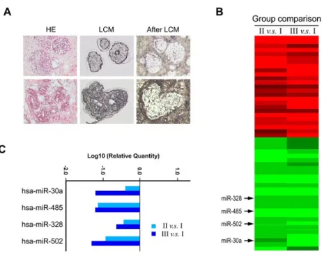
To search for miRNAs that might play a role in determining breast cancer progression, we used a miRNA chip to compare the miRNA expression profiles of three groups with differential expressions of CCND1 and CTNNB1 genes and different LNM status. We compared the profiles of 5 patients with Stage I/II cancer (LNM-) plus normal expression of CCND1 and CTNNB1 (group I), 4 with Stage I/II (LNM-) plus enhanced expression of CCND1 and CTNNB1 (group II), and 5 with Stage III/IV (LNM+) plus enhanced expression of CCND1 and CTNNB1 (group III). None of these fourteen patients had received neoadjuvant treatment before surgery, thus avoiding any confounding effect on gene expression. The two genes CCND1 and CTNNB1 make up an expression signature highly associated with poor pathological features and a worse clinical outcome [submission under revision]. It should be noted that the determination of miRNA expression status was based on cancerous cells microdissected from tumors to ensure that the samples assayed consisted of >95% pure breast tumor epithelial cells separated from the resected tumor specimen (Figure 1A), supporting the validity of our measurement. As shown in Fig. 1B and 1C, using a cutoff of a greater than two-fold change, a total of 52 miRNAs were found to be differentially expressed in breast tumors with different LNM status, 25 being
upregulated (red section of Figure) and 27 downregulated (green section) in group III or II compared to group I, five of the downregulated miRNAs being 502, miR-485, miR-519e, miR-328, and miR-30a. To gain an insight into the functional consequences of differential miRNA expression, we used the search program TargetScan 5.1 and a computational algorithm to explore whether the expression of any EMT-associated genes was regulated by these 52 miRNAs and the results suggested that expression of Vim gene, encoding an intermediate filament normally
expressed in cells of mesenchymal origin, might be associated with decreased expression of miR-502, miR-485, miR-519e, miR-328, and miR-30a. Vimentin is an important EMT-associated marker predicting patient prognosis [16, 18] and has a function in the overall organization of the cytoskeleton and is thus implicated in increased invasiveness or migration of cells [17, 19].
To test the in silico prediction that vimentin expression might be regulated by specific miRNAs, we first measured vimentin levels by immunoblotting after transient transfection of the human breast cancer cell lines Hs578T and MDA-MB-231 with the precursors of the above five miRNAs predicted to affect vimentin expression. As shown in Figures 2A and 2B, vimentin expression was dramatically inhibited by 50% by ectopic introduction of miR-30a, whereas the other four miRNAs had only a minor, or non-significant, effect. To validate the inhibitory effect of miR-30a on vimentin expression in vivo, Hs578T cells transfected with miR-30a were subjected to immunofluorescence staining and confocal microscopy. Consistent with the in vitro results, the intensity of the red fluorescence for both membrane-bound and cytoplasmic vimentin was significantly decreased following transfection with pCDNA/miR-30a (Fig. 2C). Interestingly, miR-30a is located in a region of 6q13 that is frequently affected by genetic lost in breast cancer [20, 21], suggesting that miR-30a has a causal role in breast tumorigenesis.
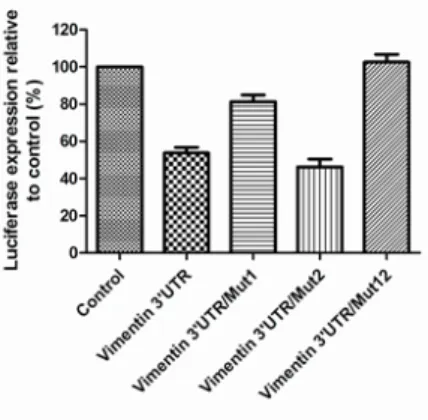
In an attempt to map putative interaction sequences, TargetScan 5.1 was used to predict possible targeting motifs and identified two seed regions within the 3’ UTR of Vim to which miR-30a might bind (Figure 3A). To investigate whether Vim mRNA was directly targeted by miR-30a and to determine the relative importance of the two regions, we cloned the full-length 3’UTR of Vim into the pGL4.13_1-luciferase reporter (Vim 3’UTR-luc) and examined the effect of miR-30a on the expression of the reporter gene. In addition, we prepared three 3’UTR mutants, two of which, Vim 3’UTR/mut1-luc and
Vim 3’UTR/mut2-luc, harbored a mismatch sequence complementary to miR-30a within either seed region 1 or seed region 2, and Vim 3’UTR/mut12-luc, harboring both
mismatch sequences (Figure 3B). Cotransfection of miR-30a and Vim 3’UTR-luc into cells led to a significant reduction (47%) in luciferase activity compared to transfection with Vim 3’UTR-luc alone (Figure 3C), supporting the notion that miR-30a can regulate vimentin expression by binding to its 3’UTR. Similarly, a significant reduced luciferase activity (54%) affected by miR-30a was observed in cells carrying the Vim 3’UTR/mut2 clone, but not those carrying Vim 3’UTR/mut1 or Vim 3’UTR/mut12 (Figure 3C), suggesting that the critical region composed of six nucleotides (GTTTAC)
complementary to miR-30a was site 2, located at the 5’ seed sequence within the 3′UTR of Vim (Figure 3A).
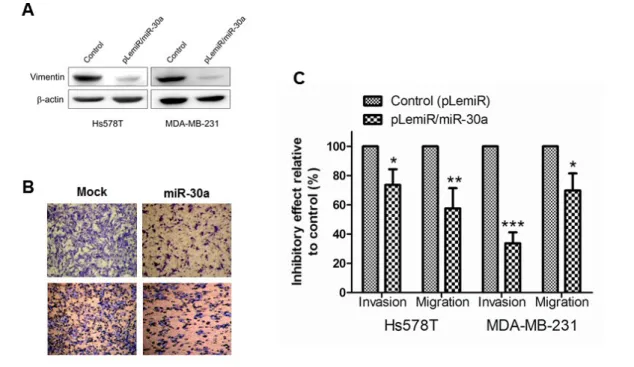
Because EMT is a trans-differentiation program which converts adherent epithelial cells into individual migratory cells, changes the cell phenotype into more loose mesenchymal-like cells, and leads to invasion of the extracellular matrix, it is critical for tumor metastasis [22, 23]. Thus, our finding that vimentin expression was inhibited by miR-30a prompted us to examine phenotypes of migration and
invasiveness affected by this microRNA. As expected from the data presented here, introduction of miR-30a into the cell lines Hs578T and MDA-MB-231 was
accompanied by reduced vimentin expression (Figure 4A) and by reduced motility (migration) and invasiveness (Figures 4B and 4C).
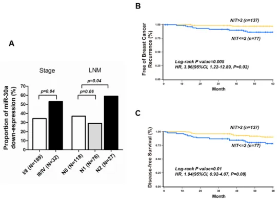
These results allow us to propose a model in which expression of miR-30a is progressively reduced during cancer progression and the loss of miR30a, in turn, results in poor cancer progression. Our observations in breast cancer patients were in line with this expectation and showed that the percentage of patients with reduced miR-30a levels, measured by a two-fold decrease in tumor tissue compared to the
adjacent non-tumor breast epithelium in which cells were micro-dissectedfrom the primary tumor site, was significantly higher in tumors of advanced stage or with LNM (Figure 5A). Finally, we examined whether decreased miR-30a levels were associated with a worse outcome. Figures 5B and 5C show that, in a cohort of breast cancer patients followed up for five years, there was a trend toward a high rate of breast cancer recurrence and a decreased disease-free survival in patients expressing lower miR-30a levels in tumor cells compared to their corresponding non-tumor parts. When patients’ stage and ER status were taken into consideration in the Cox
regression model, all breast cancer patients with decreased miR-30a levels in primary cancerous sites demonstrated an increased hazard ratio (HR) for recurrence or
recurrence plus mortality during the follow-up period (Figures 5B and 5C).
This study demonstrates that miR-30a inhibits vimentin expression and regulates invasiveness and metastasis phenotypes of breast cancer cells, allowing that low miR-30a levels are associated with poor breast cancer progression. It is thought that levels of vimentin need to be optimal for maintaining the architecture of the cytoplasm [24], aberrances of which are frequently observed during EMT and are suggested to be essential for driving cancer metastasis [18, 23, 25]. Also, similar to the effect seen with miR-30a in the present study, impaired adhesion and migration was shown to reduce their in vitro adhesion and motility in vimentin-ablated cells, emphasizing the functional contribution of vimentin to epithelial cell invasion/migration [19]. As a result, it is possible that the phenotypes seen in the breast cancer cells and the clinical outcomes observed in our patients were linked to decreased vimentin expression mediated by miR-30a. However, it should be noted that other metastasis-associated genes might be also caused by reduced miR-30a, leading to these invasive phenotypes and poor outcomes of breast cancer. Furthermore, the signaling pathways regulating
miR-30 expression are an important issue to be explored in future study. Together, these results help in understanding the tumorigenic contribution of miR-30a to breast cancer development and may result in the clinically practical advantage of mirR-30a as a marker for predicting cancer progression and as a possible therapeutic target to prevent cancer metastasis.
Materials and methods
Cell culture and miRNA transfectants.
Human cell lines were cultured as described in supplementary methods.
Transfections for different miRNA mimics and plasmid constructs were done using DOTAP and TurbofectTM in vitro transfection reagents as described in the
Supplementary Methods.
MicroRNA microarray and quantitative real-time PCR analysis.
IDC tissue samples consisting of 5 patients with Stage I/II cancer (i.e. LNM-) plus normal expression of CCND1 and CTNNB1 (group I), 4 with Stage I/II (i.e. LNM-) plus increased expression of CCND1 and CTNNB1 (group II), and 5 with Stage III/IV (i.e. LNM+) plus increased expression of CCND1 and CTNNB1 (group III) was used for miRNA microarray. Comparatively quantitative analysis for human miRNAs were done by using the TaqMan® low density array (LDA) on an Applied Biosystems 7900HT Fast Real-Time PCR System (Foster City, CA, USA) as described in the Supplementary Methods.
Plasmid constructs, luciferase assay, and Western blotting.
Target regions of miR-30a in the mRNA 3′UTR of Vim were cloned downstream of a pGL4.13-luciferase reporter. The control and various 3’UTR reporter constructs were cotransfected along with miR-30a as described in Supplementary Methods. The primers used for mRNA expression are listed in Supplementary Table S1. Luciferase assays and Western blotting were done as described in the Supplementary Methods.
Invasion, migration, and confocal studies.
Quantitative analysis of invasiveness and migration for tumor cell were done using Boyden chamber assay. Representative image of miR-30a mediated vimentin
down-regulation on the biology of breast cancer cells was done using confocal studies as detailed in the Supplementary methods.
Tissue samples and laser captured microdissection.
Primary tumor tissue samples were obtained from the Surgery Department of the Tri-Service General Hospital, Taipei, Taiwan, during mastectomy of previously untreated IDC patients. The histological diagnosis of all specimens was reviewed by a certified pathologic physician as described in the Supplementary methods and the clinico-pathological findings were summarized in the Supplementary Table S2. In addition, for molecular miRNA analysis, paired tumor and non-tumor cells from each patient were collected using laser captured microdissection technique as described in the Supplementary methods.
Quantitative real-time PCR analysis of miRNA
Total RNA and microRNA were isolated using RNAqueous®-Micro Kit and mirVana miRNA isolation kit (Ambion Inc., Austin, TX), respectively. Comparative quantitation of miR-30a levels between paired LCM-dissected tumor and non-tumor (NT) cells in each IDC tumor tissues were measured using the standard two-step TaqMan real-time PCR assay (Applied Biosystems) with2-ddCt analysis as described in
the Supplementary methods.
Statistical analysis
Data are represented as mean ± S.D. Quantitation of the miRNA transcript was determined according to formal definitions. The expression level of miR-30a in each subgroup of patients was presented as the mean of T/NT ratios of each paired breast tissues, and the difference between different subgroup of patients was examined by two-sided chi-square test as described in the Supplementary methods.
Supplementary Methods.
Study population and specimen collection
The present study is part of an ongoing cooperative study aimed at discovering markers for the evaluation of breast cancer progression in Taiwan, where breast cancer is characterized by low incidence [26], early tumor onset [27], reproductive hormone dependency [28, 29], and novel genomic alteration [29-32]. This study was approved by the Ethics Committees of the Institutional Review Boards of the Tri-Service General Hospital, Taipei, and of the Chung Shan Medical University
Hospital, Taichung, Taiwan. All breast cancer patients had pathologically confirmed primary invasive ductal carcinoma (IDC) of the breast. The enrolled female IDC patients were a subset of women randomly selected from the larger, ongoing hospital-based breast cancer cohort collected in the Surgery Department of the Tri-Service General Hospital. Informed consent was obtained from each participant prior to specimen acquisition. The resected breast cancer tissues were immediately frozen in liquid nitrogen until analysis. Tumor grade in each patient was categorized as I, II, or III according to the Nottingham modification of the Scarff-Bloom-Richardson system and the pathological assessments of these tumors were classified according to the sixth edition of the AJCC CancerStaging Manual and categorized as I, II, III, or IV. None of the patients received neoadjuvant treatment before their primary surgery, thus avoiding any effects on gene expression.
Laser capture microdissection (LCM)
To ensure that the tissue samples assayed consisted of >95% pure breast tumor epithelial cells separated from the resected tumor specimen, LCM was performed on routinely immunostained slides usinga PixCell laser capture microscope (Arcturus Engineering, MountainView, CA) as described previously [33, 34]. The dehydrated tissue section was overlaid with a thermoplasticfilm mounted on an optically transparent
cap and the visually selectedareas (tumor cells) were bound to the membrane by short, low-energylaser pulses, resulting in focal melting of the polymer. Onaverage, 2,500-3,000 LCM shots were performed on a single tumor to obtain sufficient tumor cells for comparative qRT-PCR analysis. The laser-captured tumor cells were immersed in 50-100 µl of digestion buffer (10 mM Tris-HCl, pH8.0, 1 mM EDTA, 400 µg/ml of proteinase K, and 1% Tween20) and digested at 55°C overnight. After digestion, theenzyme was heat inactivated (95°C for 10 min) and the extractused directly for RNA isolation. RNA isolation
Total RNA from paired LCM-dissected tumor and non-tumor cells from each patient was isolated using an RNAqueous®-Micro Kit (Ambion Inc., Austin, TX) and the yield of RNA determined by spectrophotometry at 260 nm. MicroRNAs from tumor and non-tumor cells were extracted from tissue sections using a mirVana miRNA isolation kit according to the manufacturer’s instructions (Ambion Inc., Austin, TX) and theRNA concentration in each sample quantified on a NanoDrop 1000 spectrophotometer (NanoDrop Technologies, Waltham, MA).
MicroRNA microarray and comparatively quantitative real-time PCR analysis A human miRNA microarray was used to detect the microRNA expression pattern in invasive ductal carcinomas. The microarray contained probes for 381 human miRNAs from the TaqMan® low density array (LDA) Human MicroRNA Panel v1.0 (Applied Biosystems) and levels of mature miRNAs were quantified on an Applied Biosystems 7900HT Fast Real-Time PCR System (Foster City, CA, USA). The expression of each target miRNA was measured using the same quantity of template RNA in each well, using small nuclear RNA U48 (RNU48) as an endogenous negative control and RNU6B as a positive control. Relative
(Applied Biosystems, Foster City, CA, USA). Ct values for all targets were determined using the automatic threshold in RQ Manager v1.1 analysis software.
In addition, LCM was performed on 221 breast cancer tissue slides, and the single-tube TaqMan miRNA assay (Applied Biosystems, Foster City, CA, USA) was used to detect and quantify the mature miRNA on an Applied Biosystems
instruments. The level of expression of the miRNA biomarker was determined using the TaqMan real-time PCR assay and normalized to that for RNU6B. Triplicate qPCR experiments were performed on each breast carcinoma to determine the target mRNA levels in the isolated tumor and non-tumor cells. The comparative CT method (-ddCt) was used to estimate the relative expression (fold change) of the miR-30a transcript in the tumor and non-tumor cells in each case (2− ddCt where ddCt = Ct
miR-30a − Ct RNU6B).
Cell culture
The breast cancer cell lines Hs578T and MD-MB-231 were obtained from the American Type Culture Collection (ATCC, Manassas, VA) and were cultured in Dulbecco’s modified Eagle’s medium (DMEM) (Life Technologies) containing 0.1 mM sodium pyruvate, 10% fetal bovine serum (FBS), 2 mmol/l of L-glutamine, 100 IU/ml of penicillin, and 100 mg/ml of streptomycin (Biosource, Rockvile MD) in a humidified 5% CO2 atmosphere at 37 °C. In some experiments, transfection of these cell lines was performed using DOTAP (Biontex, Laboratories, GmbH) and
TurbofectTM (Fermentas, Germany) in vitro transfection reagents according to the manufacturers’ recommendations. The transfection efficiency (>80%) was determined using an equal amount of a plasmid encoding the green fluorescent protein under the control of the CMV promoter. Transfectants were cultured and selected for 2 weeks in the presence of 3.0 ug/ml of puromycin. Each stable clone was also maintained in medium containing 3.0 ug/ml of puromycin.
The 3’UTR sequence of the human vimentin gene (Vim) was cloned into plasmid pGL4.13 (Promega, Madison, WI) to obtain a recombinant vector, pGL4.13_1, containing the firefly luciferase open reading frame under the control of the SV40 promoter. Two miR-30a complementary sites with the sequence GTTTAC in the Vim 3’UTR were mutated singly or together to remove complementarity to miR-30a using a QuikChange II XL site-directed mutagenesis kit (Stratagene, La Jolla, CA) with pGL4.13_1/Vim-WT as the template and the mutants were named Vim 3’UTR/mut1, Vim 3’UTR/mut2, and Vim 3’UTR/mut12. Figure 3 shows the mutated nucleotides capitalized, and the sequences of the mismatch primers used to generate the Vim 3’UTR mutants are shown in Supplementary Table S1. Hs578T cells were cotransfected with the reporter construct, the control or mutated Vim 3’UTR constructs, and pCDNA/miRNA-30a, then the cells were lysed 24 h later and the firefly luciferase/Renilla luciferase activity ratio of each sample measured in a dual-luciferase assay (Promega, Madison, WI).
Cell lysis and Western blotting
Total cell extracts were prepared in ice-cold lysis buffer (0.5% NP-40, 50 mM Tris-HCl, 150 mM NaCl, 1 mM EDTA, and 10% glycerol, pH 7.5) containing protease inhibitors (1 μg/ml of aprotinin, 0.5 μg/ml of leupeptin, and 100 μg/ml of 4-(2-aminoethyl)-benzenesulfonyl fluoride). Equivalent amounts of cell lysates were resolved by 8-12% SDS polyacrylamide gel electrophoresis and transferred onto polyvinylidene difluoride membranes, which were then incubated for 30 min at 37ºC with blocking buffer (5% fat milk in phosphate-buffered saline) to block non-specific binding and probed overnight at 4ºC with a monoclonal antibody against human vimentin (Santa Cruz Biotechnology) in blocking buffer. The membrane was treated with an appropriate horseradish peroxidase-conjugated second antibody for 1
h. Immunoreactive proteins were visualized by the enhanced chemiluminescence assay (Western blotting luminal reagent; Santa Cruz Biotechnology) and the intensities of the bands quantified by densitometry (Digital Protein DNA
Imagineware, Huntington Station, NY). Anti-β-actin antibody (Sigma-Aldrich Corp., St. Louis, MO) was used to normalize for protein loading.
Establishment of breast tumor cells stably expressing miR-30a
Using a lentivirus expression system (Thermo Fisher Scientific; Waltham, MA), human Hs578T breast cancer cells were transduced with plasmid pLemiR expressing primary-miR-30a (pri-miR-30a) transcripts under the control of the CMV promoter (Open Biosystems; Huntsville, AL). The expression of the pri-miRNA transcripts allows interaction with the endogenous processing that will subsequently yield mature miR-30a. The pLemiR/miR-30a vector was transfected into HEK 293T cells using the Trans-Lentiviral™ GIPZ Packaging System (Open Biosystems) to produce a VSV-G pseudotyped virus according to the manufacturer’s protocol. Stepwise, breast tumor cell lines (Hs578T and MD-MB-231 cells) were transduced by use of the lentivirus containing the miR-30a. TurboRed Fluorescent Protein (TurboRFP) and a puromycin-resistance selectable marker were used to allow screening for exclusion of non-transduced cells. Generation of stable cell lines that stably expressed miR-30a was established.
Cell invasiveness and migration assay
Inhibitory effects of miR-30a on the invasiveness and migration of breast cancer cells were evaluated using a modified Boyden chamber invasion assay . Matrigel (Collaborative Biomedical Products, Bedford, MA) was diluted to 25 mg/50 ml with distilled water and applied to 8 μm pore size polycarbonate membrane filters, then 1.0×10⁵ cells/well of miR-30a transfected cells was seeded into the upper section of
Invitrogen Corp., Carlsbad, CA), with the same medium containing10% FBS in the lower chamber, and incubated overnight at 37 °C, then all remaining cells were removed from the interior of the insert using a cotton-tip applicator and cells that had migrated to the lower surface of the membrane were fixed with methanol and stained with Giemsa. Invading cells were quantified by counting 5 random high-powered fields using a Olympus Ckx41 light microscope. For the migration assay, cells were seeded into the Boyden chamber on membrane filters that were not coated with Matrigel and incubated for 16 h at 37 °C, then non-migrating cells were removed from the upper membrane surface and the invading cells on the lower membrane surface quantified as described above.
Confocal microscopic analysis
Tumor cells transfected with control or miR-30a vector were cultured on coverslips in a 60 mm Petri dish for 24 h, then were washed with isotonic phosphate-buffered saline (PBS), pH 7.4, and fixed with 4% paraformaldehyde solution in PBS for 1 h at 37 °C. The coverslips were then washed three times with PBS and non-specific binding sites blocked in PBS containing 10% FBS, 0.3% Triton X-100 for 1 h at 37ºC. The cells were then incubated overnight at 4 °C with mouse anti-human vimentin antibody (Santa Cruz) (1:100 dilution in PBS containing 10% FBS), then for 2.5 h at 37 °C with Cy3-labeled goat anti-mouse antibodies (Biorad) (1:50 dilution in PBS containing 10% FBS). The nuclei were stained with Hoechst 33258, then the samples were examined under a Leica confocal laser scanning microscope
(Mannheim, Germany) equipped with a UV laser (351/364 nm), an Ar laser (457/488/514 nm), and an HeNe laser (543 nm/633 nm).
Statistical analysis
of tumor stage and LNM status. The miR-30a transcript was quantified using the comparative CT method as described previously [35, 36]. Disease-free survival was measured as the time from surgery to recurrence or the end of the study. To examine whether miRNA-30a could be used as a prognostic biomarker in IDC patients, Kaplan-Meier survival (p value of the log-rank test) and Cox regression analyses (hazard ratio (HR) and 95% confidence interval (95%CI)) were used to explore the association between the 6-year disease-free survival rate and decreased miR-30a levels in IDC patients.
Figure 1. Decreased expression of miR-30a in breast tumors with lymph node metastasis (LNM). Total RNA was extracted from laser-capture-microdissected tumor cells (A) from 5 low-stage (i.e. Stage I/II) breast tumors showing normal expression of CCND1 and CTNNB1 and no LNM (Group I), 4 low-stage tumors showing increased expression (two-fold higher in tumors than in the corresponding non-tumor cells) of CCND1 and CTNNB1, but no LNM (Group II), and 5 high-stage (i.e. Stage III/IV) tumors showing increased expression of CCND1 and CTNNB1 and LNM (Group III). RNA from tumors in the same group was pooled and subjected to TaqMan-LDA micro-RNA microarray analysis. Fig. 1B shows a comparison of the results for group II and I (left panel) and that for group III and I (right panel), particular attention being focused on microRNAs displaying a greater than 2-fold change, resulting in a total of 52 micro-RNAs associated with tumor stage and LNM, of which 25 were upregulated (panel in red) and 27 downregulated (panel in green) during tumor progression, with 4 of the downregulated ones being miR-328, miR-485, miR-502, and miR-30a. Fig 1C shows the quantitative data for these 4 miRs.
Figure 2. miR-30a mediates downregulation of vimentin in vitro and in vivo. (A and B) Micro-RNAs that might impact vimentin expression were transfected into two breast cancer cell lines Hs578T and MD-MB-231, then, 24 h later, vimentin expression was measured by Western blotting compared to that in cells transfected with control vector (A and B). All experiments were performed three times. (C) Confocal analysis of vimentin expression in miR-30a-transfected MDA-MB-231 cells.
Figure 3. miR-30a as a Vimentin-targeting microRNA. (A) The sequence of the 3’-untranslated region (3’-UTR) of vimentin mRNA with the two predicted seed regions for binding of miR-30a underlined. (B) The site 1 and 2 sequences of the three mutants harboring mismatch sequences complementary to miR-30a within site 1 (Vim 3’UTR/mut1) or site 2 (Vim 3’UTR/mut2) or both (Vim 3’UTR/mut12-luc). (C)
Constructs containing the reporter gene luciferase linked to a mutant or wild-type 3’-UTR of vimentin (luc-Vim 3’3’-UTR/wt) were cotransfected with miR-30a into MD-MB-231cells and the luciferase activity of the reporter measured and normalized to that of the internal Renilla luciferase control The values are the mean +/- standard deviation for three independent experiments..
Figure 4. miR-30a compromises vimentin-related functions. (A) miR-30a suppresses vimentin expression in the breast cancer cell lines Hs578T and MD-MB-231, known to have a high metastasis potential, stably transfected with miR-30a. (B and C) Reduced vimentin levels caused by miR-30a are associated with lower invasion and migration ability, detected using the Matrigel assay. B, Representative micrographs of invasion filter membranes after crystal violet staining. C, Quantitative analysis of invasiveness and migration. The experiments were performed three times. Columns, mean; bars, standard deviation. *, P<0.05, **, P<0.01, ***, P<0.005.
Figure 5. The reduction in vimentin protein levels caused by miR-30a affects the invasiveness and migration phenotypes of breast cancer cell lines, and decreased expression of miR-30a is associated with poor clinical features and worse tumor
progression. A. The percentage of patients showing decreased tumor miR-30a expression, defined as a two-fold decrease in expression in micro-dissected cancerous tissue (T) compared to adjacent non-cancerous breast epithelium (N) in tumors of different lymph node metastasis (LNM) status or different stages. B and C; Differences in the 6-year disease-free survival rate examined using the Kaplan-Meier method and the two-tailed log-rank test in all patients (B) or in early-stage (Stage I/II) patients (C). The harzard rations (HRs) and 95% confidence intervals (95%CIs) shown in B and C were estimated using the Cox regression model, in which the effects of age, tumor stage and estrogen receptor status were adjusted.
References
1. Orlando FA, Brown KD: Unraveling breast cancer heterogeneity through transcriptomic and epigenomic analysis. Ann Surg Oncol 2009, 16(8):2270-2279.
2. Polyak K: Breast cancer: origins and evolution. J Clin Invest 2007, 117(11):3155-3163.
3. Guarino M, Rubino B, Ballabio G: The role of epithelial-mesenchymal transition in cancer pathology. Pathology 2007, 39(3):305-318.
4. Micalizzi DS, Farabaugh SM, Ford HL: Epithelial-mesenchymal transition in cancer: parallels between normal development and tumor progression. J Mammary Gland Biol Neoplasia, 15(2):117-134.
5. Trimboli AJ, Fukino K, de Bruin A, Wei G, Shen L, Tanner SM, Creasap N, Rosol TJ, Robinson ML, Eng C et al: Direct evidence for
epithelial-mesenchymal transitions in breast cancer. Cancer Res 2008, 68(3):937-945. 6. Marsit CJ, Posner MR, McClean MD, Kelsey KT: Hypermethylation of
E-cadherin is an independent predictor of improved survival in head and neck squamous cell carcinoma. Cancer 2008, 113(7):1566-1571.
7. Prasad CP, Mirza S, Sharma G, Prashad R, DattaGupta S, Rath G, Ralhan R: Epigenetic alterations of CDH1 and APC genes: relationship with
activation of Wnt/beta-catenin pathway in invasive ductal carcinoma of breast. Life Sci 2008, 83(9-10):318-325.
8. Yates DR, Rehman I, Abbod MF, Meuth M, Cross SS, Linkens DA, Hamdy FC, Catto JW: Promoter hypermethylation identifies progression risk in bladder cancer. Clin Cancer Res 2007, 13(7):2046-2053.
9. Graziano F, Humar B, Guilford P: The role of the E-cadherin gene (CDH1) in diffuse gastric cancer susceptibility: from the laboratory to clinical practice. Ann Oncol 2003, 14(12):1705-1713.
10. Cho WC: OncomiRs: the discovery and progress of microRNAs in cancers. Mol Cancer 2007, 6:60.
11. Negrini M, Nicoloso MS, Calin GA: MicroRNAs and cancer--new
paradigms in molecular oncology. Curr Opin Cell Biol 2009, 21(3):470-479. 12. Ortholan C, Puissegur MP, Ilie M, Barbry P, Mari B, Hofman P: MicroRNAs and lung cancer: new oncogenes and tumor suppressors, new prognostic factors and potential therapeutic targets. Curr Med Chem 2009,
16(9):1047-1061.
14. Braun J, Hoang-Vu C, Dralle H, Huttelmaier S: Downregulation of
microRNAs directs the EMT and invasive potential of anaplastic thyroid carcinomas. Oncogene, 29(29):4237-4244.
15. Vetter G, Saumet A, Moes M, Vallar L, Le Bechec A, Laurini C, Sabbah M, Arar K, Theillet C, Lecellier CH et al: miR-661 expression in
SNAI1-induced epithelial to mesenchymal transition contributes to breast cancer cell invasion by targeting Nectin-1 and StarD10 messengers. Oncogene, 29(31):4436-4448.
16. Iwatsuki M, Mimori K, Fukagawa T, Ishii H, Yokobori T, Sasako M, Baba H, Mori M: The clinical significance of vimentin-expressing gastric cancer cells in bone marrow. Ann Surg Oncol, 17(9):2526-2533.
17. Mendez MG, Kojima S, Goldman RD: Vimentin induces changes in cell shape, motility, and adhesion during the epithelial to mesenchymal transition. FASEB J, 24(6):1838-1851.
18. Usami Y, Satake S, Nakayama F, Matsumoto M, Ohnuma K, Komori T, Semba S, Ito A, Yokozaki H: Snail-associated epithelial-mesenchymal transition promotes oesophageal squamous cell carcinoma motility and progression. J Pathol 2008, 215(3):330-339.
19. McInroy L, Maatta A: Down-regulation of vimentin expression inhibits carcinoma cell migration and adhesion. Biochem Biophys Res Commun 2007, 360(1):109-114.
20. Chappell SA, Walsh T, Walker RA, Shaw JA: Loss of heterozygosity at chromosome 6q in preinvasive and early invasive breast carcinomas. Br J Cancer 1997, 75(9):1324-1329.
21. Noviello C, Courjal F, Theillet C: Loss of heterozygosity on the long arm of chromosome 6 in breast cancer: possibly four regions of deletion. Clin Cancer Res 1996, 2(9):1601-1606.
22. Sarrio D, Palacios J, Hergueta-Redondo M, Gomez-Lopez G, Cano A, Moreno-Bueno G: Functional characterization of E- and P-cadherin in invasive breast cancer cells. BMC Cancer 2009, 9:74.
23. Vuoriluoto K, Haugen H, Kiviluoto S, Mpindi JP, Nevo J, Gjerdrum C, Tiron C, Lorens JB, Ivaska J: Vimentin regulates EMT induction by Slug and oncogenic H-Ras and migration by governing Axl expression in breast cancer. Oncogene, 30(12):1436-1448.
24. Franke WW, Grund C, Kuhn C, Jackson BW, Illmensee K: Formation of cytoskeletal elements during mouse embryogenesis. III. Primary mesenchymal cells and the first appearance of vimentin filaments.
25. Dutsch-Wicherek M, Lazar A, Tomaszewska R: The Potential Role of MT and Vimentin Immunoreactivity in the Remodeling of the
Microenvironment of Parotid Adenocarcinoma. Cancer Microenviron, 4(1):105-113.
26. Yang PS, Yang TL, Liu CL, Wu CW, Shen CY: A case-control study of breast cancer in Taiwan--a low-incidence area. Br J Cancer 1997, 75(5):752-756.
27. Lo YL, Yu JC, Huang CS, Tseng SL, Chang TM, Chang KJ, Wu CW, Shen CY: Allelic loss of the BRCA1 and BRCA2 genes and other regions on 17q and 13q in breast cancer among women from Taiwan (area of low
incidence but early onset). Int J Cancer 1998, 79(6):580-587.
28. Cheng TC, Chen ST, Huang CS, Fu YP, Yu JC, Cheng CW, Wu PE, Shen CY: Breast cancer risk associated with genotype polymorphism of the catechol estrogen-metabolizing genes: a multigenic study on cancer susceptibility. Int J Cancer 2005, 113(3):345-353.
29. Ming-Shiean H, Yu JC, Wang HW, Chen ST, Hsiung CN, Ding SL, Wu PE, Shen CY, Cheng CW: Synergistic effects of polymorphisms in DNA repair genes and endogenous estrogen exposure on female breast cancer risk. Ann Surg Oncol, 17(3):760-771.
30. Ding SL, Sheu LF, Yu JC, Yang TL, Chen BF, Leu FJ, Shen CY:
Abnormality of the DNA double-strand-break checkpoint/repair genes, ATM, BRCA1 and TP53, in breast cancer is related to tumour grade. Br J Cancer 2004, 90(10):1995-2001.
31. Hsu HM, Wang HC, Chen ST, Hsu GC, Shen CY, Yu JC: Breast cancer risk is associated with the genes encoding the DNA double-strand break repair Mre11/Rad50/Nbs1 complex. Cancer Epidemiol Biomarkers Prev 2007, 16(10):2024-2032.
32. Shen CY, Yu JC, Lo YL, Kuo CH, Yue CT, Jou YS, Huang CS, Lung JC, Wu CW: Genome-wide search for loss of heterozygosity using laser capture microdissected tissue of breast carcinoma: an implication for mutator phenotype and breast cancer pathogenesis. Cancer Res 2000, 60(14):3884-3892.
33. Lo YL, Shen CY: Laser capture microdissection in carcinoma analysis. Methods Enzymol 2002, 356:137-144.
34. Petroff BK, Phillips TA, Kimler BF, Fabian CJ: Detection of biomarker gene expression by real-time polymerase chain reaction using amplified
302.
35. Cheng CW, Yu JC, Wang HW, Huang CS, Shieh JC, Fu YP, Chang CW, Wu PE, Shen CY: The clinical implications of MMP-11 and CK-20 expression in human breast cancer. Clin Chim Acta, 411(3-4):234-241.
36. Pfaffl MW: A new mathematical model for relative quantification in real-time RT-PCR. Nucleic Acids Res 2001, 29(9):e45.