• 沒有找到結果。

Kaohsiung Medical University Institutional Repository:Item 310902000/13775

N/A
N/A
Protected

Academic year: 2021

Share "Kaohsiung Medical University Institutional Repository:Item 310902000/13775"

Copied!
3
0
0

加載中.... (立即查看全文)

全文

(1)

I

NTERESTING

I

MAGE

The Characteristics of FDG PET/CT Imaging in Pulmonary

Langerhans Cell Sarcoma

Yu-Wen Chen, MD,*† Chin-Chuan Chang, MD,* Po-Nian Hou, MD,* Sin-Lin Yin, MD,‡

Yung-Chang Lai, PhD,*§ and Ming-Feng Hou, MD¶

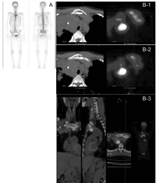
Abstract: A 53-year-old man, who was a heavy smoker, presented with recent severe cough. Radiography demonstrated a large pulmonary mass in the right upper lung. FDG PET/CT demonstrated heterogeneous high-grade activity in the pulmonary mass located in the right upper lung (standardized uptake value of 20), with central necrosis, bilateral upper mediastinal lymphadenopathy, right supraclavicular lymphadenopathy, direct left sternal manubrium invasion, and distal bilateral peripheral lung metastasis. Histol-ogy revealed significant malignant cytologic features and CD1a- and S-100-positive cells by immunohistochemistry staining, typical for Langerhans cell sarcoma.

Key Words: pulmonary, langerhans cell sarcoma, FDG, PET, CT (Clin Nucl Med 2012;37: 495– 497)

REFERENCES

1. Favara BE, Feller AC, Pauli M, et al. Contemporary classification of histio-cytic disorders. The WHO committed on histiocystic/reticulum cell prolifer-ations. Reclassification working group of the histiocyte society. Med Pediatr

Oncol. 1997;29:157–166.

2. Wood C, Wood GS, Deneau DG, et al. Malignant histiocytosis X. Report of a rapidly fatal case in an elderly man. Cancer. 1984;54:347–352. 3. Elleder M, Fakan F, Hula M. Pleiomorphous histiocytic sarcoma arising in a

patient with histiocytosis X. Neoplasma. 1986;33:117–128.

4. Itoh H, Miyaguni H, Kataoka H, et al. Primary cutaneous Langerhans cell histiocytosis showing malignant phenotype in an elderly woman: report of a fatal case. J Cutan Pathol. 2001;28:371–378.

5. Delabie J, De Wolf-Peeters C, De Vos R, et al. True histiocytic neoplasm of Langerhans’ cell type. J Pathol. 1991;16:217–223.

6. Lauritzen AF, Delsol G, Hansen NE, et al. Histiocytic sarcomas and mono-blastic leukemias. A clinical, histologic, and immunophenotypical study.

Am J Clin Pathol. 1994;102:45–54.

7. Pileri SA, Grogan TM, Harris NL, et al. Tumours of histiocytes and accessory dendritic cells: an immunohistochemical approach to classification from the International Lymphoma Study Group based on 61 cases. Histopathology. 2002;41:1–29.

8. Tani M, Ishii N, Kumagai M, et al. Malignant Langerhans cell tumour. Br J

Dermatol. 1992;126:398 – 403.

9. Julg BD, Weidner S, Mayer D. Pulmonary manifestation of a Langerhans cell sarcoma: case report and review of the literature. Virchows Arch. 2006;448: 369 –374.

10. Nakayama M, Takahashi K, Hori M, et al. Langerhans cell sarcoma of the cervical lymph node: a case report and literature review. Auris Nasus Larynx. 2010;37:750 –753.

11. Travis WD, Brambilla E, Mu¨ller-Hermelink HK, et al. WHO, Pathology &

Genetics. Tumours of the Lung, Pleura, Thymus and Heart. Lyon, France:

IARC Press; 2004.

12. Benz MR, Dry SM, Eilber FC, et al. Correlation between glycolytic pheno-type and tumor grade in soft-tissue sarcomas by 18F-FDG PET. J Nucl Med. 2010;51:1174 –1181.

13. Djouad F, Bouffi C, Ghannam S, et al. Mesenchymal stem cells: innovative therapeutic tools for rheumatic diseases. Nat Rev Rheumatol. 2009;5:392– 399.

14. Ruan K, Bao S, Ouyang G. The multifaceted role of periostin in tumorigen-esis. Cell Mol Life Sci. 2009;66:2219 –2230.

15. Bouffi C, Djouad F, Mathieu M, et al. Multipotent mesenchymal stromal cells and rheumatoid arthritis: risk or benefit? Rheumatology (Oxford). 2009;48: 1185–1189.

16. Goda T, Shimo T, Yoshihama Y, et al. Bone destruction by invading oral squamous carcinoma cells mediated by the transforming growth factor-beta signaling pathway. Anticancer Res. 2010;30:2615–2623.

17. Roodman GD. Mechanisms of bone metastasis. N Engl J Med. 2004;350: 1655–1664.

Received for publication January 17, 2011; revision accepted June 3, 2011. From the *Department of Nuclear Medicine, Kaohsiung Medical University

Hospital, Kaohsiung Medical University, Kaohsiung, Taiwan; †Department of Nuclear Medicine, School of Medicine, Kaohsiung Medical University, Ka-ohsiung, Taiwan; ‡Department of Pathology, Kaohsiung Medical University Hospital, Kaohsiung Medical University, Kaohsiung, Taiwan; §Department of Public Health, College of Health Sciences, Kaohsiung Medical Univer-sity, Kaohsiung, Taiwan; and ¶Cancer Center, Kaohsiung Medical Uni-versity Hospital, Kaohsiung Medical UniUni-versity, Kaohsiung, Taiwan. Conflicts of interest and sources of funding: none declared.

Reprints: Yung-Chang Lai, PhD, Department of Nuclear Medicine, Kaohsiung Medical University, No. 100, Tzyou 1st Rd, Kaohsiung City, 80756 Taiwan. E-mail: [email protected].

Copyright © 2012 by Lippincott Williams & Wilkins ISSN: 0363-9762/12/3705-0495

(2)

FIGURE 1. A, Coronal views of FDG PET imaging: Based on sequential imaging, it is shown that multiple high-grade FDG-avid

lesions distribute above the diaphragm. A large extremely high-grade FDG-avid pulmonary mass (standardized uptake value of

20) with central necrosis is found in the anterior right upper lobe (as marker). The upper mediastinum fills with conglomerated

high-grade FDG-avid masses (lymphadenopathy). Focally high grade of right supraclavicle and several discrete axillary lymph nodal

activities are simultaneously seen beyond thorax. Multiple roundish peripheral pulmonary nodules with low-grade FDG-avidity

scatter in bilateral lung fields. B, Transverse section of FDG PET/CT imaging: Depending on CT localization, 3 major sections of

fusion imaging of FDG PET/CT also demonstrate aggressive behavior of Langerhans cell sarcoma.

Chen et al

Clinical Nuclear Medicine • Volume 37, Number 5, May 2012

(3)

FIGURE 2. A,

99m

Tc MDP whole-body bone scan: Regionally increased activity in left sternal manubrium corner is only seen

on whole-body imaging. B, Transverse section of FDG PET/CT imaging: We can find a tiny linear cortical defect of inner side

of left sternum on CT and high FDG-avid sarcoma cellular activity from left anterior mediastinal lymphadenopathy infiltrating

into adjacent marrow cavity of left sternum on sequential FDG PET/CT imaging. Therefore, the destruction of sternum is not

traditionally the result of hematologic spreading. This fusion imaging of FDG PET/CT demonstrates and localizes unusually

detailed characteristics of Langerhans cell sarcoma locally spreading into adjacent left sternum via periosteum. Neoplasms of

dendritic or histiocytic cells are extremely rare.

1

Based on literature review, the first case of Langerhans cell sarcoma was

published by Wood et al in 1984.

2

Only 14 cases of Langerhans cell sarcoma have been reported.

2–10

Langerhans cell

sarcoma usually shows multiple organ involvement, including lymph nodes, skin, pulmonary, liver, spleen, and bone. Similar

to the last case report published in 2006,

9

we present another case of pulmonary Langerhans cell sarcoma based on FDG

PET/CT findings. The World Health Organization classification of histiocytic and dendritic cell neoplasms differentiates between

histiocytic sarcoma, Langerhans cell histiocytosis, Langerhans cell sarcoma, interdigitating dendritic cell sarcoma/tumor, follicular

dendritic cell sarcoma/tumor, and dendritic cell sarcoma, not otherwise specified.

11

To distinguish the different types, a

combination of morphologic and immunophenotypic characterization is necessary. Langerhans cells are positive for CD1a

and S-100 protein; negative for CD21, CD35, and CD68; and show Birbeck granules ultrastructurally. In our patient,

immunohistochemistry stain is consistent with positive for CD1a and S-100 protein. The first imaging of FDG PET/CT on

Langerhans cell sarcoma demonstrates the high FDG-avid aggressive behavior of this sarcoma.

12

In the imaging, distal

hematogeneous spreading into bilateral pulmonary fields is demonstrated. However, there is 1 lesion in left hemisternum seen

on

99m

Tc MDP bone scan. Based on FDG PET/CT, focally increased uptake is because of direct invasion of sarcoma via

periosteum effect, and not because of hematologic spreading. In recent literature review, periosteum owns mesenchymal

stem cell.

13–15

We propose a certain mechanism of sarcoma direct invasion of bony structure via periosteum.

16,17

Clinical Nuclear Medicine • Volume 37, Number 5, May 2012

The Characteristics of FDG PET/CT Imaging

數據

FIGURE 1. A, Coronal views of FDG PET imaging: Based on sequential imaging, it is shown that multiple high-grade FDG-avid
FIGURE 2. A, 99m Tc MDP whole-body bone scan: Regionally increased activity in left sternal manubrium corner is only seen

參考文獻

相關文件

The hypothesis of heterotopia may explain the origin of sali- vary tissue in parotideal lymph nodes, in the periparoti- deal region and the upper neck, but fails to explain the

- The hypothesis of heterotopia may explain the origin of salivary tissue in parotideal lymph nodes, in the periparotideal region and the upper neck, but fails to

In this report, we describe a rare case of bilateral symmetric ectopic oral tonsillar tissue located at the ventral surface of the tongue along with two solitary cases arising from

Dental variations like dens invaginatus in the upper left central incisor (Figure 5) and taurodontism in posterior molars (Figure 6) are associated with talon cusps.. On

CT images showed a low-density lesion in the periapical areas of the right mandible, which accompanied a well-de fined, high-density lesion, and a mixed low/high-density lesion with

The current study found that high-grade MECs had the highest incidence of strong MUC1 expression in mucous cells, with intermedi- ate- and low-grade tumours showing the

In this report, we present a case of a 44-year-old female patient who presented with a swelling in her upper right buccal region following dental treatment of her second maxillary

High school may be divided into two parts: junior high school and senior high school (or middle school and upper school in the UK), but North Americans often simply say