Oxygenated Terpenoids from a Formosan Soft Coral Sinularia gibberosa
Atallah F. Ahmed,†,‡Yao-Haur Kuo,§Chang-Feng Dai,⊥and Jyh-Horng Sheu*,†Department of Marine Resources, National Sun Yat-Sen University, Kaohsiung 804, Taiwan, Republic of China, Department of Pharmacognosy, Faculty of Pharmacy, Mansoura University, Mansoura 35516, Egypt, National Research Institute of Chinese Medicine, Taipei 112, Taiwan, Republic of China, and Institute of Oceanography, National Taiwan University, Taipei 106, Taiwan, Republic of China
Received April 4, 2005
Three new oxygenated sesquiterpenoids, gibberodione (1), peroxygibberol (2), and sinugibberodiol (3), along with sarcophytol L (4) were isolated from a Formosan soft coral, Sinularia gibberosa. The structures of the new metabolites were determined on the basis of extensive spectroscopic analyses and by comparison of NMR data with those of related metabolites. Metabolites 2 and 4 were found to exhibit moderate cytotoxicity toward a human liver carcinoma cell line.
During the course of our screening for bioactive metabo-lites from marine organisms,1-5several bioactive steroids have been isolated and identified from the soft coral Sinularia gibberosa Tixier-Durivault growing in Taiwanese waters.6Further investigation on the chemical constituents of the dichloromethane extract of the Formosan soft coral S. gibberosa has afforded three new oxygenated sesquit-erpenoids, which were identified as (1S,10S,6E)-4,5-sec-oguai-6-en-4,5-dione (gibberodione, 1), (1S*,4R*,7S*,10S*)-4-hydroxyguai-5-en-1,7-endoperoxide (peroxygibberol, 2), and (3S*,7S*,9S*)-3,9-dihydroxygermacra-4(15),10(14),11-(12)-triene (sinugibberodiol, 3), along with the known diterpenoid sarcophytol L (4). The structures of the new metabolites were determined on the basis of extensive NMR spectroscopic analyses, by comparison of spectro-scopic data with those of related metabolites, and by chemical reaction. The relative configuration of 1-3 was established by careful NOESY spectroscopic analyses. The cytotoxic activity of 1, 2, and 4 against the growth of Hepa59T/VGH (human liver carcinoma) and KB (human oral epidermoid carcinoma) was also evaluated.
Results and Discussion
The organism S. gibberosa was exhaustively extracted with EtOH. The concentrated crude alcoholic extract was successively partitioned with n-hexane and dichloromethane to afford the n-hexane and dichloromethane fractions. Chromatographic fractionation of the dichloromethane fraction, using normal-phase MPLC and HPLC, yielded metabolites 1-4 (see Experimental Section).
Gibberodione (1) was obtained as a colorless oil. Its HREIMS (m/z 236.1781, [M]+) and NMR data (Table 1) suggested the molecular formula C15H24O2, implying four degrees of unsaturation. The IR spectrum revealed the presence of a carbonyl functionality (νmax1715 cm-1) and the absence of any hydroxy groups. The13C NMR spectrum of 1, measured in pyridine-d5, displayed 15 carbon signals assigned to four methyl, four sp3 methylene, three sp3 methine, one sp2 methine, and three sp2 quaternary carbons. The carbon signals at δ 128.3 (CH), 169.6 (C), and 202.6 (C) were assigned to an R,β-unsaturated ketone, which was further supported by the appearance of a downfield shifted olefinic proton resonating at δ 6.04 (1H, br s) in the1H NMR spectrum of 1. One additional ketone was also revealed from the carbon signal appearing at δ 208.0 (C). Metabolite 1 was thus established as a mono-cyclic sesquiterpenoid. The proton signals displayed in the 1H NMR at δ 0.75, 0.91, and 0.92 (each 3H, d, J ) 6.6 Hz) were assigned as three secondary methyl groups, in which two methyls were found to be correlated in the 1H-1H COSY spectrum with the allylic methine appearing at δ 2.22 (1H, t, J ) 6.6 Hz), implying the presence of an isopropyl group. Moreover, a 3-oxobutyl group was also deduced as a side chain from the 1H/13C long-range cor-relations observed from both of the acetyl protons at δ 2.03 (3H, s) and the four protons of two methylenes (δ 1.69, 2.34, 2.37, and 2.51, each 1H, m) to the carbonyl carbon at δ 208.0. Furthermore, the fragment ion peaks appearing in the EIMS spectrum at m/z 193 [M - C3H7]+and 165 [M -C4H7O]+were considered to arise from the elimination of an isopropyl and an oxobutyl moiety, respectively, from the molecular ion of 1. The above observations together with 1H-1H COSY and HMBC data (Table 1 and Figure 1) indicated that compound 1 should possess a conjugated cycloheptenone ring substituted with an isopropyl, a 3-oxo-butyl, and a secondary methyl. These substituents were found to be linked to the cycloheptenone ring at C-7, C-1, and C-10, respectively, as interpreted from the correlations found in the 1H-1H COSY and HMBC spectra of 1. Comparison of the13C NMR data of 1 measured in CDCl
3 * To whom correspondence should be addressed. Tel: 886-7-5252000, ext.
5030. Fax: 886-7-5255020. E-mail: [email protected]. †National Sun Yat-Sen University.
‡Mansoura University.
§National Research Institute of Chinese Medicine. ⊥National Taiwan University.
10.1021/np050114u CCC: $30.25 © 2005 American Chemical Society and American Society of Pharmacognosy Published on Web 07/20/2005
Downloaded by NATIONAL TAIWAN UNIV on November 2, 2009 | http://pubs.acs.org
(see Experimental Section) with those of 4,5-seco-guaiane
5 isolated from a liverwort7revealed that 1 has the same carbon skeleton as that of 5 except that the hemiketal and sp3 oxygenated quaternary carbons (δ 98.6 and 91.8, respectively)7 of the tetrahydrofuran ring in 5 were re-placed by a carbonyl (δ 206.0) and a methine (δ 52.8) carbon in 1. On the basis of the above findings, the planar structure of gibberodione (1) was thus established as 4,5-secoguai-6-ene-4,5-dione.
The relative configuration of 1 was deduced from NOE interactions (Figure 2) observed in the NOESY spectrum and by comparison of NMR spectroscopic data with those of related compounds. The NOE correlations observed between H-1 and H-10 but not between H-1 and H3-14 reflected the cis orientation of H-10 and H-1. Furthermore, the NOE interactions found between the olefinic proton H-6 and the protons of the isopropyl methyl groups assigned the Z-configuration of the 6,7-double bond. By further inspection of NMR data of 2-cycloheptenone-derived com-pounds, it was found that clavularin A (6), with a 1S,10S-configuration, showed marked upfield shifts at the second-ary methyl (δH0.83 and δC15.7 in CDCl3) relative to those of a 1R,10S-configured isomer, clavularin B (7) (δH 1.09 and δC19.9 in CDCl3).8,9Therefore, the chemical shifts of H3-14 in 1 (δH0.79 and δC 16.2 in CDCl3) could indicate the same relative configuration in 1 as that of 6. The same sign of optical rotations of 1 ([R]D+20.8°, CHCl3) and 6
([R]D+59.2°, CHCl3) suggests that 1 should have the same 1S,10S absolute configuration as that of 6. Therefore, the structure of gibberodione was assigned as (1S,10S,6E)-4,5-secoguai-6-en-4,5-dione.
The new metabolite peroxygibberol (2) was also obtained as a colorless oil. The molecular formula of 2 was estab-lished as C15H24O3by HRESIMS (m/z 275.1621, [M + Na]+) Table 1. 1H (300 MHz),13C (75 MHz), and 2D NMR Dataafor 1
atom δC δH COSYd HMBCe 1 53.0 (CH)b 2.90 ddd (9.6, 5.2, 5.2)c H 2-2, H-10 C-2, C-3, C-5, C-9, C-10, C-14 2 22.8 (CH2) 2.34 m H-1, H2-3 C-1, C-3, C-4, C-5, C-10 1.69 m 3 41.9 (CH2) 2.37 m H2-2 C-1, C-2, C-4 2.51 m 4 208.0 (C) 5 202.6 (C) 6 128.3 (CH) 6.04 br s H2-8, H-11 C-1, C-7, C-8, C-11 7 169.6 (C) 8 29.6 (CH2) 2.16 dd (17.5, 6.5), R H-6, H2-9 C-6, C-7, C-9, C-10, C-11 2.33 m, β 9 36.6 (CH2) 1.99 m, R H2-8, H-10 C-1, C-7, C-8, C-10 0.93 m, β 10 33.5 (CH) 1.95 m H-1, H2-9, H3-14 C-2, C-5, C-8, C-9, C-14 11 38.0 (CH) 2.22 t (6.6) H-6, H3-12, H3-13 C-6, C-7, C-8, C-12, C-13 12 20.5 (CH3) 0.91 3H, d (6.6) H-11 C-7, C-11, C-13 13 20.9 (CH3) 0.92 3H, d (6.6) H-11 C-7, C-11, C-12 14 16.3 (CH3) 0.75 3H, d (6.6) H-10 C-1, C-9, C-10 15 29.6 (CH3) 2.03 3H, s C-3, C-4 aSpectra recorded in C
5D5N.bAttached protons were determined by DEPT experiments.cThe J values are in Hz in parentheses. dCorrelations HfH.eCorrelations HfC.
Figure 1. 1H-1H COSY and HMBC correlations for 1-3.
Figure 2. Key NOESY correlations of 1.
Downloaded by NATIONAL TAIWAN UNIV on November 2, 2009 | http://pubs.acs.org
and NMR spectroscopic data (Table 2). Thus, the structure of 2 possesses four degrees of unsaturation. The presence of the hydroxy functionality in 2 was revealed from the IR absorption band at νmax3598 cm-1. The13C NMR spectrum of 2 showed signals of 15 carbons, including those of four methyls (δ 25.2, 17.0, 17.1, and 13.7) and three quaternary sp3oxycarbons (δ 89.3, 83.2, and 78.5), as assigned by the DEPT spectrum, suggesting the oxygenated sesquiterpe-noid nature of 2 (Table 2). Furthermore, the carbon signals at δ 120.0 (CH) and 152.4 (C) indicated the presence of one trisubstituted double bond. Thus, the tricyclic structure of
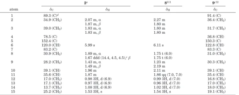
2 was revealed. From the 1H-1H COSY and HMBC correlations, the guaiane-type skeleton of 2 was established (Figure 1). The hydroxy group should be positioned at a methyl-bearing carbon C-4, as designated by the HMBC correlations observed from the tertiary methyl (δH1.53) to the quaternary carbons at δ 78.5 (C-4) and 152.4 (C-5). Therefore, the two additional oxygen atoms should be used to form an endoperoxide bridge in the cycloheptene moiety of the molecule. This was further supported by the intense ion peak in the mass spectrum appearing at m/z 220 [M -O2]+. The 1H/13C long-range correlations found from the olefinic proton H-6 (δ 5.99, s) to C-1 (δ 89.3, C) and C-7 (δ 83.2, C) further established the 1,7-endoperoxide linkage. It was found that the1H NMR data of (1S*,4R*,7S*,10R*)-4-hydroxyguai-5-en-1,7-endoperoxide (8),11 isolated from Liabum floribundum, were highly similar to those of 2 (Table 2), except for the significant downfield shift observed at one of the H2-9 protons (∆δΗ+0.70 ppm) relative to that of 2. These findings suggested that 2 might be the C-10 epimer of 8. Due to the lack of13C data for 8, we further compared the13C NMR spectroscopic data of 2 with those of 9,12a compound with a structure closely related to that of 2 and 8. We observed that 9 also exhibited downfield shifts at C-1, C-9, and C-14 (∆δC+2.1, +2.1, and +4.3 ppm, respectively) relative to those of 2, and thus further supported our assumption that the methyl group attached at C-10 should be β-oriented. The significant upfield shifted C-14 in 2 can be explained by the strong γ-effect13arising from the steric compression of a gauche interaction between 10-CH3and both the C1-C2and C1-O single bonds. The above observations together with the NOE correlations (Figure 3) observed in the NOESY spectrum supported the
relative structure of peroxygibberol (2) as (1S*,4R*,7S*,-10S*)-4-hydroxyguai-5-en-1,7-endoperoxide.
The sesquiterpenoid metabolite sinugibberodiol (3) was also obtained as a colorless oil. Its HRFABMS spectroscopic data (m/z 237.1853) suggested the molecular formula C15H24O2, requiring 4 degrees of unsaturation. Two hydroxy groups were found from the IR (νmax 3397 cm-1) and FABMS (m/z 219 [M - H2O + H]+and 201 [M - 2H2O + H]+) data. The1H NMR data of 3 (Table 3) revealed the presence of three exomethylenes (δ 5.19, 5.11, 4.70, 4.68, each 1H, s and 5.06, 2H, s), one olefinic methyl (δ 1.69, 3H, s), and two hydroxy-bearing methines (δ 4.21, 1H, t, J ) 6.6 Hz and δ 4.00, 1H, dd, J ) 11.4, 3.0 Hz). The 13C NMR spectrum of 3 displayed 15 carbon signals, which indicated the presence of three 1,1-disubstituted double bonds (δ 150.2, 149.7, 148.0, each qC and 114.7, 114.0, 110.2, each CH2), one methyl (δ 19.1), two oxymethines (δ 76.7 and 74.6), and five methylene and one nonoxygenated methine (Table 3). The above observations together with the 1H-1H COSY and HMBC correlations (Figure 1) established the planar structure of 3 and revealed that 3 is a germacrane-based sesquiterpene.14
The relative configurations at C-3, C-7, and C-9 were determined by careful and detailed investigation of NOE interactions (Figure 4) displayed in the NOESY spectra of
3 and its diacetate 3a. Assuming the R-orientation of H-7,15 the NOE correlations observed between the oxymethine protons H-9 and H-7 with H-8R indicated the β-orientation of the 9-OH. Since no other useful NOE correlations that could be used to establish the relative configuration at C-3 Table 2. 1H and13C NMR Chemical Shifts for Compounds 2, 8,11and 912
2a 8b11 9c12 atom δC δH δH δC 1 89.3 (C)d 91.4 (C) 2 34.9 (CH2) 2.07 m, R 2.27 m 36.4 (CH2) 1.87 m, β 1.80 m 3 39.0 (CH2) 1.83 m, R 1.80 m 31.7 (CH2) 1.83 m, β 1.80 m 4 78.5 (C) 36.8 (CH) 5 152.4 (C) 150.3 (C) 6 120.0 (CH) 5.99 s 6.11 s 122.8 (CH) 7 83.2 (C) 83.3 (C) 8 30.9 (CH2) 1.89 m, R 1.75 t (6.0) 31.0 (CH2) 1.67 ddd (14.4, 4.5, 4.5),eβ 1.75 t (6.0) 9 28.2 (CH2) 1.43 m, R 1.23 m 30.3 (CH2) 1.49 m, β 2.19 m 10 39.1 (CH) 1.96 m 2.11 m 39.1 (CH) 11 35.6 (CH) 1.87 m 1.86 qq (7.0, 7.0) 35.4 (CH) 12 17.0 (CH3) 0.98 3H, d (6.9) 0.99 3H, d (7.0) 16.8 (CH3) 13 17.1 (CH3) 0.97 3H, d (6.9) 0.96 3H, d (7.0) 17.0 (CH3) 14 13.7 (CH3) 1.08 3H, d (6.9) 1.02 3H, d (7.0) 18.0 (CH3) 15 25.2 (CH3) 1.53 3H, s 1.54 3H, s 19.1 (CH3) aSpectra recorded at 300 MHz in CDCl
3for1H and 75 MHz for13C.bSpectra recorded at 400 MHz for1H in CDCl3(see ref 11).cSpectra
recorded at 125 MHz for13C in CDCl
3(see ref 12).dAttached protons were determined by DEPT experiments.eThe J values are in Hz
in parentheses.
Figure 3. Observed NOESY correlations of 2.
Downloaded by NATIONAL TAIWAN UNIV on November 2, 2009 | http://pubs.acs.org
were found, the diacetate 3a was prepared for further NOE analysis. It was found that both H-9 and H-3 of 3a exhibited NOE correlations with H-7. Thus, H-3, either in
3 or 3a, was found to be positioned on the R face. Therefore,
the relative structure of sinugibberodiol (3) was established as (3S*,7S*,9S*)-3,9-dihydroxygermacra-4(15),10(14),11-(12)-triene.
It should be noted that the structure of 3a is the same as that of chrysanthediacetate B, which was isolated pre-viously from Chrysanthemum morifolium.15However, the optical rotation of 3a ([R]D-36.3°, MeOH) has the opposite sign relative to that of chrysanthediacetate B ([R]D+45.53°, MeOH)15 and thus must be the enantiomer. Also, it was reported that a similar sesquiterpene, eleganodiol, has been purified by acetylation of a crude sesquiterpene-containing fraction obtained from Gonospermum elegans.16 The af-forded diacetyleleganodiol possesses1H and13C NMR data nearly identical to those of 3a and chrysanthediacetate B,15 but with an optical activity ([R]D+9°, CHCl3) deviating markedly in magnitude, suggesting the possibility that it might be a mixture of 3a and chrysanthediacetate B.
The sesquiterpene metabolites (1, 2, and 4) were evalu-ated for their in vitro cytotoxic activity against Hepa59T/ VGH and KB cancer cell lines. The results showed that 2 and 4 exhibited moderate cytotoxicity against the growth of the Hepa59T/VGH cell line (ED50’s 8.2 and 12.6 µg/mL, respectively), while they were not cytotoxic against KB cell
lines. Gibberodione 1 was found to be inactive (ED50> 30 µg/mL) against both cancer cell lines.
Experimental Section
General Experimental Procedures. Optical rotations
were measured on a Jasco DIP-1000 digital polarimeter. IR spectra were recorded on a Jasco FT-5300 infrared spectro-photometer. EIMS was obtained with a VG Quattro GC/MS spectrometer. HRMS spectra were recorded on a Finnigan MAT-95XL mass spectrometer. The NMR spectra were re-corded on a Bruker AVANCE DPX300 FT-NMR at 300 MHz for1H and 75 MHz for13C or on a Varian Unity INOVA 500 FT-NMR at 500 MHz for1H and 125 MHz for13C, respectively, in CDCl3using TMS as internal standard. Si gel (Merck, 230-400 mesh) was used for column chromatography. Precoated Si gel plates (Merck, Kieselgel 60 F-254, 0.2 mm) were used for analytical TLC.
Organism. The soft coral S. gibberosa, Alcyoniidea (0.6 kg,
wet wt), was collected by hand via scuba at the coast of Kenting, in December 2000, at a depth of 10-15 m, and stored in a freezer until extraction. A voucher sample was deposited at the Department of Marine Resources, Sun Yat-Sen Uni-versity (specimen no. NHSC 002).
Extraction and Isolation. The organism, S. gibberosa,
was exhaustively extracted with EtOH. The EtOH extract was filtered and concentrated under reduced pressure, and the aqueous residue was successively partitioned with n-hexane and dichloromethane to afford the n-hexane (4 g) and dichlo-romethane (11 g) fractions, respectively.6The dichloromethane fraction was chromatographed on Si gel 60 using MPLC, and elution was carried out by EtOAc-n-hexane (stepwise, 0:10, 1:9, ..., 9:1, 10:0) then by MeOH-EtOAc (stepwise, 0:10, 1:9, ..., 9:1, 10:0) to yield 55 fractions. Fraction 25 eluted with EtOAc-n-hexane (2:8) was further purified by normal-phase HPLC using acetone-n-hexane (gradient, 1:9 to 2:8) to yield
1 (11 mg). Fraction 30 eluted with n-hexane-EtOAc (2:8) was
separated by MPLC on Si gel 60 and MeOH-dichloromethane (gradient, 0:100 to 1:99) to afford 2 (25 mg). Fractions 43 and 44 eluted with EtOAc-n-hexane (4:6) were purified separately by normal-phase HPLC using MeOH-dichloromethane (gradi-Table 3. 1H and13C NMR Chemical Shifts for 3 and 3a
3a 3ab atom δC δH δC δH 1 24.3 (CH2)c 2.10 m 24.7 (CH2) 2.09 m 2.30 dd (18.0, 9.0)d 2.30 m 2 32.7 (CH2) 2.08 m, R 29.5 (CH2) 2.11 m 2.08 m, β 2.11 m 3 74.6 (CH) 4.21 t (6.6) 76.0 (CH) 5.40 dd (8.5, 5.0) 4 149.7 (C) 144.9 (C) 5 30.6 (CH2) 1.55 m, R 30.4 (CH2) 1.70 ddd (14.5, 7.5, 4.5) 2.34 ddd (14.8, 5.5, 5.5), β 2.37 ddd (14.5, 7.5, 4.5) 6 32.1 (CH2) 1.59 m, R 31.1 (CH2) 1.56 m 1.64 m, β 1.77 m 7 41.2 (CH) 2.11 m 40.5 (CH) 2.22 qq (4.0) 8 37.1 (CH2) 1.64 m, R 34.3 (CH2) 1.67 m 1.85 ddd (14.0, 11.4, 3.0), β 1.90 ddd (13.5, 12.0, 4.0) 9 76.7 (CH) 4.00 dd (11.4, 3.0) 78.4 (CH) 5.09 dd (12.0, 4.0) 10 150.2 (C) 145.0 (C) 11 148.8 (C) 148.1 (C) 12 110.2 (CH2) 4.68 s 110.6 (CH2) 4.72 s 4.70 s 4.73 s 13 19.1 (CH3) 1.69 3H, s 19.3 (CH3) 1.69 3H, s 14 114.7 (CH2) 5.06 s 117.0 (CH2) 5.18 s 5.11 s 5.21 s 15 114.0 (CH2) 5.06 s 116.8 (CH2) 5.16 s 5.19 s 5.26 s 3-Ac 21.4 (CH3) 2.00 3H, s 169.9 (C) 9-Ac 21.4 (CH3) 2.05 3H, s 170.4 (C) aSpectra recorded at 300 MHz in CDCl
3for1H and 75 MHz for13C.bSpectra recorded at 500 MHz in CDCl3for1H and 125 MHz for 13C.cAttached protons were determined by DEPT experiments.dThe J values are in Hz in parentheses.
Figure 4. Observed NOESY correlations for 3, R ) H (solid arrows),
and 3a, R ) Ac (dashed and solid arrows).
Downloaded by NATIONAL TAIWAN UNIV on November 2, 2009 | http://pubs.acs.org
ent, 1:99 to 5:95) to yield 4 (8.5 mg) and 3 (7.0 mg), respectively.
Gibberodione (1): colorless oil, [R]25
D +20.8° (c 0.72, CHCl3); IR (neat) νmax 3020, 2975, 2932, 2872, 1715, 1603, 1453, 1379, 1217, 1096 cm-1;1H NMR (C 5D5N, 300 MHz) and 13C NMR (C 5D5N, 75 MHz), see Table 1;1H NMR (CDCl3, 300 MHz) δ 5.89 (1H, br s, H-6), 2.83 (1H, ddd, J ) 10.6, 5.4, 4.5 Hz, H-1), 2.54 (1H, dd, J ) 10.8, 9.0 Hz, H-3), 2.46 (1H, m, H-3), 2.33-2.41 (2H, m, H-2, H-8), 2.31 (1H, m, H-11), 2.15 (1H, m, H-8), 2.11 (3H, s, H3-15), 2.10 (2H, m, H-9, H-10), 1.61 (1H, m, H-2), 1.08 (1H, m, H-9), 1.06 (6H, d, J ) 6.9 Hz, H3 -12, H3-13), 0.79 (3H, d, J ) 6.9 Hz, H3-14);13C NMR (CDCl3, 75 MHz) δ 206.0 (C-4), 203.4 (C-5), 170.2 (C-7), 128.0 (C-6); 52.8 (C-1), 42.0 (C-3), 38.0 (C-11), 36.3 (C-9), 33.6 (C-10), 29.9 15), 29.9 8), 22.3 2), 20.9 13), 20.6 12), 16.2 (C-14); EIMS m/z 236 (2.0, [M]+), 193 (4.0), 179 (10.5), 165 (9.1), 151 (8.5), 135 (8.8), 123 (30.6), 109 (16.0), 107 (27.5), 95 (47.5), 81 (25.5), 71 (10.5), 67 (36.5); HREIMS m/z 236.1781 (calcd for C15H24O2, 236.1777).
Peroxygibberol (2): colorless oil, [R]25
D +22.4° (c 1.25, CHCl3); IR (neat) νmax 3598, 3013, 2967, 2934, 2878, 1632, 1456, 1379, 1294, 1232, 1190, 1105, 1001 cm-1; 1H NMR (CDCl3, 300 MHz) and13C NMR (CDCl3, 75 MHz), see Table 2; EIMS m/z 252 (0.2, [M]+), 220 (93.3, [M - O
2]+), 205 (58.4), 202 (18.2, [M - O2- H2O]+), 187 (16.9), 177 (40.4), 162 (37.7), 149 (42.0), 125 (25.9); HRESIMS m/z 275.1621 (calcd for C15H24O3+ Na, 275.1623).
Sinugibberodiol (3): colorless oil, [R]25
D -5.0° (c 0.20, CHCl3); IR (neat) νmax 3397, 2970, 2930, 2870, 1711, 1644, 1445, 1382, 1024 cm-1;1H NMR (CDCl
3, 300 MHz) and13C NMR (CDCl3, 75 MHz), see Table 3; FABMS m/z 259 (3.0, [M + Na]+
), 237 (2.7, [M + H]+), 219 (7.9, [M - H2O + H]+), 201 (12.1, [M - 2H2O + H]+), 154 (55.3), 137 (58.5), 136 (65.0), 117 (39.6); HRFABMS m/z 237.1853 (calcd for C15H25O2, 237.1855).
Acetylation of 3. A solution of sinugibberodiol (3) (2.4 mg,
0.009 mM) in pyridine (0.2 mL) was mixed with Ac2O (0.2 mL), and the mixture was stirred at RT for 24 h. After evaporation of excess reagent, the residue was purified by normal-phase HPLC to give the diacetyl derivative 3a (acetone-n-hexane, 1:4, 2.8 mg, 0.009 mmol, 87.5%).
Sinugibberodiol diacetate (3a): colorless oil, [R]25 D-31.3° (c 0.8, CHCl3), -36.3° (c 0.8, MeOH); IR (neat) νmax2968, 2928, 2857, 1738, 1643, 1445, 1372, 1238, 1018 cm-1; 1H NMR (CDCl3, 500 MHz) and13C NMR (CDCl3, 125 MHz), see Table 3; EIMS m/z 320 (0.1, [M]+), 200 (8.8, [M - 2AcOH]+), 185 (37.8), 171 (28.1), 159 (100.0), 143 (56.3), 131 (79.3), 117 (56.3).
Compound 4: colorless oil, [R]25
D-52.5° (c 0.80, CHCl3); IR (neat) νmax3360, 2961, 2913, 2868, 1640, 1603, 1460, 1379, 1215, 1010 cm-1;1H NMR (CDCl 3, 500 MHz) δ 5.15 (1H, br t, J ) 7.5 Hz, H-11), 5.00 (1H, d, J ) 10.0 Hz, H-2), 4.85 (1H, s, H-19), 4.78 (1H, s, H-19), 4.63 (1H, dd, J ) 10.5, 3.0 Hz, H-14), 2.52 (1H, septet, J ) 7.0 Hz, H-3), 2.48 (1H, t, J ) 10.0 Hz, H-3), 2.40 (1H, dd, J ) 12.0, 9.0 Hz, H-7), 2.26 (1H, m, H-10R), 2.18 (1H, m, H-10β), 2.19 (2H, m, H-5R, H-13R), 2.10 (1H, dd, J ) 12.0, 10.5 Hz, H-13β), 1.76 (1H, dd, J ) 16.0, 12.0 Hz, H-5β), 1.73 (1H, m, H-6R), 1.73-1.69 (2H, m, H2-9), 1.68 (3H, s, H3-20), 1.63 (1H, dd, J ) 17.0, 9.0 Hz, H-6β), 1.13 (3H, d, J ) 7.0, H3-17), 1.11 (3H, s, H3-18), 1.07 (3H, d, J ) 7.0, H3-16); 13C NMR (CDCl 3, 125 MHz) δ 149.3 (C, C-8), 148.6 (C, C-1), 130.9 (C, C-12), 126.5 (CH, C-11), 125.3 (CH, C-2), 111.5 (CH2, C-19), 81.3 (C, C-4), 71.3 (CH, C-14), 54.8 (CH, C-7), 50.1 (CH, C-3), 44.3 (CH2, C-13), 40.2 (CH2, C-9), 27.9 (CH2, C-5), 27.3 (CH2, C-10), 26.6 (CH, C-15), 26.5 (CH3, C-17), 25.5 (CH2, C-6), 24.7 (CH3, C-16), 23.5 (CH3, C-18), 18.0 (CH3, C-20); EIMS m/z 304 (1.1, [M]+), 286 (0.9, [M - H2O]+), 268 (0.5, [M - 2H2O]+), 261 (1.5), 243 (2.1), 203 (3.0), 193 (4.3), 175 (4.0), 161 (4.3), 135 (11.3).1H and13C NMR spectroscopic data were found to be in full agreement with those reported previously for sarcophytol L (4).17
Cytotoxicity Testing. Cell lines were purchased from the
American Type Culture Collection (ATCC). Cytotoxicity assays of the test compounds 1, 2, and 4 were performed using the MTT [3-(4,5-dimethylthiazole-2-yl)-2,5-diphenyltetrazolium bro-mide] colorimetric method.18,19
Acknowledgment. This work was supported by a grant
from the National Science Council of the Republic of China (Contract No. NSC-92-2323-B-110-002) awarded to J.-H.S.
Note Added after ASAP Publication: The NOE
interaction between H-8(R) and the methyl at C-11 in Figure 3 was omitted in the version published on the Web on July 20, 2005. The corrected figure appears in the version posted on July 25, 2005.
References and Notes
(1) Ahmed, A. F.; Su, J.-H.; Shiue, R.-T.; Pan, X.-J.; Dai, C.-F.; Kuo, Y.-H.; Sheu, J.-H. J. Nat. Prod. 2004, 67, 592-597.
(2) Sheu, J.-H.; Ahmed, A. F.; Shiue, R.-T.; Dai, C.-F.; Kuo, Y.-H. J. Nat.
Prod. 2002, 65, 1904-1908, and references therein.
(3) Sung, P.-J.; Su, J.-H.; Duh, C.-Y.; Chiang, M. Y.; Sheu, J.-H. J. Nat.
Prod. 2001, 64, 318-323.
(4) Sheu, J.-H.; Chen S.-P.; Sung, P.-J.; Chiang, M. Y.; Dai, C.-F.
Tetrahedron Lett. 2000, 41, 7885-7888.
(5) Sheu, J.-H.; Sung, P.-J.; Su, J.-H.; Duh, C.-Y.; Chiang, M. Y.
Tetrahedron 1999, 55, 14555-14564.
(6) Ahmed, A. F.; Dai, C.-F.; Kuo, Y.-H.; Sheu, J.-H. Steroids 2003, 68, 377-381.
(7) Cullmann, F.; Becker, H. Phytochemistry 1998, 47, 237-245. (8) Endo, M.; Nakagawa, M.; Hamamoto, Y.; Nakanishi, T. J. Chem. Soc.,
Chem. Commun. 1983, 322-323 and 980.
(9) Hiroya, K.; Zhang, H.; Ogasawara, K. Synlett. 1999, 5, 529-532. (10) Tamura, R.; Watabe, K.-i.; Ono, N.; Yamamoto, Y. J. Org. Chem. 1993,
58, 4471-4472.
(11) Jakupovic, J.; Schuster, A.; Bohlmann, F.; Dillon, M. O.
Phytochem-istry 1988, 27, 1771-1775.
(12) Hirota, H.; Okino, T.; Yoshimura, E.; Fusetani, N. Tetrahedron 1998,
54, 13971-13980.
(13) Wang, G.-H.; Ahmed, A. F.; Kuo, Y.-H.; Sheu, J.-H. J. Nat. Prod. 2002,
65, 1033-1036.
(14) Vieira, L. M.; Kijjoa, A.; Pereira, J. A.; Gedris, T. E.; Herz, W.
Phytochemistry 1997, 45, 111-115.
(15) Hu, L.; Chen, Z. Phytochemistry 1997, 44, 1287-1290.
(16) Triana, J.; Lo´pez, M.; Rico, M.; Gonza´lez-Platas, J.; Quintana, J.; Este´vez, F.; Leo´n, F.; Bermejo, J. J. Nat. Prod. 2003, 66, 943-948. (17) Osabe, K.; Kobayashi, M. Chem. Pharm. Bull. 1989, 37, 1192-1196. (18) Alley, M. C.; Scudiero, D. A.; Monks, A.; Hursey, M. L.; Czerwinski, M. J.; Fine, D. L.; Abbott, B. J.; Mayo, J. G.; Shoemaker, R. H.; Boyd, M. R. Cancer Res. 1988, 48, 589-601.
(19) Scudiero, D. A.; Shoemaker, R. H.; Paull, K. D.; Monks, A.; Tierney, S.; Nofziger, T. H.; Currens, M. J.; Seniff, D.; Boyd, M. R. Cancer
Res. 1988, 48, 4827-4833.
NP050114U
Downloaded by NATIONAL TAIWAN UNIV on November 2, 2009 | http://pubs.acs.org