I
NTERESTING
I
MAGE
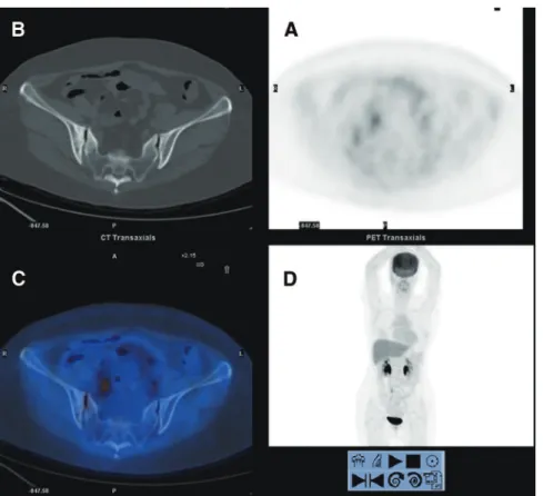
FDG Uptake in Sacroiliac Joint Due to Osteitis Condensans Ilii
Shown on PET/CT in a Patient With Breast Cancer
The Value of Coregistered CT in Avoiding Misinterpretation
Yang-Cheng Lee, MD,* Kuan-Yung Chen, MD,† Jainn-Shiun Chiu, MD,‡ Chia-Hung Kao, MD,§
and Guang-Uei Hung, MD*§
Abstract: A 56-year-old woman with breast cancer underwent FDG PET/CT
at follow-up. The PET images showed increased FDG uptake along right sacroiliac joint. The coregistered CT images showed diffuse sclerosis around the sacroiliac joints, but no bony destruction, periarticular erosion, or joint space narrowing. She had been complaining of intermittent lower back pain since her last pregnancy. The radiologic pictures and history of postpartum back pain were considered as typical characteristics for osteitis condensans ilii. This case reminds us that careful inspection of the coregistered CT images is important to avoid potential misinterpretation because of osteitis condensans ilii.
Key Words: false-positive, sacroiliac joint, osteitis condensans ilii, FDG,
PET/CT
(Clin Nucl Med 2012;37: e121– e123)
REFERENCES
1. Wong CL, Mansberg R. Solitary plasmacytoma of bone: an unusual cause of severe sacral pain in a young man. Clin Nucl Med. 2005;30:612– 624. 2. Halac¸ M, Mut SS, So¨nmezoglu K, et al. Avoidance of misinterpretation of an
FDG positive sacral insufficiency fracture using PET/CT scans in a patient with endometrial cancer: a case report. Clin Nucl Med. 2007;32:779 –781. 3. Patel CN, Smith JT, Rankine JJ, et al. F-18 FDG PET/CT can help differentiate
SAPHO syndrome from suspected metastatic bone disease. Clin Nucl Med. 2009;34:254 –257.
4. Thompson M. Osteitis condensans ilii and its differentiation from ankylosing spondylitis. Ann Rheum Dis. 1954;13:147–156.
5. Percy JS, Russell AS, Lentle VS. Letter: Osteitis condensans ilii. Lancet. 1975;1:1191–1192.
6. Olivieri I, Ferri S, Barozzi L. Osteitis condensans ilii. Br J Rheumatol. 1996;35:295–297.
7. Loneragan R, Archer K, Perry A, et al. Scintigraphy in osteitis condensans ilii.
Clin Nucl Med. 2004;29:320 –321.
8. Hsu HK, Huang CK, Bai YL, et al. False-positive bony FDG accumulations due to fractures in a patient with lung cancer: the value of integrated infor-mation of PET/CT. Ann Nucl Med Sci. 2009;22:183–187.
Received for publication December 13, 2009; revision accepted January 21, 2012. From the *Department of Hemato-oncology, Tainan Municipal Hospital, Tainan, Taiwan; Departments of †Radiology and ‡Nuclear Medicine, Chang Bing Show Chwan Memorial Hospital, Changhua, Taiwan; and §Department of Biomedical Imaging and Radiological Science, China Medical University, Taichung, Taiwan.
Conflicts of interest and sources of funding: none declared.
Reprints: Guang-Uei Hung, MD, Department of Nuclear Medicine, Chang Bing Show Chwan Memorial Hospital, 6 Lukon Road, Lukong Town, Changhua Shien, Taiwan 505, Taiwan. Email: [email protected].
Copyright © 2012 by Lippincott Williams & Wilkins ISSN: 0363-9762/12/3705-0121