Expression of protein kinase C isoforms in cancerous breast tissue and
adjacent normal breast tissue
Yueh-Min Lin1, Cheng-Chuan Su2,3, Wei-Wen Su4, Jin-Ming Hwang5, Hsi-Hsien Hsu6, Chang-Hai Tsai7, Fuu-Jen Tsai8, Chih-Yang Huang8, 9, 10,*, Jer-Yuh Liu11,12,*, and
Li-Mien Chen13,14,*
1
Department of pathology, Changhua Christian Hospital, Changhua 2
Departments of Clinical Pathology and Anatomic Pathology, Buddhist Dalin Tzu Chi General Hospital, Chiayi
3
Department of Pathology, School of Medicine, Tzu Chi University, Hualien 4
Comprehensive Breast Cancer Center, Changhua Christian Hospital, Changhua 5
School of Applied Chemistry, Chung-Shan Medical University, Taichung 6
Division of Colorectal Surgery, Mackay Memorial Hospital, Taipei 7
Department of Healthcare Administration, Asia University, Taichung 8
Graduate Institute of Chinese Medical Science,9Graduate Institute of Basic Medical Science, China Medical University, Taichung
10
Department of Health and Nutrition Biotechnology, Asia University, Taichung 11
Center for Molecular Medicine, China Medical University Hospital, Taichung 12
Graduate Institute of Cancer Biology, China Medical University, Taichung 13
Departments of Internal Medicine, Armed Force Taichung General Hospital, Taichung and
14
Center of General Education, Central Taiwan University of Science & Technology, Taichung, Taiwan, R.O.C.
* These authors contributed equally to this paper.
Corresponding author: Li-Ming Chen MD, Departments of Internal Medicine, Armed Force Taichung General Hospital, Taichung, Taiwan. Tel: +886-4-; Fax: +886-4-. E-mail address:.
Abstract
The role of PKC in the carcinogenesis of human breast cancer has been studied at the molecular level for more than two decades. In this study, we employed Western blotting to determine the existence of PKC isoforms in cancerous breast tissue and normal breast tissue. The results contained significant expressions of a conventional PKC (PKCα)and two atypicalPKCs(PKC ζand λ/ι)in both breasttumorsand adjacentnormalbreast tissue. For the isoformsα,ζ,and λ/ι,theexpression ofindividualisoformswashigherin the breast tumors than in the adjacent normal breast tissue. Although the correlation coefficient was low, significant linear correlation was found among the activities of the isoforms. This data suggests a new direction in cancer chemotherapy, namely the blockage of the signal transduction pathway of the specific isoforms.
Introduction
Protein kinase C (PKC), a lipid-regulated and calcium-dependent protein kinase, has 10 isoforms. According to their cofactors, the isoforms can be divided into three main classes: Ca++-dependent or conventional PKCs (α, βI, βII, and γ), Ca++ -independent or novel PKCs (δ, ε, ηand θ), and Ca++-independent and DAG and phosphatidylserine activated PKCs (ζand λ/ι) (3, 12). These isoforms may exist in some organs or universally in all organs. In different organs, they may exhibit differences in structure and also differ in cell signaling function.
It has been reported that overexpression of PKC may lead to disorders in cell proliferation and differentiation (8, 9). The expression of PKC in human breast tumor biopsies has been found to be significantly higher than that in adjacent normal breast tissue. Overexpression of c-myc, Ha-ras, erb13, and HER-2/neu in breast cancer have been attributed to the overexpression of this enzyme (14). In addition, PKC mediates phosphorylation of membrane-associated oncogene products such as pp60src and Ki-ras protein, and activates nuclear pro-oncogenes such as TPA (4, 7). These findings suggest that PKC may play an important role in the regulation of proto-oncogenes and oncogenes involved in the carcinogensis of human breast cancer.
Although the role of PKC in the carcinogensis of human breast cancer has been studied at the molecular level, there is no available information concerning the changes in the activities of the isoforms in the breast tissue. In this study, we employed Western blotting to determine the existence of the isoforms of PKC in cancerous breast tissue
and normal breast tissue. The relationships between the isoforms of PKC and various demographic and clinical factors were also investigated.
Materials and Methods
Specimen Collection
Written consent was obtained from each patient. Surgical specimens of breast tumor (infiltrating ductal carcinoma) and normal human breast tissue were obtained by mastectomy from the operating rooms of Chang Hua Christian Hospital in Chang Hua and Chung Shan Medical University Hospital in Taichung, both of which are in Taiwan. After resection, these specimens were stored at -70°C for the analysis.
PKC Extraction
PKC extraction was performed according to Nishizuka (13) with slight modifications. All operations were carried out at 4°C. The specimen (80 mg) was sectioned into small pieces and washed with homogenized buffer A (pH 7.5; 20 mM tris(hydroxymethyl)-aminomethane hydrochloride (Tris-HCl), 2 mM ethylenedianmine-tetraacetic acid (EDTA), 50 mM phenylmethylsulfonyl fluoride (PMSF), 10% glycerol, 50 mM β-mercaptoethanol). The tissue was then mixed with 200 µl buffer B (pH 7.5; 20 mM Tris-HCl, 2 mM EDTA, 50 mM PMSF, 10% glycerol, 0.1% Triton X-100). After homogenization, 200 µl homogenized buffer B was added to the homogenates before carrying out homogenization again. The homogenates were then transferred to another vial and then a further 330 µl of homogenized buffer B were added. This mixture was incubated for 1 h with stirring at intervals of 5-10 min. The homogenates were then centrifuged at 15,000 g for 3 hs. The supernatant (1.5 ml) was then transferred into vials and stored at -70°C for further experiments.
Determination of Protein Contents
Protein contents of the sample preparation were determined by the Bradford protein assay (6). The protein assay reagents were purchased from Bio-Rad Lab (Richmond, CA, USA). Coomassie brilliant blue G-250 was used for staining and bovine serum albumin (BSA) was employed as the standard. Changes in optical density were monitored at 595 nm.
Western Blotting
PKC isoforms in the samples were analyzed by the sodium dodecyl sulfate-polyacrylamide gel electrophoresis (SDS-PAGE) (11). The extracts were standardized to the same volume with PBS. The extracts were standardized to the same volume with PBS. After adding a treatment buffer in a ratio of 1 to 5 to the extract, the mixture was boiled for 10 min and then rapidly placed in an ice bath. The mixture was then spin-down in a centrifuge and loaded onto slab polyacrylamide gels, using a 4% stacking gel (pH 6.8) and 10% separating gel (pH 8.8). Electrophoresis was run on a mini vertical slab gel unit (Biorad Scientific Instruments, Richmond, California, USA) at 140 V and 35 mA for 3.5 h.
After electrophoresis, the gels were equilibrated in a cold transfer buffer and proteins were transferred onto nitrocellulose papers (Amersham, Hybond-C Extra Supported, 0.45 m) using a Hoefer Scientific Instruments Transphor Unit at 100 mA overnight. The nitrocellulose papers were washed with a washing buffer and incubated in 50 ml 3% FBS blocking buffer (3% FBS, 10 mM Tris-HCl, pH 8.0, 150 mM NaCl, 0.05% Tween 20) at room temperature for 1 h. Primary antibodies against individual PKC isoforms (1:100 dilute) in 20 ml 3% FBS blocking buffer was then added and incubated at room
temperature for 3 h. The nitrocellulose papers were washed in the washing buffer for 10 min in triplicate and then immersed in the secondary antibody of the corresponding isoform (1:1,000 dilute) containing 20 ml 3% FBS blocking buffer. The papers was washed in triplicate with the washing buffer for 10 min. Color was developed using 20 ml of a color developing substrate in color reagents (700 µl nitrobluetrazolium and 780 µl 5-bromo-4-chloro-3-indolyl-phosphate (10 mg/ml)). The reaction was terminated by addition of deionized water. Color changes on the nitrocellulose papers were determined using a destimetor (Alphalmager 2000 version 3.2, Alpha Innotech Corp., San Leandro, CA, USA).
Results
To determine whether PKC isoforms were associated with the breast cancer development, we scanned for ten types of PKC isoforms (α,βI,βII, γ, δ,ε,η, θ,ζand λ/ι) using the Transduction Laboratory antibodies in both the adjacent normal breast tissue and breast tumors taken from the mixed tissue samples of 10 patients (Fig. 1). Analysis revealed significant immunostaining in both adjacent normal breast tissue and cancerous breast tissue and PKCαand γantibodies at -80 KDa, and PKC ζand λ/ι antibodies at -72 KDa, whereas the PKC β, δ, ε,η,and θantibodies did not show any immunoreactivity. A band at 48 KDa was detected with all types of PKC isoforms as compared with the marker of brain cell lysates (data not shown). From this data, the following Western blotting tests focused on the detection of the 3 most abundant isoforms in the breast cancer tissues.
Significant expressions of a conventional PKC αand two atypical PKCs (PKC ζ, λ/ι) were detected in both breast tumors and adjacent normal breast tissue (Fig. 1A and 1B, Table 1). Since PKCγexists only in the brain and may cross-react with the antibody which acts against PKCα, the positive finding for this isoform in these tissues is questionable.
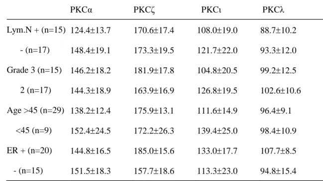
For the isoforms α, ζ, and λ/ι,the expression of individual isoforms was higher in the breast tumors than in the adjacent normal breast tissue (data not shown). Although the correlation coefficient was low, significant linear correlation was found among the activities of the isoforms (Table 2). However, there were no significant correlation in the activities of individual isoforms attributable to lymphatic metastasis, stage of carcinoma, age of the patient, or the presence of estrogen receptors (Table 3).
Discussion
It has been reported that the activity of PKC elevates significantly in human breast tumor than in the adjacent normal breast tissue (14). Using Western blotting, we not only confirmed these reports but also found that four endogenous isoforms (α,ζ,λ/ι) exist in the breast tumor. These findings suggest that PKC isoforms are important in the regulation of tumor cells and carcinogensis of human breast cancer.
In this study, the expression of PKCαin breast tumor is overexpressed, similar to that of PKCα-transfected breast cancer cell line MCF-7 (16). The finding is due to the fact that overexpression of this isoform may increase the expression of PKCβand decrease the amount of PKCηand PKCδ.This change in PKCαactivity may also lead to a more aggressive neoplastic phenotype. Therefore, overexpression of PKCα may be an important factor in the carcinogensis of human breast tumor.
Overexpression of PKC isoforms α, βI, δ, ε, and ζhave also been reported in uterine cancer cells where they increase the rate of proliferation of uterine tumor cells (5). In this study, we demonstrated thattheexpression ofPKCζwassignificantly higherin the breast tumor than in the adjacent normal breast tissue. This finding suggests the role of PKCζasan important enzyme in the proliferation of breast cancer cells.
PKCλand PKCζbelong to theatypicalPKC isoformsin human breast tumors and have a 72% similarity in their structures. These two isoforms exist universally in all kinds of tissue. The existence of PKCλhas been demonstrated in an undifferentiated mouse embryonal carcinoma cell line P19. In the P19 cells, the expressions of PKCα and PKCεare elevated and that of PKCλis significantly decreased after differentiation
(1). Overexpression of this isoform not only leads to a higher degree of undifferentiation in breast cancer cells but can also be used as a marker for the prognosis of patients.
Although PKCιdoes not affect the proliferation of cells, this isoform has been considered to have a significant association with drug resistance. This phenomenon has been demonstrated in the apoptosis of leukemia cells where inhibiting the PKCι lessened the drug resistance (10). The existence of PKCιin our specimens indicates that breast tumors may have chemopreventative effects against anti-cancer drugs. However, this suggestion requires further investigation.
Although PKCα, δ, ζ, and λare age-dependent in a developing kidney (15), we did not find significant differences in the expression of PKC isoforms in breast tumors by age group. Moreover, there were no significant associations between the levels of expression of PKCα, ζ, and λ/ιin the breast tumor at the stage of carcinoma, nor at the stage of lymphatic metastasis. There were also no significant associations regardless of the existence or absence of estrogen or progesterone receptors. PKCα,βI and βIIhave already been reported to be associated with human breast tumors (2). The identification of PKC α, ζ,and λ/ι in human breast tumors in this study suggests that chemotherapeutically blocking the signal transduction pathway of these specific isoforms may be a new direction in the treatment of breast cancer.
Acknowledgements
This work was supported by the grants from the National Science Council, Republic of China (NSC 98-2320-B-039-042-MY3), as well as by the Taiwan Department of Health Clinical Trial and Research Center of Excellence (DOH99-TD-B-111-004) and in part by the Taiwan Department of Health Cancer Research Center of Excellence (DOH99-TD-C-111-005).
References
1. Akimoto K, Mizuno K, Osada S, Hirai S, Tanuma S, Suzuki K, Ohno S. A new member of the third class in the protein kinase C family, PKC lambda, expressed dominantly in an undifferentiated mouse embryonal carcinoma cell line and also in many tissues and cells. J. Biol. Chem. 269: 12677-12683, 1994.
2. Ali, S., Al-Sukhun, S., El-Rayes, B.F., Sarkar, F.H., Heilbrun, L.K. and Philip, P.A. Protein kinases C isozymes are differentially expressed in human breast carcinomas. Life Sci. 84: 766-771, 2009.
3. Assoka, Y., Nakamura ,S.I. Yoshida ,K.and Nishizuka, Y. Protein kinase C, calcium
and phospholipid degradation. Trends Biochem. Sci. 17: 414-417, 1992..
4. Balleste, R., Furth, M.E. and Rosen, O.M. Phobol ester and protein kinase C mediated phosphorylation of cellular kirsten ras gene product. J. Biol. Chem. 262: 2688-2695, 1987.
5. Bamberger, A.M. Bamberger, C.M. Wald,M., Kratzmeier, M. and Schulte, H.M. Protein kinase C (PKC) isoenzyme expression pattern as indicator of proliferative activity in uterine tumor cells. Mol. Cell. Endocrinol. 123: 81-88, 1996.
6. Bradford, M.M. A rapid and sensitive method for the quantitation of microgram quantities of protein utilizing the principle of protein-dye binding. Anal. Biochem. 72: 248-254, 1976.
7. Gould, K.L. Woodgett, J.R. Cooper ,J.A. Buss, J.E. Shalloway ,D. and Hunter T. Protein kinase C phosphoylation pp60srcat novel site. Cell 42: 849-857, 1985.
8. Housey, G.M. Johnson, M.D. Hsiao, W.L. O'Brian, C.A. Murphy, J.P, Kirschmeier, P. and Weinstein ,I.B. Overproduction of protein kinase C causes disordered growth control in rat fibroblast. Cell 52: 343-354, 1988.
9. Krauss, R., Housey ,G., Johnson ,M. and Weinstein, I.B. Disturbances in growth control and Gene expression in a C3H10T1/2 cell line that stably overproduces protein kinase C. Oncogene 4: 991-998, 1989.
10. Murray, N.R. and Frield, A.P. Atypical protein kinase C iota protects human leukemia cells against drug apoptosis. J. Biol. Chem. 272: 27521-27524, 1997.
11. Newton, A.C. Protein kinase C: Structure, function, and regulation. J. Biol. Chem. 270: 28495-28498, 1995.
12. Nishizuka, Y. Intracellular signaling by hydrolysis of phospholipids and activation of protein kinase C. Science 258: 607-614, 1992.
13. Nishizuka, Y. Protein kinase C and lipid signaling for sustained cellular responses.
Faseb J. 9: 484-496, 1995.
14. O’Brian,C.A.Vogel,V.G.Singletary ,S.E. and Ward ,N.E. Elevation protein kinase C expression in human breast tumor biopsies relative to normal breast tissue.
Cancer Res. 49: 3215-3217, 1989.
15. Serlachius ,E., Svennilson, J., Schalling, M. and Aperia ,A. Protein kinase C in the developing kidney : Isoform expression and effects of ceramide and PKC inhibitors.
16. Ways, D.K. Kukoly, C.A. deVente, J., Hooker, J.L. Bryant ,W.O., Posekany, K.J. and Fletcher, D.J. Cook PP Parker PJ MCF-7 breast cancer cells transfected with protein kinase C-αexhibitaltered expression ofotherprotein kinaseC isoformsand display a more aggressive neoplastic phenotype. J. Clin. Invest. 95: 1906-1915, 1995.
Table 1 Clinical characteristics of the patients with breast tumor (infiltrating ductal
carcinoma) analyzed in this study
ID PKCα PKCζ PKCλ PKCι LN Grade ER PR Age (years) 1 256 163 159 166 3 - -2 235 163 196 181 3 3 246 124 153 154 3 4 214 163 116 133 2 + + 63 5 107 101 104 133 1 + + 56 6 256 147 294 217 2 - - 58 7 150 202 165 136 2 46 8 150 240 116 102 3 + + 74 9 65 63 18 30 1 3 - - 46 10 175 188 200 144 1 2 + + 63 11 104 169 36 52 1 3 - - 46 12 91 200 100 106 1 3 + + 75 13 78 94 5 29 1 3 - - 48 14 58 19 5 16 0 2 - - 67 15 150 194 232 163 0 3 + 16 100 100 100 100 1 2 + + 82 17 71 88 45 57 0 2 + 57 18 136 119 141 88 0 2 + - 49 19 80 125 255 132 1 2 + 43 20 129 131 86 57 1 2 - - 58 21 85 169 67 88 1 2 + 40 22 85 219 153 101 51 23 85 183 92 105 0 2 - 45 24 111 219 55 92 0 2 - - 66 25 170 261 123 123 1 2 + 64
26 196 233 233 167 0 2 - 55 27 179 268 86 107 63 28 60 106 61 78 0 3 + 49 29 119 219 37 53 0 3 + 55 30 94 176 4 25 0 3 + 55 31 196 226 110 94 1 3 - 76 32 94 247 104 96 1 2 + + 48 33 238 261 199 136 1 3 - - 31 34 162 220 164 99 1 2 - - 56 35 264 318 216 123 0 3 + + 26 36 153 153 150 102 0 3 - - 38 37 102 148 66 45 0 2 58 38 221 294 258 163 0 1 + 72 39 187 85 94 60 0 2 - 23 40 340 245 251 154 0 2 + 62 41 179 148 129 93 0 3 + 40 42 102 107 52 45 1 2 43
Table2. Correlations among activities of PKCα、PKCζ、PKCι、PKCλin the tissue of
human breast tumor
PKC PKC PKC
PKC 0.001
PKC 0.001 0.0039
Table 3. Comparison of activities of PKCα、PKCζ、PKCι、PKCλby lymphatic
metastasis, grade of carcinoma, age of the patient, and the presence of estrogen receptor
PKCα PKCζ PKCι PKCλ Lym.N + (n=15) 124.413.7 170.617.4 108.019.0 88.710.2 - (n=17) 148.419.1 173.319.5 121.722.0 93.312.0 Grade 3 (n=15) 146.218.2 181.917.8 104.820.5 99.212.5 2 (n=17) 144.318.9 163.916.9 126.819.5 102.610.6 Age >45 (n=29) 138.212.4 175.913.1 111.614.9 96.49.1 <45 (n=9) 152.424.5 172.226.3 139.425.0 98.410.9 ER + (n=20) 144.816.5 185.015.6 133.017.7 107.78.5 - (n=15) 151.518.3 157.718.6 113.323.0 94.815.4
Legend
Fig. 1. Immunoblot analysis of protein kinase C iosoforms in both adjacent normal breast tissue (A) and breast tumors (B). Specimens was prepared as described in “Materialsand Methods”.Aliquotsofthehomogenateswereseparated on denaturing polyacrylamide gels and transferred to nitrocellulose paper. The blots were stained with PKC isoenzyme-specific antibodies. M, the molecular weights are indicated on the left