Wide-field light scattering imaging of laser trapping dynamics
of single gold nanoparticles in solution
Takayuki Uwada,*
a,bTeruki Sugiyama,
bAtsushi Miura,
aHiroshi Masuhara
a,ba
Department of Applied Chemistry and Institute of Molecular Science, National Chiao Tung
University, Hsinchu 30010, Taiwan;
b
Graduate School of Materials Science, Nara Institute of Science and Technology, Ikoma, Nara
630-0192, Japan
ABSTRACT
We present direct observation of particle transfer and assembling upon laser irradiation under a microscope. We employed gold nanoparticles (60 nm) dispersed in water as optical markers and studied laser trapping and accompanying phenomenon by wide-field Rayleigh scattering microscopy. At the focal spot of the near IR laser, laser trapping of gold was observed. Simultaneously, we observed that the particle migration toward the focal spot from all the directions within several tens micrometer. We consider that thermocapillary effect due to laser heating can assist the particle migration from far away, resulting in concentration increase not only at the focal point but also near the surrounding area.
Keywords: gold nanoparticles, light scattering, laser trapping, laser heating, wide-field illumination
1. INTRODUCTION
When laser light is tightly focused close to the diffraction limit, photon momentum transfer results in three dimensional trapping potential, which is called as laser trapping or optical tweezer due to photon pressure.1, 2 This photon
pressure has received much attention as an established tool for trapping and manipulating micrometer sized object from polymer particle to biological cell. Recently, the target of photon pressure is expanding to nanomaterial. When photon pressure potential is sufficiently larger than the thermal energy of nanomaterials, they can be trapped stably. The depth of photon pressure potential is determined by the polarizability of the trapped material and laser power and the polarizability of such nanomaterial is linear to the volume, so that combination of intense laser light with high numerical aperture lens becomes more important for nanomaterial to overcome the thermal energy. In this size region, a number of nanomaterials can be trapped in a single focal point simultaneously, because the laser spot is much larger than nanometer scale. Interestingly, the trapped nanomaterials can be assembled in the focal point, which indicates that photon pressure can modify an electrostatic interaction among nanomaterials in the focal point. Our group has reported some applications of this photon pressure to assembling of nanomaterial such as metallic nanoparticles,3 latex nanoparticles,4 polymers,5-7
molecular J-aggregates,8, 9 and so on. And quite recently, our group also achieved photon pressure-induced crystallization
and crystal growth of amino acids in solution,10-12 which clearly shows further potential application of photon pressure.
Despite development of the photon pressure application to nanomaterials, the dynamic processes of their migration to the focal point and their following aggregation there have been rarely considered due to the lack of mature methods. To our knowledge, only a few works using confocal microscope have been reported. Even though confocal microscope posses high spatial resolution three dimensionally, the detection area is spatially limited in a specific point, in the case of laser trapping research, in the focal point of trapping laser. This means that only particle/molecule assembly dynamics at the focal point is paid attention until now. However, in the view point of assembly and crystallization, it can be supposed
__________________________________
*[email protected]; phone 886(Taiwan) 3 571 2121 ext. 56595; fax 886(Taiwan) 3 571 2121 ext. 56593 http://raisha.nctu.edu.tw
Optical Trapping and Optical Micromanipulation VII, edited by Kishan Dholakia, Gabriel C. Spalding, Proc. of SPIE Vol. 7762, 77620N · © 2010 SPIE · CCC code: 0277-786X/10/$18 · doi: 10.1117/12.860433
Proc. of SPIE Vol. 7762 77620N-1
that an equilibrium state is realized not only at the focal point but also at its surrounding area. Total picture including the transportation of nanomaterials toward the focal point and their assembling there should be clarified. Therefore, direct observation by two dimensional imaging and spectroscopic analysis not only at the focal point but also including the immediate area, i.e., In situ observation of a sequence from transportation to assembling with wide visual field as shown in Fig. 1 is strongly required to clarify underlying dynamics
In this work, migration and assembling of gold nanoparticle in solution by a focused near infrared laser beam are investigated by wide-field light scattering microspectroscopy. Light scattering microscope is well known as high-sensitive and universal observation technique of small objects because it is easily escapable from stray light, and because Rayleigh scattering does not depend on fluorophore. And Rayleigh scattering spectra are closely related to electronic absorption ones, so that this can be an approach to understand electronic structures,13 thus it can be expected that the
wide field light scattering microspectroscopy can realize both nanomaterials recognition and spectroscopic assignment at single particle level.14-16 Moreover, gold nanoparticle is now collecting much attention because of the electromagnetic
enhancement,17 so that study of their assemble will be helpful for surface enhanced Raman spectroscopy because the
assemble can enhance light efficiently compared to isolated particle. We propose a methodology to show two dimensional transportation of particles to the focal point of trapping laser and their assembling simultaneously based on wide filed light scattering microspectroscopy, and derivative phenomena induced by the trapping laser are discussed.
Figure 1. Schematic illustration of (a) Confocal illumination vs. (b) wide field illumination under a laser trapping condition.
2. EXPERIMENTAL
We purchased gold colloidal solution (meandiameter 60 nm, EMGC60, British Biocells) and diluted it with pure water by 10 %. Finally the concentration of gold nanoparticles roughly reached to 5.0 × 10⁹ particles/ml. The prepared
sample solution was putted on a glass substrate and was sealed with a CoverWell perfusion chamber (Depth: 1.0mm, Diameter: 20mm, GraceBio-Labs) to avoid evaporation, then it was settled and fixed on a sample stage of an inverted optical microscope (IX71, Olympus). For comparison, we prepared same concentration sample solution but the solvent is replaced with heavy water (Aldrich) by centrifuge separation several times.
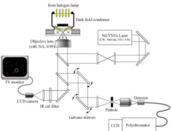
A schematic experimental setup is depicted in Fig. 2. A TEM00-mode, Gaussian beam with linear polarization from a CW Nd:YVO4 laser (wavelength: 1064 nm, J20I-BL-106C, Spectra Physics) was used as a light source for optical trapping and was introduced into the microscope from its backport. The laser beam was reflected by dichroic mirror for near IR reflection-visible transmittion and was focused into the sample solution via a high numerical objective lens (UPLSAPO 40X2, 40×, N.A 0.95, Olympus). The focal height was adjusted mechanically by using stepping motor driven objective lens positioner. The sample on the inverted microscope was illuminated with white light from a 100-W halogen lamp through an oil-immersion dark-field condenser lens (U-DCD, NA 1.35-1.20, Olympus). The scattered light from sample was collected with a same objective lens for laser trapping and was sent to a monochrome digital CCD camera (CV-A50IRC, JAI) and movie was recorded with video rate (30 frame/sec.). As spectroscopic measurement, the scattered light was spatially selected by using a pinhole (300 μm radius) via a pair of scanning mirror in the confocal unit (FV300, Olympus) and sent to a polychromator (SpectraPro 2300i, Princeton) coupled CCD camera (PIXIS 400, Princeton) to achieve high spatial resolution. The scanning mirror enables to measure light scattering spectra from a specific point even out of the focal point.
Proc. of SPIE Vol. 7762 77620N-2
Figure 2. Wide field light scattering microspectroscopic imaging system
3. RESULTS AND DISCUSSION
Video 1 presents a sequence upon NIR laser irradiation. The laser focal height is fixed at 100 μm above the surface of glass substrate. At the first stage, many bright points show micromotion in dark background. Because of the high light scattering efficiency of gold nanoparticles and the dark background realized by dark field illumination, it becomes possible to detect single gold nanoparticles. Thus, the bright points are corresponding to individual gold nanoparticles though spectroscopic confirmation is eventually required, and the micromotions correspond to Brownian motion of gold nanoparticles. It should be noted that the dark field condenser lens illuminate widely not only in lateral dimension (~several hundreds μm width) but also in vertical dimension (~10 μm depth), so that the light scattering from the defocused area can be detected. The vertical spatial resolution of microscope is determined by focal depth of objective lens and is theoretically predicted as 1.77λn/(NA)2, where λ, n, and NA correspond to wavelength of light, refractive
index of medium, and numerical aperture of objective, respectively. Thus we can observe gold nanoparticles in a tiny vertical dimension (~ 1.5 μm). With defocusing a particle, it becomes a multiple ring with negligible weak scattering light. As a consequence, the wide field light scattering microscope can visualize a film event clearly with real time despite the lower resolution compared to confocal microscope. Needless to say, confocal microscope is not capable of real-time imaging.
After starting 700 mW NIR laser irradiation (~5 sec. after playing Video 1), brighter point was fixed at the focal spot of the laser, which indicates gold nanoparticle trapping and assembling at the position. In addition to this laser trapping, transportation of particles to the focal point from all the direction within several hundreds micrometer was observed. This migration process starts immediately after laser irradiation and reaches to steady state within seconds. Because photon pressure potential is spatially limited in the diffraction limit of light, the full width of the working distance is about 1 μm in considering the experimental condition. Laser trapping related phenomena cannot explain this particle migration. It is also confirmed that the migration speed is strongly dependent on laser power and the height of the focal point from substrate surface, which will be reported in future publications for details. This indicates that another reason but whose origin is a focused laser should be considered. One possible explanation is local heating of solvent by trapping laser, since the overtone of OH bond exists near 1064 nm wavelength.18 Duhr and Braun reported that a focused
laser could manipulate DNA in solution not by photon pressure but by local heating at the focal point, causing thermodiffusion. They succeeded in deposition of DNA on glass substrate as a ring shape, which may reflect the diffusion and following electrostatic adsorption of DNA around the focal point. It can be considered that particle migration can be induced by the thermodiffusion, i.e., convection flow.19, 20 And the flow should be inhomogeneous
because of temperature gradient around the focal point. Concerning the focal position dependence, Peterman et al. showed experimentally that the heating coefficient depends on the distance of the focal point from the glass substrate, because the glass can act as a heat sink.21 This means, temperature gradient may be three dimensionally propagated and
be strongly affected by the geometric condition. And they also reported that temperature elevation at the focal point is
Proc. of SPIE Vol. 7762 77620N-3
Video 1. Gold nanoparticle motion before and after NIR laser beam focusing into solution. The laser power was 700 mW after passing through objective lens. The focal height was adjasted at 100 μm above the glass substrate surface. The shown picture is an accumulation of the movie around the focal position. http://dx.doi.org/12.860433.1
revealed as Δ14 K/W at the steady state. In our case, the elevation can be estimated as ~10 K, which seems enough to induce thermal convection flow. To confirm the role of laser heating of solvent, we performed the same experiment in which H2O is replaced by heavy water, D2O. It is well known that the absorption at 1064 nm wavelength in D2O is less
than 1/10 of that in H2O. We have confirmed that such particle migration from far away is not observed and only
particles near the focal point (~ 2 μm) can be gathered and trapped, thus only photon pressure can make sense in D2O
solvent. It can be assumed that the trigger is temperature elevation and gradient around the focal point due to light absorption by solvent. Needless to mention, only the wide field imaging enables to observe such two dimensional phenomena, which are impossible to be confirmed by confocal microscope as previous works.
In order to discuss the migration speed of particles quantitatively, we performed the particle tracking analysis of Video 1 using algorithm developed in a plug-in of Image J.22 Thanks to two dimensionally homogeneous illumination of
the wide-field light scattering imaging technique, we can follow the trajectories occurring in the two dimensional view. From when a particle visits the observation field, the position at each frame is recorded. We count particles which show
Figure 3. Relation between particle migration speed and distance from trapping laser focal point analyzed by single particle tracking method. The original data was taken from Video 1 shown above. Each black point corresponds to a trajectory of single particle. The line is a calculated result based on least squares method.
Proc. of SPIE Vol. 7762 77620N-4
constant light scattering intensity while the tracking, otherwise the particle motion may contain vertical motion. In Fig. 3, the result of particle tracking analysis is shown. Each point indicates particle speed against the initial position where the particle was observed. All the trajectories direct to the focal point from all the direction in two dimensional view. Particles migration speed is different for each particle, but there is a tendency that the speed decreases with decreasing the distance from the focal point. If it is supposed that the photon pressure potential can propagate far away up to several tens micrometers, the migration speed should be increased with decreasing the distance from the focal point, however, the particles show opposite behavior. This suggests that laser trapping is not dominant for the particle migration as considered above. And the tracking analysis revealed that the travel length of particle tend to be shorter with decreasing the distance from the focal point (not shown here). Based on Navie-Stokes theory describing thermal convection flow, fluid decreases the density by heating and flow up to upper side against the viscosity and gravity.23 Thus the thermal convection flow reflects the spatial gradient of temperature
directly. In the present system, near the focal point the gradient of temperature should be shaper than that out of the focus. At the focal point, the temperature distribution is linear to light intensity distribution. Out of the focal point, the gradient is inversely proportional to the distance from the focal point. But the spatial limit of the thermal propagation, i.e., the boundary condition is determined by the distance between the focal point and the glass surface as mentioned before. As the results, the convection flow driven by local heating may be asymmetric shaped one, and this is predicted in literatures based on Navie-Stokes equation.24 So, the convection flow driven by local heating
should contain vertical movement in nature. Because we selected particle trajectories keeping constant light intensity, it is impossible to follow the vertical movement in the detectable region. Especially near the focal point, the effect of the vertical movement will be more dominant. We can safely assume that the particle migration from far away is due to thermal convection driven by local heating of solvent and the convection flow around the focal point shows three dimensional motion. As shown here, the detailed analysis of two dimensional event can give us suggestions of three dimensional motion.
Fig. 4 (a) shows an example of light scattering spectra at the trapping laser focal point. The acquisition time for each spectrum is 500 ms. The spectra always show peak band around 550 nm, which can be assigned by localized surface plasmon resonance band of gold nanoparticles.25 Note that light at the background is recorded in advance and subtracted
from the signal, so a contribution of defocused particles is ignorable. Depending on the number of trapped particles, the intensity fluctuates drastically and the peak wavelength shift slightly. While it is well known that gold nanoparticle assembly exhibits additional strong band due to electromagnetic interaction,3, 26, 27 we observe a weak secondly band at
680 nm. One explanation of this is strong electric repulsion among particles. Photon pressure potential cannot overcome
Figure 4. (a) Three examples of light scattering spectra at the trapping laser focal point taken at different time points. (b) Averaged light scattering intensity at 550 nm vs. distance from the focal point with and without laser irradiation. The minus region of the horizontal axis correspond to mirror image of the positive region.
-6 -4 -2 0 2 4 6 0 50 100 150 200 250 300 350 400 700 mW irradiation without irradiation L ight scatter ing in ten sity at 55 0 nm
Distance from the focal point / m
(b)
(a)
450 500 550 600 650 700 750 Scattering efficiency / a.u. Wavelength / nmProc. of SPIE Vol. 7762 77620N-5
the barrier for assembling. Moreover, temperature elevation of gold nanoparticles should be considered. Because gold nanoparticles exhibit strong light absorption, temperature should be increased upon laser irradiation, resulting in increase of thermal energy.28 To encourage gold nanoparticle assembling, adding salt into solution may be necessary to decrease
the thickness of the electric double layer.29 Setting this gold nanoparticles assembly aside, transient light scattering
spectroscopic measurement enables us to estimate particle concentration, in other word, probability of particle existence at a specific point because it should be linear to the number of particles there. The galvano mirror in the confocal unit realizes position selective measurement, so that spectroscopic investigation from the focal position to the outside with changing the distance is possible. According to this procedure, position dependence of light scattering spectra is in Fig. 4 (b). The light scattering spectra were recorded more than 200 sec. with changing the distance from the focal point. The vertical axis corresponds to average light scattering intensity at 550 nm for all the recorded spectra at the position. Around the focal point the concentration is drastically increased, which can be explained by laser trapping of particles. The half width of particle concentration around the focal point is wider than that of photon pressure potential. This may be due to scattering force pushing toward the focus from the lower position. Without laser irradiation, light intensity is almost zero because particles do not move heavily. It should be pointed out that not only at the focal point but also at the outside, particle concentration is increased extensively. In the area, the particle motion is governed by thermal convection flow, so that this concentration increase is a contribution of the flow. As shown here, particle concentration can be increased by focused laser beam and it is suggestive that photon pressure and convection flow can be coupled in a view of particle concentration increase. Assembly and crystallization of molecules can be assisted by the focusing laser induced phenomena.
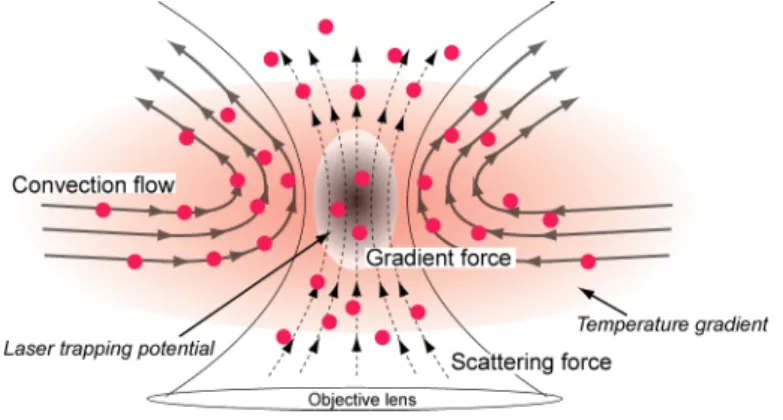
Finally, possible picture of particle diffusion and assembling process upon NIR laser focusing suggested by the experimental evidences is proposed as summarized in Fig. 5. As well known, at the laser focal point, gold nanoparticles are trapped and assembled. In a view of photon pressure, supplying particles to the focal point is assisted by two components, gradient force (1) and scattering one (2). Because the former one is governed by diffraction limit of light, the spatial propagation is roughly limited in the diffraction limit of the objective lens. The latter one works in the light cone, so it can transport particle to the focal point from the lower side but can push the particle out to the upper side. Theoretically a vertical gap between the focal point and the trapping position due to the scattering force is predicted,1
however, it is not detectable with our system since the wide field imaging is capable only of two dimensional observations. In addition to the contributions of photon pressure, we pointed out that temperature gradient around the focal point, which is generated by light absorption of solvent, should be considered (3). The local heating of solvent by laser can cause thermal gradient, which can three dimensionally propagate far away with being inversely proportional to the distance from the focal point. The three dimensional temperature gradient results in three dimensional convection flow and it can transport particles along with the flow direction. The migration speed and direction are strongly affected by solvent, laser power, and the geometric condition of sample because of the change of temperature gradient. Moreover, it should be noted that the contributions of photon pressure and that of convection flow can be coupled each other. The convection flow assists to collect particles from far away and to supply particles toward focal point continuously in addition to the gradient force. Consequently, we assume that the laser trapping phenomena should be regarded as a thermodynamically equilibrium condition especially in the case that solvent can absorb trapping laser light.
Figure 5. Possible explanation of particle diffusion and assembling process upon tightly laser focusing.
Proc. of SPIE Vol. 7762 77620N-6
4. CONCLUSION
In this paper, we have successfully demonstrated quantitative analysis of gold nanoparticle diffusion and assembling dynamics induced by focusing NIR laser beam into solution. Wide field light scattering imaging method helps us to visualize particle motion not only at the laser focal point but also the outside at single particle level. It is clearly shown that particles migration toward the focal spot from all the directions within several tens micrometer, which cannot be explained only by a contribution of laser trapping but is suggestive to convection flow generated by light absorption of solvent. And interplay of photon pressure and convection flow to increase particle concentration around the focal point is also revealed. Spatial propagation of the heat flux and its equilibrium with photon pressure should be important and indispensable to understand transportation of the small objects toward the focal point and stability of laser trapping. Theoretical approach based on Navie-Stokes equation will be effective to confirm the mechanism and dynamics, and to predict the particle behavior in a specific container. Further dynamic investigation on the initial stage to steady state will be also performed to elucidate the dynamics from non-steady state to steady state. Concerning instrument improvement, introducing dynamic light scattering (DLS) measurement will be complementary to two dimensional imaging. DLS provides information of particle motion with three dimensional resolution, so that the role of scattering force and the dynamics of three dimensional convection flow can be revealed. Utilizing higher intensity light source such as supercontinuum laser light as an illumination light is also expecting for nonfluorescence macromolecules and molecules observation.
ACKNOWLEDGEMENT
The present work was partially supported by the MOE-ATU Project (National Chiao Tung University) from the Ministry of Education of Taiwan, the National Science Council of Taiwan to T. U. (NSC 98-2113-M-009-013-MY2) and to H. M. (NSC 98-211-M-009-001), and a KAKENHI grant (a Grant-in-Aid for Scientific Research) in the Priority Area “Molecular Science for Supra Functional Systems” to T. U. and “Strong Photon-Molecule Coupling Fields” from the Ministry of Education, Culture, Sports, Science and Technology of Japan (MEXT) (21020022) to T. S., a KAKENHI (C) grant (20550136) to T.S., a KAKENHI (S) grant (18106002) to H. M. from the Japan Society for the Promotion of Science (JSPS).
REFERENCES
1. Ashkin, A., "Acceleration and Trapping of Particles by Radiation Pressure," Phys. Rev. Lett., 24, 156 (1970). 2. Ashkin, A., Dziedzic, J. M., Bjorkholm, J. E. and Chu, S., "Observation of a single-beam gradient force optical trap
for dielectric particles," Opt. Lett., 11, 288-290 (1986).
3. Yoshikawa, H., Matsui, T. and Masuhara, H., "Reversible assembly of gold nanoparticles confined in an optical microcage," Phys. Rev. E, 70, 061406 (2004).
4. Hosokawa, C., Yoshikawa, H. and Masuhara, H., "Cluster formation of nanoparticles in an optical trap studied by fluorescence correlation spectroscopy," Phys. Rev. E, 72, 021408 (2005).
5. Borowicz, P., Hotta, J., Sasaki, K. and Masuhara, H., "Chemical and Optical Mechanism of Microparticle Formation of Poly(N-vinylcarbazole) in N,N-Dimethylformamide by Photon Pressure of a Focused Near-Infrared Laser Beam," J. Phys. Chem. B, 102, 1896-1901 (1998).
6. Masuo, S., Yoshikawa, H., Nothofer, H.-G., Grimsdale, A. C., Scherf, U., Mullen, K. and Masuhara, H., "Assembling and Orientation of Polyfluorenes in Solution Controlled by a Focused Near-Infrared Laser Beam," J. Phys. Chem. B, 109, 6917-6921 (2005).
7. Smith, T. A., Hotta, J., Sasaki, K., Masuhara, H. and Itoh, Y., "Photon Pressure-Induced Association of Nanometer-Sized Polymer Chains in Solution," J. Phys. Chem. B, 103, 1660-1663 (1999).
Proc. of SPIE Vol. 7762 77620N-7
8. Tanaka, Y., Yoshikawa, H. and Masuhara, H., "Two-Photon Fluorescence Spectroscopy of Individually Trapped Pseudoisocyanine J-Aggregates in Aqueous Solution," J. Phys. Chem. B, 110, 17906-17911 (2006).
9. Tanaka, Y., Yoshikawa, H., Asahi, T. and Masuhara, H., "Laser microfixation of highly ordered J aggregates on a glass substrate," Appl. Phys. Lett., 91, 041102 (2007).
10. Sugiyama, T., Adachi, T. and Masuhara, H., "Crystallization of Glycine by Photon Pressure of a Focused CW Laser Beam," Chem. Lett., 36, 1480-1481 (2007).
11. Rungsimanon, T., Yuyama, K.-i., Sugiyama, T., Masuhara, H., Tohnai, N. and Miyata, M., "Control of Crystal Polymorph of Glycine by Photon Pressure of a Focused Continuous Wave Near-Infrared Laser Beam," J. Phys. Chem. Lett., 1, 599-603 (2010).
12. Sugiyama, T., Adachi, T. and Masuhara, H., "Crystal Growth of Glycine Controlled by a Focused CW Near-infrared Laser Beam," Chem. Lett., 38, 482-483 (2009).
13. Bohren, C. F. and Huffman, D. R., [Absorption and Scattering of Light by Small Particles], Wiley, New York, Vol. (1983).
14. Hashimoto, S., Uwada, T., Masuhara, H. and Asahi, T., "Fabrication of Gold Nanoparticle-Doped Zeolite L Crystals and Characterization by Optical Microscopy: Laser Ablation- and Crystallization Inclusion-Based Approach," J. Phys. Chem. C, 112, 15089-15093 (2008).
15. Uwada, T., Hosokawa, Y., Takizawa, N., Okano, K. and Masuhara, H., "Organic molecular sensing by single metal porphyrin nanoparticles," Proceedings of SPIE, 6865, 68650I (2008).
16. Uwada, T., Toyota, R., Masuhara, H. and Asahi, T., "Single Particle Spectroscopic Investigation on the Interaction between Exciton Transition of Cyanine Dye J-Aggregates and Localized Surface Plasmon Polarization of Gold Nanoparticles," J. Phys. Chem. C, 111, 1549-1552 (2007).
17. Kawata, S. and Masuhara, H., [Nanoplasmonics: From Fundamentals to Applications], Elsevier, Amsterdam, (2006).
18. Curcio, J. A. and Petty, C. C., "The Near Infrared Absorption Spectrum of Liquid Water," J. Opt. Soc. Am., 41, 302-302 (1951).
19. Braun, D. and Libchaber, A., "Trapping of DNA by Thermophoretic Depletion and Convection," Phys. Rev. Lett., 89, 188103 (2002).
20. Duhr, S. and Braun, D., "Why molecules move along a temperature gradient," Proc. Nat. Acad. Sci., 103, 19678-19682 (2006).
21. Peterman, E. J. G., Gittes, F. and Schmidt, C. F., "Laser-Induced Heating in Optical Traps," Biophys. J., 84, 1308-1316 (2003).
22. https://weeman.inf.ethz.ch/ParticleTracker/
23. Kitahara, K., [Nonequilibrium Statistical Physics], Iwanami, Tokyo, (1997).
24. Ichikawa, M., Ichikawa, H., Yoshikawa, K. and Kimura, Y., "Extension of a DNA Molecule by Local Heating with a Laser," Phys. Rev. Lett., 99, 148104 (2007).
25. Kreibig, U. and Vollmer, M., [Optical Properties of Metal Clusters], Springer, Berlin (1995).
26. Rechberger, W., Hohenau, A., Leitner, A., Krenn, J. R., Lamprecht, B. and Aussenegg, F. R., "Optical properties of two interacting gold nanoparticles," Optics Communications, 220, 137-141 (2003).
27. Prikulis, J., Svedberg, F., Kall, M., Enger, J., Ramser, K., Goksor, M. and Hanstorp, D., "Optical Spectroscopy of Single Trapped Metal Nanoparticles in Solution," Nano Lett., 4, 115-118 (2003).
28. Seol, Y., Carpenter, A. E. and Perkins, T. T., "Gold nanoparticles: enhanced optical trapping and sensitivity coupled with significant heating," Opt. Lett., 31, 2429-2431 (2006).
29. Enustun, B. V. and Turkevich, J., "Coagulation of Colloidal Gold," J. Am. Chem. Soc., 85, 3317-3328 (1963).
Proc. of SPIE Vol. 7762 77620N-8