Direct Synthesis of Zr-Doped Ceria Nanotubes
Ying-Chen Chen, Kuei-Bo Chen, Chi-Shen Lee,* and M. C. Lin
Department of Applied Chemistry, National Chiao Tung UniVersity, 1001 UniVersity Road, Hsinchu 30010, Taiwan
ReceiVed: NoVember 29, 2008; ReVised Manuscript ReceiVed: February 18, 2009
Zr-doped ceria nanotubes (ZrxCe1-xO2) were obtained in a high yield from a hydrothermal reaction in an aqueous solution of NaOH with Ce(NO3)3· 6H2O and ZrO2powder in a small proportion. The morphology and crystalline structure were characterized with X-ray diffraction, a scanning electron microscope, and a transmission electron microscope. Mechanisms for the growth of Zr-doped ceria nanotubes are proposed based on the Kirkendall effect; the formation of the tubular structure is strongly dependent on the precursor. This is the first report of a direct synthesis of cerium-oxide nanotubes in high yield. The surface area of the nanotubes is 76 m2/g and their average pore size is∼52.2 nm. Catalytic measurements show that the nanotubes as synthesized are active for an oxygen-storage capacity and for ethanol reforming.
The synthesis of metal-oxide nanocrystals with specific shapes has attracted considerable attention because of their distinctive properties and extensive applications that include photolumi-nescence, batteries, gas sensors, magnetic media, and catalysis.1-5 Among these materials, ceria is attractive because it exhibits a large capacity for the storage of oxygen6,7(OSC) and serves as a solid electrolyte or anode material in solid-oxide fuel cells,8-12 oxygen sensor,13ultraviolet absorber,14,15and catalyst for various reactions.16Dispersion of a catalytically active metal and cerium-oxide nanoparticles throughout an alumina support of large surface area has been reported to enhance the catalytic perfor-mance of the ethanol-reforming reaction.17,18For the utilization of cerium oxide as a catalyst, a simple and effective technique to prepare it with both a large surface area and a controlled shape is needed. Shape-controlling syntheses of cerium oxide in various forms including particle,19rod,20-23cube,18,24hollow sphere,25 wire,26 flowerlike,27,28 and nanotube29-32 have been reported. Among several methods developed recently for the synthesis of ceria nanotubes, Han et al. demonstrated the use of precipitation and an aging process to prepare CeO2nanotubes; their method requires protracted aging (45 d), and the product contains CeO2nanostructures in both rod and tube forms.31Tang and co-workers described another method to prepare CeO2 nanotubes on annealing Ce(OH)3 nanorods under reducing conditions;32the entire process requires operation under an inert atmosphere, and the major product has a nanorod instead of a nanotube form. Zhou and co-workers reported the synthesis of CeO2nanotubes using H2O2to etch Ce(OH)3nanorods,31 but the product contains CeO2nanomaterial in both tube and particle forms. Sun and co-workers reported the use of Ce(OH)CO3 nanorods as a precursor to synthesize a CeO2nanomaterial in tube form in alkaline solutions.32These approaches to synthesing cerium oxide nanotubes typically require protracted reaction, templating materials (e.g., AAO), surfactants (e.g., PEG), or multiple steps during which the reaction condition is difficult
to control. For the solid-solution phase, ZrxCe1-xO2, various authors have described methods for the synthesis of mesoporous and nanocage forms,25,33 but there seems to be no report regarding the direct synthesis of Zr-doped CeO2nanotubes with a high yield. Here we report a facile approach for the synthesis of ZrxCe1-xO2nanotubes.
In a typical synthesis, Ce(NO3)3· 6H2O (0.4 g) and ZrO2 (powder, 0.01 g) were dispersed in NaOH (15 M,∼16 mL) solution under stirring near 295 K for 10 min; the mixture was transferred into an autoclave (25 mL, diluted to 80% of the capacity with distilled water), which was sealed and maintained at 100-200 °C for 48 h, followed by cooling naturally to ambient temperature. The white precipitate was washed several times with deionized water, collected by filtration, and dried in air. The yield of the product was estimated to be∼90% based on the initial mass of Ce(NO3)3· 6H2O. The crystal phase was determined with powder X-ray diffraction (XRD, Bruker D8-Advance, Cu KR radiation). Structural and compositional information for the product material was obtained with a scanning electron microscope (SEM, Hitachi S-4700I), a transmission electron microscope, selected-area electron dif-fraction (TEM/SAED, JEM-2010F), an energy-dispersive X-ray spectrometer (HRTEM/EDX, JEM-2010F), an inductively coupled plasma-mass spectrometer (ICP-MS, Perkin-Elmer SCIEX ELAN 5000), a vis spectrophotometer (Shimadzu UV-1601). Analyses of surface area and pore size were carried out with Quantachrome. The catalytic activity was investigated by temperature-programmed reduction (TPR) with H2 and with ethanol-reforming analyses using a GC-MS system (Thermo Trace GC 2000). Reactions with various Zr precursors and reaction conditions were performed for the synthesis of Zr-doped cerria nanomaterials. The synthetic procedures were kept constant; the results show that only the precursor ZrO2yields the desired nanotube form. The duration of reaction time, temperature, and concentration of NaOH have no effect on the morphology. For reaction with high Zr/Ce ratio, the products, based on the powder XRD and SEM, showed no nanotube form
* To whom correspondence should be addressed. Tel.: 886-3-5131332. Fax: 886-3-5723764. E-mail: [email protected].
5031
10.1021/jp810492s CCC: $40.75 2009 American Chemical Society Published on Web 03/06/2009
and generally yielded a mixture of CeO2, ZrO2 with varied morphology (Table S1).
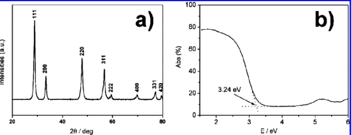
The powder XRD pattern for the product as synthesized is shown in Figure 1. In general, all diffraction features exhibit wide signals, indicative of small particles. The reflections were indexed to the cubic CeO2structure (a ) 5.411(1), Fm3jm (No. 225), cubic, JCPDS 78-0694), in close agreement with the literature value a ) 5.411 Å; no signal of an impurity phase was detected. The UV spectrum of the as-synthesized nanotubes measured in ethanol suspensions exhibited an intense absorption at 330 nm; the band gap of the nanotubes as synthesized is accordingly calculated to be 3.24 eV (Figure 1b). The XPS spectrum of the sample as synthesized exhibits the core levels of Ce(3d), Ce(4d), Zr(3s), Zr(3p), Zr(3d), and O(1s); the Ce(3d) spectrum shows small contributions of v′and u′bands from
the Ce(3d5/2) and Ce(3d3/2) ionizations of Ce3+. The result indicates that the chemical valence of cerium on the surface of
Figure 1. (a) X-ray diffraction pattern of Zr-doped ceria nanotubes, with indexes of X-ray features labeled. (b) Diffuse reflectance spectra of
Zr-doped ceria nanotubes.
Figure 2. (a) SEM and (b) TEM images of Zr-doped ceria nanotubes. (c) TEM and selective-area electron-diffraction pattern (SAED, inset) of a
nanotube. (d) HRTEM image of a portion of nanotube from (c) showing a domain of a single crystal.
SCHEME 1: Schematic Illustration of the Transformation from Nanorod to Nanotube of ZrxCe1-xO2Based on the Kirkendall Effect
the Zr-doped ceria nanotube is mainly Ce4+with Ce3+in a small proportion (Figure S1).34
The hydrothermal reaction of Ce(NO3)3· 6H2O with ZrO2in a small proportion at 150°C yielded much nanomaterial in rod form, as evident from the scanning-electron-microscope (SEM) image (Figure 2a). The TEM images (HRTEM) in Figure 2b indicate that most products dispersed on the copper TEM grids has an open-ended nanotube morphology. The nanotubes are uniform with average lengths up to several hundred nm. The nanotubes have inner diameter in a range 30-50 nm; the wall thickness is about 15 ( 5 nm. A TEM image of a nanotube with a round tip and a length greater than 200 nm is shown in Figure 2c. The granularity and porosity of the tube walls indicate that the tubes as obtained consist of many CeO2nanoparticles. The selected-area-diffraction (SAED) pattern (inset of Figure 2c) demonstrates a domain of a single-crystalline part, and the high-resolution TEM image shown in Figure 2d reveals clear layer fringes along the tube wall with a layer separation approximately 3.8 Å for the (110) planes. Elemental analyses with energy-dispersive X-ray spectra (EDS) and ICP mass tests reveal a small proportion of Zr with atomic ratios Ce/Zr 0.99/ 0.01 for EDS and 0.97/0.03 for ICP-mass (Figure S2). The results indicate that the Zr content is much smaller than the reaction composition ∼7 atom %, indicating that most Zr is
not incorporated into the nanotube. The entire morphology evolution might be derived from the Kirkendall effect (see Scheme 1).25,35,36 The reaction initially yielded Zr
xCe1-x(OH)3 in a rod form;37 the Zr4+ ions may act as the catalyst that promote the diffuse rate of Ce3+/Ce4+ions inside the nanorod; the metal hydroxide nanorods were gradually decompose to form ZrxCe1-xO2nanotubes according to a partial oxidation of Ce3+ and differential rate of diffusion between Ce4+and Ce3+ions inside the material.31
Figure 3 shows nitrogen adsorption-desorption isotherms and the corresponding distributions of pore size of the Zr-doped ceria nanotubes. The isotherm curve evidently shows a hysteresis loop for relative pressures over a wide range. The BET (Brunauer-Emmett-Teller) surface area of nanotubes as synthesized was 76 m2/g. The curve for the distribution of pore size (inset of Figure 3), obtained with the Barrett-Joyner-Halenda (BJH) method using the adsorption branches of the isotherms, is centered about∼52 nm, consistent with the observed size from TEM tests. The subsidiary maximum at∼3.8 nm may be due to the voids formed on the surface of a nanotube.38Such a large surface area and pore size is comparable to previously reported CeO2nanotube materials, obtained from Ce(OH)CO3nanorods.32 The results indicate clearly that the surface area and porosity of the nanomaterials as synthesized are significantly superior to those of bulk CeO2materials.
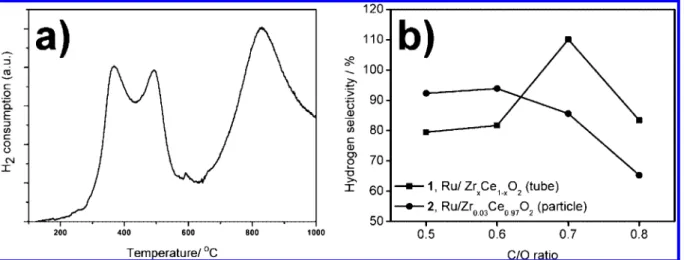
Ceria might be used as an oxygen-transfer component in redox catalysis for its excellent oxygen storage capacity (OSC).39 To characterize the OSC of the Zr-doped ceria nanotubes, we performed temperature-programmed reduction (TPR) with H2 to obtain the result shown in Figure 4a. The reduction profile shows two broad maxima and a further reduction maximum above 700 °C; the former features at 354 and 474 °C are attributed to a global process corresponding to the consumption of surface oxygen species, whereas the maximum at 825 °C reflects the bulk reduction. The area under the low-temperature curve can be used to estimate the oxygen that contributes to the reduction during the TPR operation. A quantitative evalu-ation of the low-temperature reduction feature (below 620°C, representation for OSC) reveals that hydrogen consumed by Zr-doped ceria nanotubes is 984µmol H2per g.31,40,41The effect of as-synthesized nanotubes on H2selectivity (SH2) in ethanol
reforming was tested using catalysts of 5 wt % Ru/Zr-doped ceria nanotubes (catalyst 1) and 5 wt % Ru/Zr0.03Ce0.97O2 nanoparticles (catalyst 2) using porous Al2O3 as support, according to the literature scheme. The hydrogen selectivity (SH2) Figure 3. Nitrogen adsorption-desorption isotherms and the
corre-sponding curves for the distribution of BJH pore size (inset) of Zr-doped ceria nanotubes.
Figure 4. (a) TPR profile of Zr-doped ceria nanotubes. (b) Hydrogen selectivity as a function of C/O ratio obtained over catalysts 1 and 2 (5 wt
% Ru/(Zr-doped cerria nanotubes or Zr0.03Ce0.97O2particles).
is defined as the molar ratio of the product H2to the hydrogen production per mole of ethanol. The effect of the C/O ratio on the catalytic performance of catalysts is shown in Figure 4b with the C/O ratio varied from 0.4 to 0.8. The results indicate that, for catalyst 1, SH2increased gradually on increasing C/O to attain a maximum value 110% at C/O ) 0.7, which is larger than the maximum SH2of catalyst 2 (SH2) 90% at C/O ratio ∼ 0.6). SH2of catalyst 1 decreased sharply from 110 to 60% upon altering the C/O ratio from 0.7 to 0.8, whereas SH2of catalyst 2 decreased from 90 to 70%. A catalyst with Zr-doped ceria nanotubes is evidently more active than CeO2particles for the conversion of ethanol to hydrogen, which might be due to the fact that CeO2nanotubes contain a greater exposed surface of {100} and {110} facets than CeO2nanoparticles.18
In conclusion, Zr-doped ceria nanotubes were synthesized with a direct hydrothermal reaction using ZrO2 in a small proportion to assist the formation of nanotubes. The possible mechanism may be described by the Kirkendall effect, and the formation of the tubular structure depends strongly on the precursor, rather than on the effect of the concentration of base. This reaction route significantly diminishes the duration of synthesis of Zr-doped ceria nanotubes. The product as synthe-sized exhibits a large surface area and a large pore size, and is highly active for hydrogen reduction and ethanol reforming relative to bulk material; the product might thus be used as a highly active catalyst. The results demonstrate that the direct synthesis of Zr-doped ceria nanotubes with a large surface area and a large pore size indicates potential applications in catalysis. We expect this method to be applicable to the preparation of diverse metal-oxide nanotubes from suitable precursors.
Acknowledgment. For technical assistance, we thank
Profes-sors Chia-Ming Yang for BET measurements and Professor Teng-Ming Chen for UV measurements. The Institute of Nuclear Energy Research, Atomic Energy Council, Taiwan (Contract NL940251) and National Science Council (Contracts NSC94-2113-M-009-012, 94-2120-M-009-014) supported this research. M.C.L. acknowledges Taiwan Semiconductor Manufacturing Co. for the TSMC distinguished professorship and Taiwan National Science Council for the Distinguished Visiting Profes-sorship at National Chiao Tung University.
Supporting Information Available: Details of experimental
procedures and additional XPS spectra, and SEM/TEM and EDS/ICP-mass results for Zr-doped ceria nanotubes. This material is available free of charge via the Internet at http:// pubs.acs.org.
References and Notes
(1) Ferna´ndez-Garca´, M.; Martı´nez-Arias, A.; Hanson, J. C.; Rodriguez, J. A. Chem. ReV. 2004, 104, 4063–4104.
(2) Murray, R. W. Chem. ReV. 2008, 108, 2688–2720.
(3) Laurent, S.; Forge, D.; Port, M.; Roch, A.; Robic, C.; Elst, L. V.; Muller, R. N. Chem. ReV. 2008, 108, 2064–2110.
(4) Jeong, U.; Teng, X.; Wang, Y.; Yang, H.; Xia, Y. AdV. Mater.
2007, 19, 33–60.
(5) Franke, M. E.; Koplin, T. J.; Simon, U. Small 2006, 2, 36–50.
(6) Mai, H.-X.; Sun, L.-D.; Zhang, Y.-W.; Si, R.; Feng, W.; Zhang, H.-P.; Liu, H.-C.; Yan, C.-H. J. Phys. Chem. B 2005, 109, 24380.
(7) Campbell, C. T.; Peden, C. H. F. Science 2005, 309, 713. (8) Gasteiger, H. A.; Lamm, A.; Vielstich, W. Handbook of Fuel Cells:
Fundamentals, Technology, and Applications; Wiley: Chichester, England;
New York, 2003.
(9) Hoogers, G. Fuel Cell Technology Handbook; CRC Press: Boca Raton, FL, 2003.
(10) Larminie, J.; Dicks, A. Fuel Cell Systems Explained, 2nd ed.; J. Wiley: Chichester, West Sussex, 2003.
(11) Singhal, S. C.; Kendall, K. High-Temperature Solid Oxide Fuel
Cells: Fundamentals, Design and Applications; Elsevier: Oxford; New York,
2003.
(12) Zhang, Y.; Zha, S.; Liu, M. AdV. Mater. 2005, 17, 487. (13) Jasinski, P.; Suzuki, T.; Anderson, H. U. Sens. Actuators, B 2003,
95, 73–77.
(14) Masui, T.; Fujiwara, K.; Machida, K. I.; Adachi, G. Y. Chem. Mater.
1997, 9, 2197.
(15) Li, R. X.; Yabe, S.; Yamashita, M.; Momose, S.; Yoshida, S.; Yin, S.; Sato, T. Solid State Ionics 2002, 151, 235.
(16) Trovarelli, A. Catalysis by Ceria and Related Materials; Imperial College Press: London, 2002.
(17) Deluga, G. A.; Salge, J. R.; Schmidt, L. D.; Verykios, X. E. Science
2004, 303, 993–997.
(18) Hsiao, W. I.; Lin, Y. S.; Chen, Y. C.; Lee, C. S. Chem. Phys. Lett.
2007, 441, 294–299.
(19) Niederberger, M. Acc. Chem. Res. 2007, 40, 793–800.
(20) Kuiry, S. C.; Patil, S. D.; Deshpande, S.; Seal, S. J. Phys. Chem.
B 2005, 109, 6936.
(21) Vantomme, A.; Yuan, Z.-Y.; Du, G.; Su, B.-L. Langmuir 2005,
21, 1132.
(22) Sun, C.; Li, H.; Wang, Z.; Chen, L.; Huang, X. Chem. Lett. 2004,
33, 662–663.
(23) Sun, C.; Li, H.; Zhang, H.; Wang, Z.; Chen, L. Nanotechnology
2005, 16, 1454–1463.
(24) Ho, C.; Yu, J. C.; Kwong, T.; Mak, A. C.; Lai, S. Chem. Mater.
2005, 17, 4514–4522.
(25) Liang, X.; Wang, X.; Zhuang, Y.; Xu, B.; Kuang, S.; Li, Y. J. Am.
Chem. Soc. 2008, 130, 2736–2737.
(26) Yada, M.; Sakaji, S.; Torikai, T.; Watari, T.; Furuta, S.; Katsuki, H. AdV. Mater. 2004, 16, 1222.
(27) Sun, C.; Sun, J.; Xiao, G.; Zhang, H.; Qiu, X.; Li, H.; Chen, L. J.
Phys. Chem. B 2006, 110, 13445–13452.
(28) Sun, C.; Li, H.; Chen, L. J. Phys. Chem. Solids 2007, 68, 1785– 1790.
(29) Tang, C.; Bando, Y.; Liu, B.; Golberg, D. AdV. Mater. 2005, 17, 3005.
(30) Han, W.-Q.; Wu, L.; Zhu, Y. J. Am. Chem. Soc. 2005, 127, 12814– 12815.
(31) Zhou, K.; Yang, Z.; Yang, S. E. N. Chem. Mater. 2007, 19, 1215– 1217.
(32) Chen, G.; Xu, C.; Song, X.; Zhao, W.; Ding, Y.; Sun, S. Inorg.
Chem. 2008, 47, 723–728.
(33) Yuan, Q.; Liu, Q.; Song, W.-G.; Feng, W.; Pu, W.-L.; Sun, L.-D.; Zhang, Y.-W.; Yan, C.-H. J. Am. Chem. Soc. 2007, 129, 6698–6699.
(34) Si, R.; Zhang, Y.-W.; Li, S.-J.; Lin, B.-X.; Yan, C.-H. J. Phys.
Chem. B 2004, 108, 12481–12488.
(35) Fan, H. J.; Knez, M.; Scholz, R.; Hesse, D.; Nielsch, K.; Zacharias, M.; Go1sele, U. Nano Lett. 2007, 7, 993–997.
(36) Raidongia, K.; Rao, C. N. R. J. Phys. Chem. C 2008, 112, 13366– 13371.
(37) Zhou, K.; Wang, X.; Sun, X.; Peng, Q.; Li, Y. J. Catal. 2005, 229, 206.
(38) Zhang, C. P. D.; Shi, L. J. Solid State Chem. 2008, 181, 1298– 1306.
(39) Trovarelli, A. Catalysis by Ceria and Related Materials; Imperial College Press: London, 2002.
(40) Zhou, K. B.; Wang, X.; Sun, X. M.; Peng, Q.; Li, Y. D. J. Catal.
2005, 229, 206.
(41) Andreeva, D.; Ivanov, I.; Ilieva, L.; Abrashev, M. V. Appl. Catal.,
A 2006, 302, 127–132.
JP810492S