146 1-4244-1525-X/07/$25.00 © 2007 IEEE
1
Abstract—This paper proposes an efficient framework of
high-quality image compression method for upper Gastrointestinal tract endoscopy images. The proposed DEWC coding method saves traditional image preprocessing computations, such as demosaicking and color-space transformation, and directly utilizes raw image data acquired from CMOS sensor. R, G and B band image are then separately encoded by wavelet-based SPECK coding. In a cardinal GI tract environment, the spatial frequency distribution of red component is lower than green or blue, and green component is relatively high while compared to blue and red components. DEWC coding saves more bits on red band while allocating more bits on green and blue bands. Therefore, under a fixed compression ratio, such non-uniform bit-rate allocation may earn a better image quality. To measure quality-loss in non-uniform bit-rate allocation, a quality quantified measurement called color-distortion based on CIE94 color-difference formula is also proposed. By using analytical result of color-distortion in bit-rate and bit-rate-difference analysis, an optimal/suboptimal bit-rate allocation scheme can be found by solving linear equations derived from the relationship of color-distortion and bit-rate-difference. When comparing to general JPEG2000 compression standard, the experimental result shows that proposed DEWC coding has a better image quality in color-distortion measurement and more efficient performance in execution time.
Index Terms—Gastrointestinal endoscopy, DWT, JPEG2000,
Image compression.
I. INTRODUCTION
high-quality endoscope image compression method called DEWC (Delta-E Wavelet Coding) method is proposed in this paper, where Delta-E means the CIE94
color-difference formula ∆E. Fig. 1 illustrates the flow chart for the proposed DEWC coding method. Each 512×512 8-bit GI tract raw image is directly acquired by the CMOS image sensor with the most commonly utilized Bayer color filter [1]. In each acquired raw image, each pixel contains only one of the R, G and B primary color in it. Traditionally, two missing color components are first estimated from adjacent pixels by using
demosaicking process [2,3], and the RGB-to-YCbCr color space transformation is applied before image encoding.
However, demosaicking process may induce twice redundant
data as many as original raw image [4,5], and color-space transformation also requires the calculation of inner products. It is not worth performing both image preprocessing steps unless we can really gain image quality from those redundant computations.
Unlike traditional image compression technique, proposed DEWC coding method starts from raw image in the form of Bayer pattern and processes each R, G1, G2 and B signal separately. These band images are down sampling of (2:1, 2:1) according to the row and column of original raw image. Four 256×256 8-bit images are then transformed by using reversible DWT (Discrete Wavelet Transform) separately to produce wavelet coefficients. By applying Set-Partitioned Embedded bloCK (SPECK) encoding [6] on the transformed coefficients, adjustable fractional bit-rate control is applied on each band for specific quality specification.
II. PROPOSED IMAGE COMPRESSION
In proposed DEWC coding method, image bands may have different bit-rates depending on quality specification. However, this reveals two major problems. The first problem should be considered is how to allocate bit-rates to all color bands. Inside cardinal GI tract environment, the spatial frequency distribution of the red component is lower than green or blue, and the green component is relatively high while compared to the blue and red components [7]. In Fig. 2, the bit-rate and MSE
distortion (Mean Squared Error) analysis also shows that an
Lan-Rong Dung and Tsung-Hsi Chiang National Chiao Tung University, Taiwan
High-Quality Image Compression for
Gastrointestinal Endoscope
A
Fig.1 Block diagrams of wavelet-based image compression: (a) image encoder; (b) image decoder.
147
2 appropriate bit-rate allocation scheme for GI images is to
allocate the most bits to green band and a few portions to red band. Therefore, under a fixed compression ratio, DEWC coding method reserves some bits from red band and allocates more portions to green or blue band, therefore, we may obtain a higher image quality in less color-distortion in it. However, someone may allocate too much bit-rate on green band than on others, it may result serious color-distortion on a restored image. In clinic diagnosis, color or appearance abnormality detection can help clinician to diagnose the digestive symptoms.
Therefore, color-distortion should be considered and quantified in image quality measurement. The second problem comes up when we determine measurement of the image quality. Traditionally, the quality measurement of PSNR (Peak Signal-to-Noise Ratio) [8] is commonly used in image compression. Nevertheless, PSNR is only focus on the
luminance rather on the color information, thus, it can not be
used to measure the color-distortion of a restored image. Two restored images may have the same image quality in luminance PSNR; however, they may have different degree quality loss in
color-distortion. To overcome these crucial problems, the color-distortion D∆E measurement based on CIE94 formula
∆E*
94 is proposed in this paper.
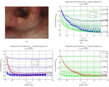
The result of bit-rate and color distortion analysis in Fig. 3(b)-3(d) shows that color-distortion provides more information than bit-rate and MSE distortion analysis. Another analysis of color-distortion and bit-rate difference in Fig. 4(a)-4(b) provides a quantified measurement of color-distortion and bit-rate-difference between two arbitrary color bands. It reflects the degree of color-distortion due to non-uniform bit-rate allocation. Based on analytical result of
color-distortion and bit-rate-difference, bit-rate allocation
problem is modeled by linear equations. By solving linear equations, a bit-rate allocation scheme for high-quality compression can be found.
Fig.4. (a) Color-distortion and bit-rate-difference for various bit-rates (sample image 04); (b) The relationship between color-distortion D∆E and bit-rate
difference.
Fig.3. The rate-distortion curves for D∆E . (a) The GI image clip#4. (b)
D∆E at rR=0.5bpp. (c) D∆E at rG=0.5bpp. (d) D∆E at rB=0.5bpp.
Fig. 2. The rate-distortion curve of the GI image clip #2. (a) The GI image #2. (b) the rate-distortion curves.
148
3
Based on the analytical result, linear equations are derived in this section in finding a better bit-rate allocation scheme. For convenience, three variables are firstly defined which indicate distinct bit-rate-differences: δBG = rB – rG, δBR = rB – rR, and δGR
= rG – rR. These variables also have current or previous prefix
depending on the notation δ or δ’ respectively. The same representation is also used on distortion functions and bit-rate variables.
According to analytical result in Fig. 4(b) and considering red and blue curves, minimal values of color-distortion are almost located at the same place. Therefore, current values of δBG and δGR can be found from previous functions β’r'R and β’r'B
in Fig. 4(b), as shown in (1). ' ' ( ) 0 R r BR d x x dx β δ = = , ' ( )rB' 0 GR d x x dx β δ = = . (1)
Then, coding bit-rates for rR, rG and rB are found by solving
linear equation as given following:
0
1 1
1 1
0
,
4
1
2
1
32 /
R BG G GR Br
r
r
δ
δ
ρ
ρ
−
−
=
≤
. (2)Note that, β is the compression ratio of an original image related to its restored image defined by (3).
CFA raw image size in bits
total encoding/decoding bits
ρ
=
(3)The acquired CFA raw image size is 512×512×8 bits, i.e. 2 Mega bits. A boundary condition of an 4:1 compression ratio to a restored image is given by (4). In an CFA raw image, each 2×2-pixel block contains four bytes (or 32 bits) including R, G1, G2 and B components in that. Therefore, for a restored image with 4:1 compression ratio, it only needs 8 bits in each 2×2-pixel block.
2
32 /
8
R G B
r
+
r
+ =
r
ρ
=
for 4:1 compression ratio. (4) If compression ratio ρ is larger than 4, the solution to Equation 3 may lead a negative bit-rate which means a non-valid solution. According to analytical result, we can see that function βrR does not have large variety while red bandranging from rR=0.1 to 3.0bpp. Herein, a modification can be
made by directly assigning rR to an average value
r
R. As aresult, another equation is given in (5) while compression ratio ρ > 4.
0
1 1
1
0
0
,
4
1
2
1
32 /
R BG G R Br
r
r
r
δ
ρ
ρ
−
=
>
. (5)Therefore, before encoding an CFA raw image, current δBG
and δGR are found from previous function β’ rR’ and β’ rB’ by
using (1). A desired bit-rate allocation scheme (rR,rG,rB) is
solved by using (2) or (5). Then, image encoding is applied to image bands with desired bit-rate. At last, function βrR, βrG and
βrB are updated for next image compression.
III. EXPERIMENTAL RESULTS
Table I compares the color-distortion and luminance PSNR obtained for GI tract images at various compression ratio with proposed DEWC coding method and general JPEG2000 [9] compression. For each ρ=4, 8 or 16 in DEWC coding, bit-rate combinations (rR,rG,rB) for GI images are computed by
calculating Equation 3 and 6. On the other hand, JPEG2000 compression is also applied on GI images with the same compression ratio as DEWC coding. We can see that DEWC coding has a better quality than JPEG200 compression in luminance PSNR while ρ=4, but DEWC coding loses a little quality in luminance PSNR while ρ=16. As mentioned in previous section, luminance PSNR does not consider quality loss in color information. Therefore, image quality of color-distortion measurement is considered.
When considering color-distortion measurement in Table 1, it shows that DEWC coding has a better restored image quality in less color-distortion than JPEG2000 compression, in average case. Although, luminance PSNR may lose a little quality while ρ=16 in DEWC coding, the values of D∆E in DEWC coding are
still low which means DEWC coding has better image quality in color-distortion than general JPEG2000 compression.
Fig. 5 compares the average encoding time obtained for sample GI images at bit-rate scheme in Table I with our DEWC coding and general JPEG2000 compression running on a 1.8-GHz Pentium 4 PC. For the JPEG2000 compressor, Pentium 4 takes longer than DWGEC coding in ρ=4, 8 and 16 in average.
TABLE I
149
4
IV. CONCLUSION
General JPEG2000 standard is an image compression providing superior compression at the expense of greater computation required. We have developed the DEWC coding method which saves image preprocessing computing and directly utilizes CFA raw image acquired from CMOS sensor on wavelet SPECK coding. R, G and B color bands are encoded separately with high-quality issue in color-distortion measurement based on CIE94 ∆E*
94 formula. The analytical
result of color-distortion and bit-rate difference provides us guidance for finding a better bit-rate allocation scheme while applying image coding. According to the experimental result, we also show that proposed DEWC coding has a better image quality while comparing to general JPEG2000 compression. Additionally, DEWC coding performance is also better than general JPEG2000 compression.
ACKNOWLEDGMENT
This work was supported in part by the National Science Council, Taiwan, under the grant number NSC 95-2221-E009-337-MY3 and Chung-Shan Institute of Science and Technology, Taiwan, under the project BV94G10P. The authors would like to thank National Chip Implementation Center (CIC) for technical support.
REFERENCES
[1] Bryce E. Bayer, Color imaging array, U.S. Patent 3971065, 1976. [2] J. E. Adams and J. E. Hamilton, Design of practical color filter array
interpolation algorithms for digital cameras, Proc. SPIE, vol.3028, pp.117-125, 1997.
[3] R. Kimmel, Demosaicing: Image reconstruction from color CCD samples, IEEE Trans. on IP, vol.8, no.9, pp. 1221-1228, 1999.
[4] C.C. Koh, J. Mukherjee and S. K. Mitra, New efficient methods of image compression in digital cameras with color filter array, IEEE Trans. on CE, vol.49, no.4, pp. 1448-1456, 2003.
[5] S.Y. Lee and A. Ortega, A novel approach of image compression in digital cameras with a Bayer color filter array, IEEE ICIP, vol.3, pp. 482-485, 2001.
[6] W.A. Pearlman, A. Islam, N.N. and A. Said, Efficient, low-complexity image coding with a set-partitioning embedded block coder, IEEE Trans. on CSVT, vol.14, no.11, pp. 1219-1228, 2004.
[7] Y. Konomura and T. Tsuruoka, Image data compressing device for endoscope, US. Patent, no. 4,845,553, 1989.
[8] M.C. Lin and L.R. Dung and P.K. Weng, An ultra-low-power image compressor for capsule endoscope, BioMedical Engineering OnLine, vol.5, no.14, 2006.
[9] OpenJPEG, Communications and remote sensing Laboratory, Universite catholique de Louvain, Belgium, http://www.openjpeg.org
![Table I compares the color-distortion and luminance PSNR obtained for GI tract images at various compression ratio with proposed DEWC coding method and general JPEG2000 [9] compression](https://thumb-ap.123doks.com/thumbv2/9libinfo/7637585.137019/3.918.476.791.76.157/compares-distortion-luminance-obtained-various-compression-proposed-compression.webp)
