Consensus sequence L/PKSSLL mimics crucial epitope on Loop III of Taiwan
cobra cardiotoxin
Ping-Chieh Wanga, Kah-Sin Loha, Shih-Ting Lina, Tzu-Ling Chiena, Jen-Ron Chiangb,
Wen-Chin Hsiehb, Bor-Lin Miaob, Cheng-Fu Suband Wen-Jen Yanga,*
a
Institute of Biotechnology, National University of Kaohsiung, Kaohsiung, 811, Taiwan
b
Vaccine Center, Centers for Disease Control, Taipei, 115, Taiwan
*Corresponding author: Wen-Jen Yang
Institute of Biotechnology, National University of Kaohsiung,
700, Kaohsiung University Road., Nanzih District, Kaohsiung, 811, Taiwan
Tel: +886-7-5919454
Fax: +886-7-5919404
Abstract
Phage display is effective in screening peptides that mimic a venom’sneutralizing epitopes. A phage display cyclized heptapeptide library (C7C library) was panned with purified divalent
antivenin IgG, which neutralizes Naja naja atra venom (NAV) and Bungarus multicinctus
venom (BMV). The selected heptapeptide sequences were aligned with known protein
sequences of NAV and BMV in GenBank. One of the four consensus sequences, L/PKSSLL,
mimicked the crucial epitope on Loop III of Taiwan cobra cardiotoxin that is associated with
the venom’slethal potency. In dot blot analysis, several clones showed varying reactivities for NAV monovalent antivenin and lesser cross-reactions with BMV monovalent antivenin. The
KSSLLRN-carrying phage occurred four times in selected clones and showed the strongest
reactivity to NAV monovalent antivenin. Furthermore, the QDSLLPS-carrying phage also
presented significant dot blot signal, indicating that the SLL sequence shared by these two
clones may be a crucial antibody binding site.
Introduction
Snakebite is a serious global public health issue, especially in numerous tropical and
subtropical countries like Taiwan. Taiwan cobra (Naja naja atra) is a crucial venomous snake
and causes about 10% of all snakebite incidents in Taiwan [1]. Snake venoms are a complex
mixture of many diverse toxins. Cobrotoxin, cardiotoxins, and phospholipase A2 (PLA2) are
the three major toxic proteins of Taiwan cobra venom [2]. Among these, cobrotoxin is the
most predominant neurotoxin. It is a small, basic protein consisting of a single polypeptide
chain with 62 amino acids, cross-linked by four disulfide bonds [3]. Cobrotoxin binds to the
nicotinic acetylcholine receptor on postsynaptic membranes with high binding affinity and
blocks neuromuscular transmission, leading to muscle contraction dysfunction [4].
Cardiotoxins are composed of 60–63 amino acids (molecular weight 6.5–7.0 kDa) in a single
-sheet polypeptide chain, cross-linked by four disulfide bonds [5]. Interestingly, although cobrotoxin and cardiotoxin are similar in their 3D structures, they present very different
biological activities. Cardiotoxin can produce depolarization of nerve and muscle cells to
affect the contraction of the cardiac muscle, induce cancer cell apoptosis, and cause lysis of
erythrocytes and epithelial cells [2; 5; 6]. To date, several cardiotoxin isoforms have been
isolated and characterized from the venom of the Taiwan cobra (Naja naja atra) [5; 7].
Comparison of the lethal potency and 3D structure(s) of these cardiotoxin isoforms from
with the presence of a nonpolar “finger-shaped”projection that comprises of hydrophobic
residues Leu47 and Leu48 at the tip of Loop III. It was predicted that this finger-shaped
projection forms a part of the putative receptor binding site of cardiotoxins [8]. Snake PLA2s
are a group of polypeptides, about 120–130 amino acids in length, and each chain is
cross-linked by seven disulfide bonds [9]. Venom PLA2 has been shown to possess various
pharmacological effects such as cardiotoxic, myonecrotic, neurotoxic, hemolytic, and
anticoagulant actions in addition to its enzymatic activity in the hydrolysis of ester bonds in
phosphoglycerides [10]. It has been shown that both His-47 and Asp-93 are essential for the
catalytic activity of PLA2from Taiwan Cobra [11].
To date, antivenin administration is the world’smajor therapy for snakebites. In Taiwan, a pepsin-digested F(ab’)2 bivalent antivenin produced from equine serum by the National Institute of Preventive Medicine has been used to treat cobra snakebites since 1986, and it has
shown a very low risk of acute adverse reactions [12]. A local antivenin injection can speed
up neutralization and reduce the spread of venom. The efficacy of antivenin is strongly
correlated with the neutralizing antibodies against the epitopes of venom that are crucial for
its lethal potency. The phage display random peptide library has emerged as a powerful tool
for analyzing antigen-antibody interactions and is successfully used to mimic epitopes of
antigens (mimotopes) [13]. Peptides mimicking two epitopes of neuwiedase from Bothrops
recognized the toxin [14].
In this study, a phage display cyclized heptapeptide library (C7C library) was used to
identify peptides that bind to protein A-purified antivenin IgGs that can neutralize Taiwan
cobra venom. A major clone that displayed KSSLLRN and a consensus sequence, L/PKSSLL,
which mimicked the epitope on the Loop III of cardiotoxin of Taiwan cobra that is associated
with its lethal potency, were identified These data of epitopes can provide valuable
Materials and methods
Materials. A phage display cyclized heptapeptide library (C7C library) was purchased
from New English Biolabs, USA. The library has a pair of cysteine residues flanking the
random heptapeptide, resulting in phage display of cyclized peptides, was fused to the minor
coat protein III at the N-terminus of M13 phage. Taiwan cobra (Naja naja atra) venom (NAV)
and Bungarus multicinctus venom (BMV) were kindly provided from the Vaccine Center,
Centers for Disease Control (CDC), Taiwan. The HRP/anti-M13 monoclonal antibody
conjugate was purchased from GE Healthcare Inc.
Animals and antivenin preparation. An equine-derived monovalent antivenin against
NAV and a bivalent antivenin against NAV and BMV were kindly provided by the Vaccine
Center, CDC, Taiwan. The BMV antivenin used in this study was prepared by subcutaneous
injection of glutaraldehyde (GA)-detoxified venom mixed with Freund’s adjuvant in mice.
Inbred specific pathogen-free (SPF) BALB/c female mice were purchased from the National
Laboratory Animal Center (Taipei, Taiwan). All experiments were performed in accordance
with institutional guidelines. The venom was detoxified by 0.125% glutaraldehyde, incubated
at room temperature for 30 min, and then at 4°C overnight. Twelve weeks old mice were immunized with 20g detoxified BMV for each animal. The Freund’scompleteadjuvantwas administered in the prime injection, and the incomplete adjuvant was used in the subsequent
titers of antibodies were measured by ELISA using serial dilutions of antisera against BMV.
The antibody titer was defined as the reciprocal of the maximum dilution factor of the test
serum that kept the OD405reading above 0.2.
Purification and characterization of equine antivenin. The equine-derived bivalent
antivenin with a potency of 80 antitoxic units against NAV was obtained from the Vaccine
Center, CDC, Taiwan. To better perform the biopanning process, IgGs of the antivenin were
purified using a protein A purification method, modified from [15]. Briefly, a protein
A-agarose resin packed column was washed with ten column-volumes of starting buffer (100
mM Tris-HCl, pH 7.5, 100 mM NaCl). The antivenin sample was mixed with an equal
volume of starting buffer before being applied to the column. The pass-through solution was
collected while measuring OD280. The column was washed with starting buffer until OD280
reduced to background levels. The IgG was eluted with 0.1 M glycine-HCl (pH 2.5),
immediately neutralized with 1 M Tris-HCl (pH 8.0), and stored at –20°C until use. The
concentration of eluted IgG was estimated by OD280 (1 OD280 = 0.75 mg/ml). Western blot
analysis and potency test were used to further characterize the eluted IgG against NAV [16].
The potency test was performed according to the standard operation procedure for antivenin
antitoxin activity measurement at Vaccine Center, CDC, Taiwan.
Biopanning, DNA sequencing, and sequence analysis. The biopanning of purified IgG
phage were used for determining the phage titer. The remaining elute was used to infect E.
coli ER2738 for phage amplification. After three rounds of biopanning, the DNA sequences of
randomly selected phage clones were determined by a fluorescence-based sequencer. The
sequences of phage-displayed heptapeptides were deduced from the DNA sequences and were
further grouped according to the amino acids that were identical at the position of alignment.
Alignments of these sequences with the known protein sequences of NAV and BMV in
GenBank were also performed by the BLAST software to understand whether the sequences
mimic the epitopes on these venom proteins The amino acid sequences of eight cardiotoxin
isoforms from Taiwan cobra venom, CTX-3 (NCBI accession number: U42585), CTX-1
(U42583), CTX-2 (U58485), CTX-4 (Y12491), CTX-5 (U58489), CTX-8 (U42586), CTX-10
(Y18957) and CTX-N (Z54230), were aligned and their different amino acids residues were
compared The 3D structure of cardiotoxin-3 (PDB:1i02) was used to represent the topology of
cardiotoxins. The structure was generated using the KiNG (Kinetic Image, Next Generation.)
3D viewer program.
Binding specificity of selected phage clones. The binding specificities of the selected
phages to the purified IgG from NAV monovalent antivenin were analyzed by ELISA
according to the phage library manufacturer’s recommendation.
Dot blot analysis. All selected phage clones were tested individually for their reactivity
NAV-specific epitope sequences. The amplified phage clones were analyzed by dot blot
according to the method described previously [17], with some modifications. Briefly, phage
clones (1011 pfu) were blotted onto nitrocellulose membrane and incubated at 37°C for 1 h.
The membrane was incubated with blocking buffer (TBST containing 5% non-fat milk) at
37°C for 1 h and then washed with TBST. The purified equine-derived NAV antivenin or
mouse-derived BMV antivenin was incubated with the membrane at 37 oC for 1 h. After
washing with TBST, the membrane was incubated with alkaline phosphatase-conjugated goat
anti-horse IgG (1:1500) or anti-mouse IgG (1:1500) antibodies at 37oC for 1 h. The blot was
developed and visualized with NBT/BCIP substrate at room temperature with agitation until a
purple spot appeared, and it was then rinsed with TBS containing 20 mM EDTA to stop the
Results
IgG purification and characterization of NAV and BMV antivenin
The equine-derived bivalent antivenin was purified with protein A to exclude the
interference from other serum components in the biopanning assay. The IgG was eluted and
the concentration of purified IgG, estimated by the absorbance at 280 nm, was 1 mg/ml. The
purification results were evaluated using 10% SDS-PAGE as shown in Fig. 1A. They
indicated that most of the components in the serum had been removed, and purified IgGs had
been obtained from the eluate Snake venoms are composed of a mixture of many different
toxic proteins. The analysis of NAV components using 15% SDS-PAGE (Fig 1B) indicated
that small molecular weight proteins were abundant in NAV. Western blot analysis using
unpurified divalent antivenin (Fig. 1C) or protein A-purified IgGs (Fig. 1D) against NAV was
used to examine whether the activity of purified IgGs was lost during the purification process.
The results indicated that the purified IgGs could still recognize most of the NAV proteins
(Fig. 1C and 1D). The potency of purified IgGs against NAV dropped to 40 antitoxic units,
compared to 80 antitoxic units with unpurified antivenin, indicating that a portion of
neutralizing IgGs failed to be captured by protein A.
Biopanning and Sequences analysis of selected heptapeptides
the epitopes of NAV. The number of pfu obtained after each round of biopanning increased
gradually (data not shown), indicating that the biopanning experiments were successful. After
three rounds of biopanning, 33 phage clones were randomly isolated and characterized by
DNA sequencing. The deduced amino acid sequences were analyzed (Table S1); thirty
different sequences were obtained from the randomly selected phage clones. Interestingly,
most of the sequences appeared once, but the KSSLLRN sequence, which was an exception,
was found in four selected clones. The phage-displayed heptapeptides were aligned with the
known protein sequences of NAV and BMV in GenBank. The KSSLLRN heptapeptide
matched with the KSSLL sequence located at the tip of Loop III of the cardiotoxin from NAV,
which has already been demonstrated to be strongly associated with the venom’s lethal potency [8]. The BLAST results showed that several heptapeptide sequences matched with
the sequences on both NAV and BMV, some heptapeptides matched with only one of the two
venoms, and a few sequences could not find any matched sequence on the NAV and BMV
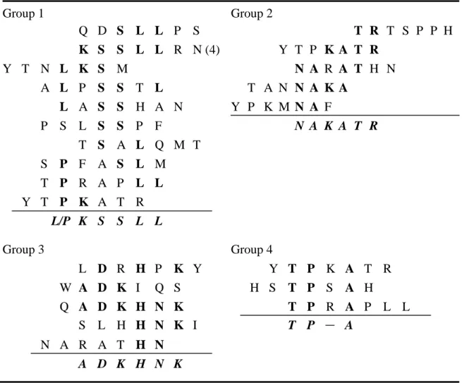
proteins (Table S1). As shown in Table 1, four consensus sequences, L/PKSSLL, NAKATR,
ADKHNK and TP_A, were found among the selected heptapeptide sequences by aligning the
identical amino acid residues that were shared between the heptapeptides. The consensus
sequence L/PKSSLL was obtained from the clone with the highest frequency and several
selected clones, indicating that it should be the major epitope which the purified IgG bound
As shown in Fig. 2A, alignment of eight amino acid sequences of N. naja atra cardiotoxin
isoforms showed that amino acid sequences were highly conserved. The major variable
regions were located at positions 5–11 in Loop I, 27–32 in Loop II, and 45–47 in Loop III of
cardiotoxin molecules. The molecular model of cardiotoxin-3 (PBD:1i02) allowed the
observation of the epitopes in the tertiary structure (Fig. 2B). The sequence at the tip of Loop
III form a distinct “finger-shaped”projection that is identical with the major consensus
epitope L/PKSSLL identified in this study.
Immunoreactivity and binding specificity of selected phage clones
In order to evaluate the binding specificity of selected phage clones to the purified IgGs
from NAV monovalent antivenin, individual clones were assayed by ELISA. Assessing the
different immunoreactivities of purified IgGs against individual phage clones, the positive
clones which can specifically bind to antibodies were identified. In contrast, the wild-type
phage showed only background immunoreactivity to IgG.s (data not shown). BLAST results
of selected heptapeptide sequences aligned with the known protein sequences of NAV and
BMV in GenBank showed that several heptapeptide sequences mimicked sequences on
various NAV and BMV proteins (Table S1). To further confirm whether the phage clones
could be recognized by BMV antivenin, the antiserum against BMV was produced in mouse
immunization three times with 20g detoxified BMV. The dot blot analysis was performed to test the binding specificity of each selected clone with purified NAV monovalent IgG or BMV
antiserum. NAV and BMV were used as positive controls and showed significant signals. Dot
blot results showed that all selected phage clones could be recognized by purified NAV
monovalent IgG with different immunoreactivities (Fig. 3A). The phage displaying the
KSSLLRN sequence showed the strongest signal in comparison to others, indicating that this
sequence has high binding affinity with purified NAV monovalent IgG and should represent a
crucial epitope on NAV. Based on the results of BLAST (Table S1) and the dot blot analysis
(Fig. 3), clone 3 (KSSLLRN) and clone 25 (QDSLLPS) showed significant dot blot signals.
This suggested that the SLL sequence, which is shared by both clones, should be an important
antibody-binding site. Furthermore, no significant signals were found in all selected phage
clones when BMV antiserum was used in the dot blot assay (Fig. 3B), suggesting that these
heptapeptides were specifically bound to NAV IgG and less cross-reactivity occurred with
Discussion
Snake venoms consist of different proteins with various biological functions and usually
contain lethal toxic proteins. This complexity of snake venoms makes it more difficult to
characterize or purify their components and to treat snakebites [18]. Therefore, the discovery
of crucial epitopes on snake venoms is extremely valuable in the development of diagnostic
methods or more effective therapies. Based on these concerns, the neutralizing epitopes of
Taiwan cobra venom were explored in our study.
Although the primary aim of this study was to investigate neutralizing epitopes of
Taiwan cobra venom, the NAV monovalent antivenin, unfortunately, was not routinely
available from the Vaccine Center, CDC, Taiwan. This is the reason why a bivalent antivenin
against NAV and BMV was used in biopanning to study NAV epitopes. The NAV monovalent
antivenin was produced by the Vaccine Center a few months later (as a result of our request),
and it was used to measure the binding specificities of selected phage clones.
Comparison of the results in Western blot analysis using unpurified bivalent antivenin
(Fig. 1C) and protein A-purified IgGs (Fig. 1D) against NAV indicated that most of the
proteins were still recognized by purified IgGs. However, the signals of some protein bands
decreased while using purified IgGs bound to NAV. It suggested that some antibodies could
have been lost during protein A purification. This issue was supported by the decrease in the
used to improve the IgG purification efficiency, no significant improvement in the antitoxin
activity of the purified antibody was observed.
Previous studies proposed that the residues of Loop III at positions 44, 46, and 50 were
crucial for the depolarization activity of cobra cardiotoxins [19]. Additionally, the residues at
the tip of Loop III of the cardiotoxin form a distinct “finger-shaped”projection, the presence
of which has been strongly correlated with the venom’slethal potency [8]. Because these heptapeptides were obtained from the biopanning of the C7C phage library with neutralizing
antibodies, these sequences could represent the neutralizing epitopes. In this study, a
consensus sequence, L/PKSSLL, was deduced from alignment with selected major phage
clones, and dot blot analysis showed significant binding affinity. These results demonstrated
that the L/PKSSLL sequence should be a crucial neutralizing epitope for antivenin binding.
Interestingly, even though some heptapeptide sequences (e.g. clone 21, 24) showed no
similarity with the NAV or BMV protein sequences, the dot blot analysis still exhibited
immunoreactivity. This suggests that such sequences maybe located at the conformational
epitopes of the proteins.
To date, antivenin has been produced in equine by immunization with detoxified snake
venom. Accordingly, antivenin contains various immunoglobulins specific for numerous
antigens in the venom. However, many of them have no neutralizing effect on the venom [20].
large amount of heterogenous immunoglobulins are administrated to snakebite victims [18].
This study should provide valuable information for the production of neutralizing antibodies
and the development of distinctive diagnosis between the snakebites of Taiwan cobra (Naja
Acknowledgments
This study was supported by the grant DOH 96-DC-1021 from the Centers for Disease
References
[1] B.L. Miao, Huang, R.J., Hu, M.S., Liau, M.Y., Venomous snakebites in Taiwan
(1988–1991), Chin. J. Public Health (Taipei) 14 (1995) 455-460.
[2] S.H. Chiou, C.C. Hung, H.C. Huang, S.T. Chen, K.T. Wang, C.C. Yang, Sequence
comparison and computer modelling of cardiotoxins and cobrotoxin isolated from Taiwan
cobra, Biochem. Biophys. Res. Commun. 206 (1995) 22-32.
[3] L.S. Chang, K.C. Chen, B.N. Wu, S.K. Lin, P.F. Wu, Y.R. Hong, C.C. Yang, Expression
and mutagenesis studies of cobrotoxin from Taiwan cobra, Biochem. Biophys. Res.
Commun. 263 (1999) 652-6.
[4] C.C. Yang, Structure and function of cobra neurotoxin, Adv. Exp. Med. Biol. 391 (1996)
85-96.
[5] T.K. Kumar, G. Jayaraman, C.S. Lee, A.I. Arunkumar, T. Sivaraman, D. Samuel, C. Yu,
Snake venom cardiotoxins-structure, dynamics, function and folding, J. Biomol. Struct.
Dyn. 15 (1997) 431-63.
[6] C.H. Tsai, S.H. Yang, C.M. Chien, M.C. Lu, C.S. Lo, Y.H. Lin, X.W. Hu, S.R. Lin,
Mechanisms of cardiotoxin lll-induced apoptosis in human colorectal cancer colo205 cells,
Clin. Exp. Pharmacol. Physiol. 33 (2006) 177-82.
[7] T. Sivaraman, T.K. Kumar, P.W. Yang, C. Yu, Cardiotoxin-like basic protein (CLBP) from
[8] G. Jayaraman, T.K. Kumar, C.C. Tsai, S. Srisailam, S.H. Chou, C.L. Ho, C. Yu,
Elucidation of the solution structure of cardiotoxin analogue V from the Taiwan cobra
(Naja naja atra)--identification of structural features important for the lethal action of
snake venom cardiotoxins, Protein Sci. 9 (2000) 637-46.
[9] M.J. Dufton, R.C. Hider, Classification of phospholipases A2 according to sequence.
Evolutionary and pharmacological implications, Eur. J. Biochem. 137 (1983) 545-51.
[10] H.M. Verheij, A.J. Slotboom, G.H. de Haas, Structure and function of phospholipase A2,
Rev. Physiol. Biochem. Pharmacol. 91 (1981) 91-203.
[11] F.M. Pan, S.C. Chao, S.H. Wu, W.C. Chang, S.H. Chiou, Characterization of
phospholipase A2 (PLA2) from Taiwan Cobra: isoenzymes and their site-directed mutants,
Biochem. Biophys. Res. Commun. 250 (1998) 154-60.
[12] J.C. Chen, M.J. Bullard, T.F. Chiu, C.J. Ng, S.J. Liaw, Risk of immediate effects from
F(ab)2bivalent antivenin in Taiwan, Wilderness Environ. Med. 11 (2000) 163-7.
[13] W.J. Yang, J.F. Lai, K.C. Peng, H.J. Chiang, C.N. Weng, D. Shiuan, Epitope mapping of
Mycoplasma hyopneumoniae using phage displayed peptide libraries and the immune
responses of the selected phagotopes, J. Immunol. Methods 304 (2005) 15-29.
[14] R. Cardoso, M.I. Homsi-Brandeburgo, V.M. Rodrigues, W.B. Santos, G.L. Souza, C.R.
Prudencio, A.C. Siquieroli, L.R. Goulart, Peptide mimicking antigenic and immunogenic
[15] W.J. Yang, D. Shiuan, Plaque reduction test: an alternative method to assess specific
antibody response to pIII-displayed peptide of filamentous phage M13, J. Immunol.
Methods 276 (2003) 175-83.
[16] B.L. Miao, M.Y. Liau, R.J. Huang, S.W. Chen, T.K. Chen, S.C. Chang, Preparation of
toxoid from Taiwan cobra (Naja naja atra) venom, Zhonghua Yi Xue Za Zhi (Taipei) 46
(1990) 1-6.
[17] R. Hawkes, The dot immunobinding assay, Methods Enzymol. 121 (1986) 484-91.
[18] S.C. Wagstaff, G.D. Laing, R.D. Theakston, C. Papaspyridis, R.A. Harrison,
Bioinformatics and multiepitope DNA immunization to design rational snake antivenom,
PLoS Med. 3 (2006) e184.
[19] S.J. Hodges, A.S. Agbaji, A.L. Harvey, R.C. Hider, Cobra cardiotoxins. Purification,
effects on skeletal muscle and structure/activity relationships [published errtum appears in
Eur J Biochem 1988 Feb 1;171(3):727], Eur. J. Biochem. 165 (1987) 373-83.
[20] P. Malasit, D.A. Warrell, P. Chanthavanich, C. Viravan, J. Mongkolsapaya, B.
Singhthong, C. Supich, Prediction, prevention, and mechanism of early (anaphylactic)
Legends
Fig. 1. Characterization of snake venoms and antivenin. (A) SDS-PAGE analysis of
bivalent antivenin against NAV and BMV. Lane M: protein marker (the molecular weight is
indicated on the left); lane 1: unpurified divalent antivenin; lane 2: the collection of unbound
fractions of divalent antivenin from protein A affinity column; lane 3: protein A-purified IgG,
the heavy chain (50 kDa) and light chain (25 kDa) of IgG are indicated with an asterisk. (B)
SDS-PAGE analysis of NAV. Lane M: protein marker; lane V: 12 g NAV protein. Western blot analysis using (C) unpurified divalent antivenin or (D) protein A purified IgG against 12
g NAV protein.
Fig. 2. Comparison of amino acid sequences of eight cardiotoxin (CTX) isoforms from Taiwan cobra (Naja naja atra) venom and 3D structure of CTX-3. (A) The amino acid
sequences of eight cardiotoxin isoforms from Taiwan cobra venom are aligned. The dash lines
indicate the conserved residues, and the regions corresponding to the three-loop structure of
mature cardiotoxin are marked. The sequence PKSSLL matched with selected phage clones is
underlined. (B) The 3D structure of cardiotoxin-3 (PDB:1i02) is used to represent the
topology of cardiotoxins. The protein is cross-linked by four disulfide bonds (shown in zigzag
lines) and forms a three-loop structure. The residues at the tip of Loop-III matched with
comprise a portion of the putative receptor binding site of cardiotoxins [8; 19].
Fig. 3. Immunoreactivity and binding specificity of selected phage clones.
Immunoreactivity was assayed using dot blot analysis with (A) purified equine-derived
anti-NAV IgGs; and (B) mouse-derived anti-BMV antiserum against selected phage clones.
NAV or BMV (snake venom; 2 ng) was used as a positive control in separate assays. The
Table 1. Classification of the deduced amino acid sequences alignment of selected
heptapeptides obtained after three rounds of biopanning.
Group 1 Group 2 Q D S L L P S T R T S P P H K S S L L R N (4) Y T P K A T R Y T N L K S M N A R A T H N A L P S S T L T A N N A K A L A S S H A N Y P K M N A F P S L S S P F N A K A T R T S A L Q M T S P F A S L M T P R A P L L Y T P K A T R L/P K S S L L Group 3 Group 4 L D R H P K Y Y T P K A T R W A D K I Q S H S T P S A H Q A D K H N K T P R A P L L S L H H N K I T P - A N A R A T H N A D K H N K
The deduced amino acid sequences of selected heptapeptides were aligned and analyzed.
These sequences could be classified into four groups according to the shared amino acid
sequences. Sequences were shown as the single-letter amino acid code. Identical amino acids
shared between the heptapeptides were shown in bold upper case and the consensus residues
were summarized as bold-italic-type letters. The number in the parenthesis indicated the
Table S1. The sequences of phage-displayed heptapeptides of the 33 recombinant phage
clones randomly selected after three rounds of biopanning and alignments of selected
heptapeptide sequences with the known sequences of NAV and BMV in GenBank.
No. Sequences NAV proteins BMV proteins
1 SPFASLM Natrin (30 SPTASNM 36)a No similar sequence
2 PSLSSPF Cytochrome b (183 SLSS 186) Cytochrome b (183 SLSS 186)
3 KSSLLRNb Cardiotoxin (65 KSSLL 69) c-mos (97 SLTRN 101)
4 TSALQMT NADH dehydrogenase subunit 6 Cytochrome b (283 TMALIM 288)
(21 ALGMT 25)
5 THKLYKN No similar sequence No similar sequence
6 YTNLKSM NADH dehydrogenase subunit 4 NADH dehydrogenase subunit 4
(146 YT-LTTSM 152) (146 YT-LTTSM 152)
7 HPTVVYG Putative serine protease Putative serine protease
(68 HPFLVY 73) (68 HPFLVY 73)
8 HHPQQRQ No similar sequence -bungarotoxin deletion precursor (54 HPKQR 58)
9 NARATHN No similar sequence c-mos (51 ARLDHN 56)
10 SHQPNTN No similar sequence No similar sequence
11 ISSIPHQ Cathelicidin-related protein No similar sequence
precursor (21 SSFPH 25)
NADH dehydrogenase subunit 5
12 QPPIGRI No similar sequence -bungarotoxin subunit SP I-B (10 PPDTRI 15)
13 HSTPSAH cytochrome c oxidase subunit I cytochrome c oxidase subunit I
(138 HSGPS 142) (138 HSGPS 142)
14 LDRHPKY No similar sequence No similar sequence
15 YPKMNAF cytochrome c oxidase subunit I putative serine protease
(467 M-AF 470) (210 YPTM 213)
NADH dehydrogenase subunit 4
(44 PKSNAY 49)
16 SLHHNKI L-amino acid oxidase No similar sequence
(415 SLIHD 419)
17 LASSHAN No similar sequence No similar sequence
18 LSPRPAM Cystatin (5 LSPR 8) No similar sequence
19 HTQISRS Cytochrome b Cytochrome b
(300 HTSYTRS 306) (300 HTSYTRS 306)
Cytochrome c oxidase subunit II
(115 TQIS 118)
20 INSQTIQ No similar sequence No similar sequence
21 QTQSHRF No similar sequence No similar sequence
22 TANNAKA NADH dehydrogenase subunit 5 NADH dehydrogenase subunit 5
(126 TANN 129) (123 TANN 126)
kappa3-bungarotoxin (28 PSST 31)
24 PQAGSRD No similar sequence No similar sequence
25 QDSLLPS Natrin (193 DSLL 196) c-mos (72 QDSL 75)
NADH dehydrogenase subunit 1
(181 LLPS 184)
26 YTPKATR No similar sequence No similar sequence
27 WADKIQS Natrin-2 (109 IQS 111) No similar sequence
28 QADKHNK Natrin-2 (39 DKHN 42) Short neurotoxin homolog NTL4
(82 DKCNK 86)
29 TRTSPPH Natrin-2 (80 SPPH 83) No similar sequence
Metalloproteinase (100 TSPP 103)
30 TPRAPLL NADH dehydrogenase subunit 5 NADH dehydrogenase subunit 5
(232 TPVSALL 238) (229 TPISALL 235)
a :The number and amino acid sequence in the parenthesis indicate the position in the
matched protein and the identical amino acids are underlined