Gelatinases and Extracellular Matrix Metalloproteinase
Inducer (EMMPRIN) are Associated with Cyclosporine-A
Induced Attenuation of Periodontal Degradation in Rats
Martin Ming-Jen Fu,*, † Earl Fu,* Po-Jan Kuo,* Hsiao-Pei Tu,*, ‡ Yu-Tang Chin,*, §
Cheng-Yang Chiang,* Hsien-Chung Chiu*
* Department of Periodontology, School of Dentistry, National Defense Medical Center and Tri-Service General Hospital, Taipei, Taiwan, ROC
† Department of Oral Medicine, Infection, and Immunity, Harvard School of Dental Medicine, Boston, MA
‡ Department of Dental Hygiene, China Medical University, Taichung, Taiwan, ROC § Institute for Cancer Biology and Drug Discovery, Taipei Medical University, Taipei,
Taiwan, ROC
Correspondence to: Dr. Hsien-Chung Chiu
Department of Periodontology School of Dentistry
National Defense Medical Center
PO Box 90048-507, Taipei, Taiwan, ROC Tel: +886-2-87927150; Fax: +886-2-87927145 Email: [email protected]
One Sentence Summary: Cyclosporine-A inhibits periodontal destruction via inhibition of gelatinases and EMMPRIN
Running Title: Cyclosporine-A inhibits gelatinases, EMMPRIN, and periodontitis
Figures: 6
Tables: 0
References: 47
ABSTRACT
Background: The present study aimed to exam the inhibitory effect of Cyclosporin-A
(CsA) on periodontal destruction and to further explore the correlations of CsA-induced
attenuation of periodontal bone loss with the expressions of gelatinases (i.e. matrix
metalloproteinase-2 (MMP-2) and matrix metalloproteinase-9 (MMP-9)) and their
inducer, extracellular matrix metalloproteinase inducer (EMMPRIN).
Methods: Forty SD rats were randomly divided into four groups: control, CsA, ligature
(Lig), and ligature-plus-CsA (Lig+CsA) groups. CsA group received 10 mg/Kg/day of
CsA for 8 days. Lig group received silk ligature on selected molars. Lig+CsA group
received silk ligature and CsA treatment. The inhibitory effects of CsA on the
ligature-induced periodontal destruction was examined with micro-CT and histometric analysis to
analyze the amount of attachment loss, crestal bone loss, connective tissue attachment,
and the surface area with inflammatory cell infiltration. The effects of CsA on
ligature-induced expressions of gelatinases and EMMPRIN in gingival tissues were examined
with Western blotting and zymography, respectively.
Results: By micro-CT and histology, Lig+CsA group had significantly more periodontal
destruction than control and CsA groups, but less periodontal destruction than Lig group.
Consistent results were found for the expressions of gelatinases and EMMPRIN among
expression of gelatinases and EMMPRIN than Lig group.
Conclusion: CsA inhibited the expressions of gelatinase MMPs and EMMPRIN, and
partially prevented the periodontal destruction in ligature-induced experimental
periodontitis. The CsA-induced attenuation of periodontal bone loss was strongly
correlated positively with the expressions of MMP-2, MMP-9, and EMMPRIN in
gingiva.
KEY WORDS
Periodontitis; alveolar bone loss; gelatinases, matrix metalloproteinase 2; matrix
INTRODUCTION
Periodontitis is an immune-inflammatory disorder caused by microbial pathogens of
plaque that initiate host-mediated destruction of the periodontium. The microbial
pathogens and immune responses interact and initiate inflammatory cells to produce
pro-inflammatory mediators, such as cytokines, hydrolytic enzymes, reactive oxygen species,
and matrix metalloproteines (MMPs), and consequently leads to the destruction of the
surrounding connective tissue, periodontal attachment, and supporting alveolar bone.3-5
MMPs are a large family of proteases/peptidases characterized as key players in the
degradation of extracellular matrix and connective tissue proteins during periodontal
destruction.6 MMPs are classified into six groups, one of which is the gelatinase group.
The gelatinases include MMP-2 (gelatinase A) and MMP-9 (gelatinase B), and have been
shown to be associated with periodontitis.7-15 Increased expressions of MMP-2, MMP-9,
and their potent inducer, extracellular matrix metalloproteinase inducer (EMMPRIN, also
known as CD147, basigin, BSG, and neurothelin) have been found in the gingival
crevicular fluid7-9 and gingiva10-13 from patients with chronic periodontitis. The
over-expression of these proteins has also been found to be decreased significantly after
periodontal therapies.
EMMPRIN is a widely expressed plasma membrane-bound glycoprotein that
is an upstream inducer of several MMPs17 and stimulates the production of MMP-1, -2,
-3, and -9.18-20 In chronic periodontitis, EMMPRIN has been shown to mediate the
collagenolytic balance to favor the production and activation of MMPs.
Cyclosporin-A (CsA), a fungus-derived cyclic peptide, is a widely used
immunosuppressant which helps prevent organ transplant rejection due to its suppressive
action on specific T-cell subpopulations. One of the common side effects of the
administration of CsA is gingival overgrowth. The synthetic and catabolic effects of CsA
on gingival tissues have also been broadly observed. CsA is able to affect fibroblast
proliferation, promote abnormal accumulation of extracellular matrix components in the
gingival lamina propria, and influence the catabolic enzymes of the extracellular
matrix.25-29 The suppression mechanism of the inhibition of CsA on the activity of MMPs
in gingiva has also been investigated recently. During gingival overgrowth, CsA has been
shown in vitro to inhibit the expression of MMP-230-34 and MMP-930 by our laboratory
and others.
It is a common belief that the inflamed and overgrown gingiva induced by CsA
could become a bacterial plaque reservoir, therefore aggravating the progression of
periodontitis. However, advanced periodontal destruction in the area with CsA-induced
gingival overgrowth is not frequently observed for unknown reasons.
the initial periodontal breakdown by bringing an imbalance in the alveolar bone
homeostasis.35 The present study, which uses the model of ligature-induced experimental
periodontitis in rats, aimed to examine the inhibitory effect of CsA on periodontal
destruction and to further explore the correlations of CsA-induced attenuation of
periodontal bone loss with the expressions of gelatinases (i.e. MMP-2 and MMP-9), and
MATERIALS AND METHODS
Animal and Experimental Design
The animal research protocol was approved by the Institutional Animal Care and
Use Committee of National Defense Medical Center. In order to examine the effect of
CsA on the experimental periodontal destruction after the placement of silk ligature, forty
6-week-old male SD rats were randomly divided into four groups (i.e. control, CsA,
ligature, and ligature-plus-CsA groups) with ten rats in each group. The rats in the control
group were fed daily with mineral oil which is the solvent of CsA, whereas the rats in
CsA group received CsA∥ (10 mg/Kg in mineral oil) once a day for 8 consecutive days.
The rats in the ligature (Lig) group were fed daily with mineral oil and had 3-O silk
suture¶ placed on the cervical necks of maxillary second and mandibular first molars. The
rats in ligature-plus-CsA (Lig+CsA) group received silk ligature as those in ligature
group but also received the same CsA regimen as the CsA group did. At the end of the
experiment, which was 8 days after, all of the animals were sacrificed with carbon
dioxide inhalation. In each rat, the gingival tissues at the palatal side of maxillary second
molars and around mandibular first molars were taken and pooled for Western blot (to
examine the expression of EMMPRIN) and zymography (to examine the expressions of
MMP-2 and MMP-9). The remaining maxillary specimens were fixed in 4%
histology.
Micro-CT Imaging
All of the maxillary block biopsies were subjected to micro-CT imaging by a
multi-modality preclinical imaging system.# The x-ray tube was operated at an accelerated
potential of 75 kVp with a beam current of 120 μA. The field of view (FOV) for
micro-CT was fixed to 61.44 mm leading to 2X magnification of image. The micro-micro-CT
images were taken under fly mode with 1024 projections and 1 frame per projection
to achieve a voxel size of 120x120x120 μm3. The micro-CT data were
reconstructed** prior to being visualized and analyzed by software.†† The micro-CT
bone level, which is defined as the distance from cemento-enamel junction (CEJ) to the
coronal level of alveolar bone crest (ABC), was measured at twelve sites. These sites
included the mesio-buccal, mid-buccal, disto-buccal, mesio-palatal, mid-palatal, and
disto-palatal sites of the right and left maxillary second molars.
Gelatin Zymography
The release of gelatinases (including pro-MMP-2, MMP-2, and pro-MMP-9) from
gingival tissues was evaluated using gelatin zymography. After grinding, the proteins in
the gingival homogenates were measured using protein assay.‡‡ The proteins of the
SDS-polyacrylamide gel containing 0.1% gelatin. Equal amounts of protein were loaded into
each lane of the gel. After electrophoresis, the gel was placed into renaturing buffer
(2.5% Triton-X100), shaken gently to remove SDS, and then incubated in developing
buffer for 16 hours. The gel was stained with 2.5% Coomassie Brilliant Blue. The latent
and active forms of MMP-2 were detected as 72 kDa and 62 kDa bands, respectively,
while the latent form of MMP-9 was detected as 92 kDa band. The gel images were then
scanned and detected quantitatively.§§
Western Blotting
Protein samples were resolved on a 10% Sodium Dodecyl Sulfate (SDS) polyacrylamide
gel. Twenty micrograms of protein were loaded in each well with 4 sample buffer, and
the protein samples were resolved by electrophoresis at 100 V for 2 hours. The resolved
proteins were transferred∥∥from the polyacrylamide gel to membrane.¶¶ The membranes
were blocked with a solution with 5% nonfat milk and 1% Tween 20 in Tris-buffered
saline, and incubated with primary antibodies to EMMPRIN## and α-tubulin##
overnight. The proteins were detected using HRP-conjugated secondary antibodies and
reagent.*** The images were then visualized and recorded using autoradiography.
Histopathology and Histometry
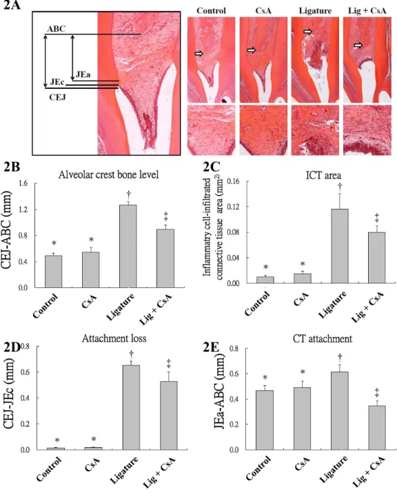
On the mesial surfaces of second molars in each of rats, the distance between the
following histometric landmarks were performed (Fig. 2A): CEJ to ABC (indicating
alveolar bone loss), CEJ to the coronal level of epithelial cells (JEc)(indicating
attachment loss), and the apical level of epithelial cells (JEa) to ABC (indicating CT
attachment). The amount of surface area with inflammatory cell-infiltrated connective
tissue (ICT) in a zone of 0.14 mm2 of sub-epithelial gingiva on the mesial surfaces of
maxillary second molars of each rat was also measured.
Statistical Analysis
Repeated-measures analysis of variance (ANOVA) with Duncan’s test for post-hoc
analysis was used to evaluate the influence of the inter-subject factor (i.e. CsA or ligature
treatment), as well as the intra-subject factors from all four groups together (including the
mesial, middle or distal site, the buccal or palatal surface, and the left or right side) on the
micro-CT bone level (the distance from CEJ to ABC). Regression analysis with
correlation coefficient was used to determine the associations of alveolar crest bone loss
(the distance from CEJ to ABC) to the expressions of MMPs and EMMPRIN in all of the
animals with four different treatments. Results are expressed as means and standard
deviations. P < 0.05 was considered as significant.
¶ Surgical silk sutures, UNIK, Taiwan
# FLEX Triumph for platformX-O, Gamma Media-Ideas, Inc., Northridge, CA ** Triumph XO, Gamma Media-Ideas, Inc., Northridge, CA
†† VIVID, Gamma Media-Ideas, Inc., Northridge, CA ‡‡ Bicinchoninic acid protein assay, Pierce, Rockford, IL
§§ Transilluminator/SPOT, Diagnostic Instruments, Sterling Heights, MI ∥∥ Hoefer semi-dry transfer system, Hoefer, Inc., Holliston, MA
¶¶ Millipore Immobilon-PSQ transfer PVDF membranes, Millipore, Billerica, MA ## Abcam, Cambridge, MA
RESULTS
Cyclosporine-A Impedes Periodontal Destruction
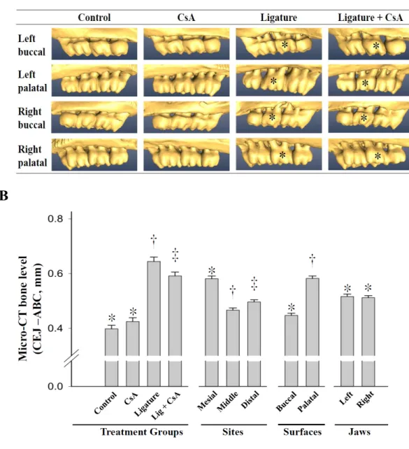
By micro-CT, more alveolar bone loss was observed at the ligature sites of the Lig
and the Lig+CsA groups, compared to those at the same sites of the control or CsA group
(Fig. 1A). However, the bony destruction was less severe in the Lig+CsA group than in
the Lig group. By the repeated measures analysis of variance, the mean micro-CT bone
levels (CEJ-ABC) were significantly different among the four animal groups and among
the examining sites, as well as between the buccal and palatal/lingual locations (Fig. 1B).
Statistically similar bone crests levels were recorded in the control and CsA groups after
the post-hoc analysis, whereas the distance from CEJ to ABC in Lig+CsA group was
statistically longer than the control and CsA groups but shorter than the Lig group.
By histology, gingival inflammation and alveolar bone loss were easily observed in
the ligature group, but were not as severe in the control and CsA groups (Fig. 2A). Less
severe periodontal inflammation and destruction was also noted in the Lig+CsA group
when compared with the Lig group. Histometric data demonstrated a consistent findings
showing that significantly less alveolar bone loss, attachment loss, and inflammation
(ICT area) were noted in the Lig+CsA group when compared with the Lig group (Figs.
2B, 2C, 2D). However, significantly less connective tissue attachment (JEa-ABC) was
Cyclosporine-A-induced Attenuation of Periodontal Destruction was Correlated with Expressions of Gelatinases, and Their Inducer, EMMPRIN.
By zymography, the gelatinolytic activities of pro-MMP-9, pro-MMP-2 and MMP-2
among four animal groups were examined (Figs. 3A-3D). The activities of the examined
MMPs were all significantly increased in the Lig and Lig+CsA groups compared with the
control and CsA groups. The MMPs activities in Lig+CsA group were significantly lower
those in Lig group, but higher than those in the control and CsA groups. Correlation
analysis also demonstrated very strong positive correlations of the activities of
pro-MMP-9, pro-MMP-2 and MMP-2 with alveolar bone loss (CEJ-ABC) in both micro-CT (Figs.
4A-4C) and histometrical analysis (Figs. 4D-4F).
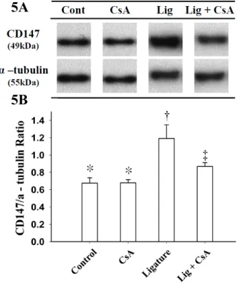
By Western blotting, the protein expression of EMMPRIN was significantly
stronger in gingiva obtained from the Lig and Lig+CsA groups when compared with
those from the control and CsA groups; whereas the expression of EMMPRIN in the
Lig+CsA group was significantly weaker when compared to that in the Lig groups (Fig.
5). The correlation analysis also showed a strong positive correlation of the activities of
EMMPRIN with the amount of alveolar bone loss (CEJ-ABC) in both micro-CT (Fig.
DISCUSSION
The local tissue breakdown of periodontitis is caused by a series of
plaque-associated immuno-inflammatory processes in the periodontium. The inflamed and
overgrown gingiva induced by CsA could become a bacterial plaque reservoir and
aggravate the progression of periodontitis. However, the severity of periodontal
breakdown is often inconsistent with the severity of enlargement, inflammation, and
plaque accumulation observed in the patients with CsA-induced gingival overgrowth. As
seen in a previous radiographic observation,35 this present study found a correspondingly
inhibitory effect of CsA on the periodontal destruction using micro-CT and histometric
analysis. The present study, which uses a ligature-induced periodontitis rat model, was
the first in vivo study demonstrating that CsA-induced attenuation of periodontal bone
loss is correlated positively with the expressions of MMP-2, MMP-9, and their inducer,
EMMPRIN. It is also the first study illustrating an inhibitory effect of CsA on
EMMPRIN in gingiva.
MMPs are known to play a major role in the degradation of extracellular matrix,
basal membrane, and connective tissue proteins. MMPs has been shown to be associated
with embryonic development, wound healing, and some pathophysiological responses
such as arthritis, fibrosis, and inflammation.38 MMP-2 and MMP-9 belong to the
synthesized by fibroblasts, keratinocytes, endothelial cells, macrophages, platelets,
osteoblasts, and chondroblasts, whereas MMP-9 is usually produced by neutrophils,
keratinocytes and macrophages.3 Increased expressions of MMP-211-13 and MMP-9 have
been found in patients with chronic periodontitis which, is consistent with the findings of
the present study (Fig. 3 negative and positive controls and Fig. 4).
EMMPRIN is a transmembrane glycoprotein originally identified on the surfaces of
tumor cells. Its expression on tumor cells may induce tumor progression and invasion by
triggering the production or release of MMPs. EMMPRIN is expressed on all leukocytes,
keratinocytes, endothelial cells, and platelets,42 and is now best known as an inducer of
MMPs (including MMP-2 and MMP-9).20 The ability of EMMPRIN to stimulate MMP
production suggests the possibility that this molecule is associated with several
pathophysiological tissue modulatory processes and tissue remodeling, including
asthma-mediated lung inflammation, rheumatoid arthritis, sclerosis, myocardial infarction, and
ischemic stroke.42-44 In periodontitis, increased expression of EMMPRIN has also been
shown in patients with chronic periodontitis, and in a ligature-induced periodontitis
model. The results of the present study showing a significantly increased expression of
EMMPRIN (Fig. 5 negative and positive controls and Fig. 6) were also consistent with
the findings in the previous studies.
of EMMPRIN in their gingival tissues has been examined once.47 However in that study,
both of the CsA and healthy control patients showed a very strong intensity of peroxidase
staining for EMMPRIN. Our laboratory has also found that the total protein expression of
EMMPRIN in gingival fibroblasts which received CsA was unchanged when compared
to that without CsA in vitro; however, its membranous expression was decreased after
CsA stimulation (unpublished data). In the current study, we demonstrated that the
expression of EMMPRIN did decrease when CsA was given in the ligature-induced
periodontitis (Figs. 5 and 6).
Recent in vitro studies from our laboratory and others have also shown that CsA can
inhibit the productions of MMP-230-34 and MMP-930 in gingival fibroblasts. The in vivo
results in the present study (Figs. 3 and 4) explain the attenuated effect of CsA on
periodontal bone loss, and are consistent with the inhibitions of MMP-2 and MMP-9 seen
in the previous in vitro studies. In addition, a less severe inflammatory cell infiltration
was observed in the CSA+Lig group compared to the Lig group (Fig. 2C). It might imply
that in the patients with CsA-induced gingival overgrowth and periodontitis, the
interaction between the gingival fibroblasts and inflammatory cells involves not only the
synergic activities from CsA but also the metabolic activities caused by the CsA. It has
been suggested that CsA may inhibit the initial periodontal breakdown, because in the
imbalance in the alveolar bone homeostasis in rats.35
In order to elucidate the possible role of gelatinase MMPs and EMMPRIN on
CsA-induced attenuation of periodontal destruction, the expressions of MMP-2, MMP-9, and
EMMPRIN in gingiva, as well as their correlations with the micro-CT and histologic
tissue losses were examined (Figs. 3 to 6). The results suggest that the CsA-induced
attenuation of periodontal destruction might be involved with the regulation of
MMP/CD147 pathway. However, the roles of EMMPRIN and its activities on MMPs in
CsA-induced gingival overgrowth are still unclear and require further detailed
investigations.
CONCLUSIONS
The current study demonstrated inhibitory effects of CsA on gelatinases (i.e.
MMP-2 and MMP-9), EMMPRIN, and periodontal destruction using a ligature-induced
periodontitis rat model. The study results also showed a strong positive correlation of
CsA-induced attenuation of periodontal bone loss with the expressions of 2,
MMP-9, and EMMPRIN in gingiva.
ACKNOWLEDGMENTS
National Defence (DOD-C101-12-4) and the National Science Council
(NSC-100-2314-B-039-053), Republic of China. The authors report no conflicts of interest related to this
REFERENCES
1. Listgarten MA. The role of dental plaque in gingivitis and periodontitis. J Clin
Periodontol 1988;15:485-487.
2. Cochran DL. Inflammation and bone loss in periodontal disease. J Periodontol 2008;79:1569-1576.
3. Birkedal-Hansen H. Role of cytokines and inflammatory mediators in tissue destruction. J Periodontal Res 1993;28:500-510.
4. Lindhe J, Nyman S. Clinical trials in periodontal therapy. J Periodontal Res 1987;22:217-221.
5. Listgarten MA. Nature of periodontal diseases: pathogenic mechanisms. J
Periodontal Res 1987;22:172-178.
6. Sapna G, Gokul S, Bagri-Manjrekar K. Matrix metalloproteinases and periodontal diseases. Oral Dis 2013;doi: 10.1111/odi.12159.
7. Marcaccini AM, Meschiari CA, Zuardi LR, et al. Gingival crevicular fluid levels of MMP-8, MMP-9, TIMP-2, and MPO decrease after periodontal therapy. J Clin
Periodontol 2010;37:180-190.
8. Emingil G, Tervahartiala T, Mantyla P, Maatta M, Sorsa T, Atilla G. Gingival crevicular fluid matrix metalloproteinase (MMP)-7, extracellular MMP inducer, and tissue inhibitor of MMP-1 levels in periodontal disease. J Periodontol 2006;77:2040-2050.
9. Rai B, Kaur J, Jain R, Anand SC. Levels of gingival crevicular metalloproteinases-8 and -9 in periodontitis. Saudi Dent J 2010;22:129-131. 10. Wang J, Yang D, Li C, Shang S, Xiang J. Expression of extracellular matrix
metalloproteinase inducer glycosylation and caveolin-1 in healthy and inflamed human gingiva. J Periodontal Res 2014;49:197-204.
matrix metalloproteinase inducer is associated with matrix metalloproteinase-1 and -2 in gingival tissues from patients with periodontitis. J Periodontal Res 2009;44:125-132.
12. Korostoff JM, Wang JF, Sarment DP, Stewart JC, Feldman RS, Billings PC. Analysis of in situ protease activity in chronic adult periodontitis patients: expression of activated MMP-2 and a 40 kDa serine protease. J Periodontol 2000;71:353-360.
13. Makela M, Salo T, Uitto VJ, Larjava H. Matrix metalloproteinases (MMP-2 and MMP-9) of the oral cavity: cellular origin and relationship to periodontal status. J
Dent Res 1994;73:1397-1406.
14. Emingil G, Atilla G, Sorsa T, Tervahartiala T. The effect of adjunctive subantimicrobial dose doxycycline therapy on GCF EMMPRIN levels in chronic periodontitis. J Periodontol 2008;79:469-476.
15. Marcaccini AM, Novaes AB, Jr., Meschiari CA, et al. Circulating matrix metalloproteinase-8 (MMP-8) and MMP-9 are increased in chronic periodontal disease and decrease after non-surgical periodontal therapy. Clin Chim Acta 2009;409:117-122.
16. Biswas C, Zhang Y, DeCastro R, et al. The human tumor cell-derived collagenase stimulatory factor (renamed EMMPRIN) is a member of the immunoglobulin superfamily. Cancer Res 1995;55:434-439.
17. Foda HD, Rollo EE, Drews M, et al. Ventilator-induced lung injury upregulates and activates gelatinases and EMMPRIN: attenuation by the synthetic matrix metalloproteinase inhibitor, Prinomastat (AG3340). Am J Respir Cell Mol Biol 2001;25:717-724.
18. Sun J, Hemler ME. Regulation of MMP-1 and MMP-2 production through CD147/extracellular matrix metalloproteinase inducer interactions. Cancer Res
2001;61:2276-2281.
19. Zucker S, Hymowitz M, Rollo EE, et al. Tumorigenic potential of extracellular matrix metalloproteinase inducer. Am J Pathol 2001;158:1921-1928.
20. Huet E, Gabison EE, Mourah S, Menashi S. Role of emmprin/CD147 in tissue remodeling. Connective tissue research 2008;49:175-179.
21. Borel JF. Mechanism of action of cyclosporin A and rationale for use in nephrotic syndrome. Clinical nephrology 1991;35 Suppl 1:S23-30.
22. Kaufmann Y, Chang AE, Robb RJ, Rosenberg SA. Mechanism of action of Cyclosporin A: inhibition of lymphokine secretion studied with antigen-stimulated T cell hybridomas. J Immunol 1984;133:3107-3111.
23. Rateitschak-Pluss EM, Hefti A, Lortscher R, Thiel G. Initial observation that cyclosporin-A induces gingival enlargement in man. J Clin Periodontol 1983;10:237-246.
24. Wysocki GP, Gretzinger HA, Laupacis A, Ulan RA, Stiller CR. Fibrous hyperplasia of the gingiva: a side effect of cyclosporin A therapy. Oral Surg Oral
Med Oral Pathol 1983;55:274-278.
25. Ono M, Hatamochi A, Arakawa M, Ueki H. Effects of cyclosporin A on cell proliferation and collagen production by human skin fibroblasts. J Dermatol Sci 1991;2:274-280.
26. Willershausen-Zonnchen B, Lemmen C, Schumacher U. Influence of cyclosporine A on growth and extracellular matrix synthesis of human fibroblasts.
J Cell Physiol 1992;152:397-402.
27. Ayanoglou CM, Lesty C. Cyclosporin A-induced gingival overgrowth in the rat: a histological, ultrastructural and histomorphometric evaluation. J Periodontal Res 1999;34:7-15.
Ultrastructural and histochemical features of the ground substance in cyclosporin A-induced gingival overgrowth. J Periodontol 1996;67:21-27.
29. Thomason JM, Sloan P, Seymour RA. Immunolocalization of collagenase (MMP-1) and stromelysin (MMP-3) in the gingival tissues of organ transplant patients medicated with cyclosporin. J Clin Periodontol 1998;25:554-560.
30. Kuo PJ, Tu HP, Chin YT, et al. Cyclosporine-A inhibits MMP-2 and -9 activities in the presence of Porphyromonas gingivalis lipopolysaccharide: an experiment in human gingival fibroblast and U937 macrophage co-culture. J Periodontal Res 2012;47:431-438.
31. Chiu HC, Lu YT, Chin YT, et al. Cyclosporine A inhibits the expression of membrane type-I matrix metalloproteinase in gingiva. J Periodontal Res 2009;44:338-347.
32. Sobral LM, Aseredo F, Agostini M, et al. Molecular events associated with ciclosporin A-induced gingival overgrowth are attenuated by Smad7 overexpression in fibroblasts. J Periodontal Res 2012;47:149-158.
33. Kim JY, Park SH, Cho KS, et al. Mechanism of azithromycin treatment on gingival overgrowth. J Dent Res 2008;87:1075-1079.
34. Abe M, Yokoyama Y, Syuto T, Ishibuchi H, Ishikawa O. Interleukin-6 counteracts effects of cyclosporin A on extracellular matrix metabolism by human dermal fibroblasts. Cell Tissue Res 2008;333:281-288.
35. Nassar CA, Nassar PO, Abi Rached RS, Holzhausen M, Marcantonio E, Jr., Spolidorio LC. Effect of cyclosporin A on alveolar bone homeostasis in a rat periodontitis model. J Periodontal Res 2004;39:143-148.
36. Bode W, Gomis-Ruth FX, Stockler W. Astacins, serralysins, snake venom and matrix metalloproteinases exhibit identical zinc-binding environments (HEXXHXXGXXH and Met-turn) and topologies and should be grouped into a
common family, the 'metzincins'. FEBS Lett 1993;331:134-140.
37. Stocker W, Grams F, Baumann U, et al. The metzincins--topological and sequential relations between the astacins, adamalysins, serralysins, and matrixins (collagenases) define a superfamily of zinc-peptidases. Protein Sci 1995;4:823-840.
38. Stamenkovic I. Extracellular matrix remodelling: the role of matrix metalloproteinases. J Pathol 2003;200:448-464.
39. Rai B, Kharb S, Jain R, Anand SC. Biomarkers of periodontitis in oral fluids. J
Oral Sci 2008;50:53-56.
40. Bordador LC, Li X, Toole B, et al. Expression of emmprin by oral squamous cell carcinoma. Int J Cancer 2000;85:347-352.
41. Kanekura T, Chen X, Kanzaki T. Basigin (CD147) is expressed on melanoma cells and induces tumor cell invasion by stimulating production of matrix metalloproteinases by fibroblasts. Int J Cancer 2002;99:520-528.
42. Zhu X, Song Z, Zhang S, Nanda A, Li G. CD147: a Novel Modulator of Inflammatory and Immune Disorders. Curr Med Chem 2014;21:2138-2145. 43. Gabison EE, Hoang-Xuan T, Mauviel A, Menashi S. EMMPRIN/CD147, an
MMP modulator in cancer, development and tissue repair. Biochimie 2005;87:361-368.
44. Guo H, Zucker S, Gordon MK, Toole BP, Biswas C. Stimulation of matrix metalloproteinase production by recombinant extracellular matrix metalloproteinase inducer from transfected Chinese hamster ovary cells. J Biol
Chem 1997;272:24-27.
45. Yang D, Wang J, Ni J, et al. Temporal expression of metalloproteinase-8 and -13 and their relationships with extracellular matrix metalloproteinase inducer in the development of ligature-induced periodontitis in rats. J Periodontal Res
2013;48:411-419.
46. Liu L, Li C, Cai X, Xiang J, Cao Z, Dong W. The temporal expression and localization of extracellular matrix metalloproteinase inducer (EMMPRIN) during the development of periodontitis in an animal model. J Periodontal Res 2010;45:541-549.
47. Bulut OE, Sokmensuer LK, Bulut S, Tasman F, Muftuoglu S. Immunohistochemical study of cyclosporin-induced gingival overgrowth in renal transplant recipients. J Periodontol 2004;75:1655-1662.
Figure 1. Effect of CsA on the ligature-induced alveolar bone loss. (A) presents the
reconstructed 3D images from the computerized tomography for maxillae viewed from the buccal or palatal direction in the control, CsA, ligature, and CsA-plus-ligature groups. Asterisks indicate the molar with the ligature. (B) shows the influence of the between-subject factors (the treatment groups) and the within-subject factors (the mesial/middle/distal site, the buccal/palatal surface, and the right/left jaw) on the micro-CT bone level. (*, †, and ‡: significantly different subsects obtained by the post hoc Duncan test) (Lig + CsA: ligature-plus-CsA group).
1A
Figure 2. Effect of CsA on ligature-induced periodontal tissue destruction. (A)
histographs present the inter-proximal periodontal tissue between the first and second maxillary molars from the control, CsA, ligature, and ligature-plus-CsA groups (H & E stain). Arrows indicates the most coronal level of alveolar bone crest (ABC). The micrographs in the bottom row (40X) represent higher magnifications of images in the top row (20X). (B to E) present the comparisons of results by histometry, including periodontal tissue loss (CEJ-JEc distance), histological bone level (CEJ-ABC distance), severity of gingival tissue inflammation (ICT), and length of connective tissue attachment (JEa-ABC distance) among the four animal groups. (*, †, and ‡: significantly different subsects obtained by the post-hoc Duncan test) (mm2)
2A
2B
2C
Figure 3. Effect of CsA on ligature-induced gelatinolytic activities of pro-MMP-2, MMP-2, and pro-MMP-9 in gingiva. (A) shows the patterns of gelatinolytic activities at
92 (pro-MMP-9), 66 (MMP-2), and 72 (pro-MMP-2) kDa. (B to D) show the comparisons of their optical intensities of the MMPs among the treatment groups. Data are expressed as means and standard deviations. The experiment was repeated four times. (*, †, and ‡: significantly different subsects obtained by the post-hoc Duncan test)(Lig: ligature group; Lig + CsA: CsA-plus-ligature group )
3A
3B
Figure 4. Correlation of the gingival expressions of gelatinase MMPs with severity of alveolar bone loss in ligature-induced periodontitis. (A to F) correlation analysis
revealed a strong, positive correlation of the activity of MMPs (in pixels) with the micro-CT distance of CEJ-ABC (A to C), and the histological distance of CEJ-ABC (D to F).
4A
4B
4C
Figure 5. Effect of CsA on ligature-induced protein expressions of EMMPRIN in gingiva. (A and B) present the protein expressions of EMMPRIN (CD-147) in gingivae
of rats obtained from four treatment groups examined by Western blotting. Data are expressed as means and standard deviations. (*, †, and ‡: significantly different subsects obtained by the post-hoc Duncan test)
5A
5B
Figure 6. Correlation of the gingival expressions of EMMPRIN with severity of alveolar bone loss in ligature-induced periodontitis. (A and B) correlation analysis
revealed a strong positive correlation of the activity of EMMPRIN (in pixels) with the micro-CT distance of CEJ-ABC (Fig. 6A), and the histological distance of CEJ-ABC (Fig. 6B).


![TraditionalMLCalgorithmsmainlytacklethebatchMLCproblem,wheretheinputdataarepresentedinabatch[24,28].Nevertheless,inmanyMLCapplicationssuchase-mailcategorization[22],multi-labelexamplesarriveasastream.Onlineanalysisistherefore dimensionreducermotivatedbyma](data:image/gif;base64,R0lGODlhAQABAIAAAP///wAAACH5BAEAAAAALAAAAAABAAEAAAICRAEAOw==)