INFORMATION FOR AUTHORS
Full details of how to submit a manuscript for publication in Natural Product Communications are given in Information for Authors on our Web site http://www.naturalproduct.us.
Authors may reproduce/republish portions of their published contribution without seeking permission from NPC, provided that any such republication is accompanied by an acknowledgment (original citation)-Reproduced by permission of Natural Product Communications. Any unauthorized reproduction, transmission or storage may result in either civil or criminal liability.
The publication of each of the articles contained herein is protected by copyright. Except as allowed under national “fair use” laws, copying is not permitted by any means or for any purpose, such as for distribution to any third party (whether by sale, loan, gift, or otherwise); as agent (express or implied) of any third party; for purposes of advertising or promotion; or to create collective or derivative works. Such permission requests, or other inquiries, should be addressed to the Natural Product Inc. (NPI). A photocopy license is available from the NPI for institutional subscribers that need to make multiple copies of single articles for internal study or research purposes.
To Subscribe: Natural Product Communications is a journal published monthly. 2010 subscription price: US$1,695 (Print, ISSN# 1934-578X); US$1,695 (Web edition, ISSN# 1555-9475); US$2,095 (Print + single site online); US$595 (Personal online). Orders should be addressed to Subscription Department, Natural Product Communications, Natural Product Inc., 7963 Anderson Park Lane, Westerville, Ohio 43081, USA. Subscriptions are renewed on an annual basis. Claims for nonreceipt of issues will be honored if made within three months of publication of the issue. All issues are dispatched by airmail throughout the world, excluding the USA and Canada.
NPC Natural Product Communications
EDITOR-IN-CHIEF
DR. PAWAN K AGRAWAL
Natural Product Inc. 7963, Anderson Park Lane, Westerville, Ohio 43081, USA
[email protected] EDITORS
PROFESSOR ALESSANDRA BRACA
Dipartimento di Chimica Bioorganicae Biofarmacia, Universita di Pisa,
via Bonanno 33, 56126 Pisa, Italy [email protected]
PROFESSOR DEAN GUO
State Key Laboratory of Natural and Biomimetic Drugs, School of Pharmaceutical Sciences,
Peking University, Beijing 100083, China [email protected]
PROFESSOR J. ALBERTO MARCO
Departamento de Quimica Organica, Universidade de Valencia, E-46100 Burjassot, Valencia, Spain [email protected]
PROFESSOR YOSHIHIRO MIMAKI
School of Pharmacy,
Tokyo University of Pharmacy and Life Sciences, Horinouchi 1432-1, Hachioji, Tokyo 192-0392, Japan [email protected]
PROFESSOR STEPHEN G. PYNE
Department of Chemistry University of Wollongong
Wollongong, New South Wales, 2522, Australia [email protected]
PROFESSOR MANFRED G. REINECKE
Department of Chemistry, Texas Christian University, Forts Worth, TX 76129, USA [email protected]
PROFESSOR WILLIAM N. SETZER
Department of Chemistry
The University of Alabama in Huntsville Huntsville, AL 35809, USA
PROFESSOR YASUHIRO TEZUKA
Institute of Natural Medicine
Institute of Natural Medicine, University of Toyama, 2630-Sugitani, Toyama 930-0194, Japan [email protected]
PROFESSOR DAVID E. THURSTON
Department of Pharmaceutical and Biological Chemistry, The School of Pharmacy,
University of London, 29-39 Brunswick Square, London WC1N 1AX, UK
ADVISORY BOARD Prof. Berhanu M. Abegaz
Gaborone, Botswana
Prof. Viqar Uddin Ahmad
Karachi, Pakistan
Prof. Øyvind M. Andersen
Bergen, Norway
Prof. Giovanni Appendino
Novara, Italy
Prof. Yoshinori Asakawa
Tokushima, Japan
Prof. Lee Banting
Portsmouth, U.K.
Prof. Julie Banerji
Kolkata, India
Prof. Anna R. Bilia
Florence, Italy
Prof. Maurizio Bruno
Palermo, Italy
Prof. Josep Coll
Barcelona, Spain
Prof. Geoffrey Cordell
Chicago, IL, USA
Prof. Cristina Gracia-Viguera
Murcia, Spain
Prof. Duvvuru Gunasekar
Tirupati, India
Prof. A.A. Leslie Gunatilaka
Tucson, AZ, USA
Prof. Kurt Hostettmann
Lausanne, Switzerland
Prof. Martin A. Iglesias Arteaga
Mexico, D. F, Mexico
Prof. Jerzy Jaroszewski
Copenhagen, Denmark
Prof. Leopold Jirovetz
Vienna, Austria
Prof. Teodoro Kaufman
Rosario, Argentina
Prof. Norbert De Kimpe
Gent, Belgium
Prof. Karsten Krohn
Paderborn, Germany
Prof. Hartmut Laatsch
Gottingen, Germany
Prof. Marie Lacaille-Dubois
Dijon, France
Prof. Shoei-Sheng Lee
Taipei, Taiwan Prof. Francisco Macias
Cadiz, Spain
Prof. Imre Mathe
Szeged, Hungary
Prof. Joseph Michael
Johannesburg, South Africa
Prof. Ermino Murano
Trieste, Italy
Prof. M. Soledade C. Pedras
Saskatoon, Cnada
Prof. Luc Pieters
Antwerp, Belgium
Prof. Om Prakash
Manhattan, KS, USA
Prof. Peter Proksch
Düsseldorf, Germany
Prof. Phila Raharivelomanana
Tahiti, French Plynesia
Prof. Satyajit Sarker
Wolverhampton, UK
Prof. Monique Simmonds
Richmond, UK
Prof. Valentin Stonik
Vladivostok, Russia
Prof. Winston F. Tinto
Barbados, West Indies
Prof. Karen Valant-Vetschera
Vienna, Austria
Prof. Peter G. Waterman
Lismore, Australia
HONORARY EDITOR
PROFESSOR GERALD BLUNDEN
The School of Pharmacy & Biomedical Sciences, University of Portsmouth, Portsmouth, PO1 2DT U.K.
Chemical Composition and Bioactivities of the Marine Alga
Isochrysis galbana from Taiwan
Chi-Cheng Yua, Hsiao-Wei Chenb, Mao-Jing Chenb, Yu-Ching Changb, Shih-Chang Chien c,
Yueh-Hsiung Kuod, Feng-Ling Yange, Shih-Hsiung Wue, Jie Chenf, Hsiao-Hui Yuf and
Louis Kuop-Ping Chaof*
aGreenlink Biotech Inc., Taipei 111, Taiwan
bChemistry and Environment Labs., Taiwan Power Research Institute. 84, Da-ani Rd., Shulin,
Taipei country 238, Taiwan
cSchool of Chinese Pharmaceutical Sciences and Chinese Medicine Resources,
China Medical University, Taichung 404, Taiwan
dTsuzuki Institute for Traditional Medicine, College of Pharmacy, China Medical University,
Taichung 404, Taiwan
eInstitute of Biological Chemistry, Academia Sinica, Taipei 115, Taiwan fDepartment of Cosmeceutics, China Medical University, Taichung 404, Taiwan
[email protected]; [email protected]
Received: August 27th, 2010; Accepted: October 6th, 2010
The present study investigated the chemical composition of Isochrysis galbana Parke, a marine microalga which is widely used as a feedstock in aquaculture. From gas chromatography/mass spectrometric analysis the mono-sugar compositions of I. galbana were 2.1% fucose, 2.5% rhamnose, 2.7% arabinose, 8.5% xylose, 15.7% mannose, 32.7% galactose and 35.8% glucose. The polysaccharides of I. galbana were able to induce prointerleukin-1β (pro-IL-1β) protein expression within murine macrophages. Furthermore, five kinds of chlorophyll and one sterol were separated from the ethanolic extracts, including pheophorbide-a, ethyl pheophorbide-a, 10S-10-hydroxypheophytin-a, 10R-10-hydroxypheophytin-a, (132-R)-pheophytin-a, and
brassicasterol. In addition, the major soluble components of the ethanol/n-hexane extract were 9-octadecenoic acid (E) (38.4%), hexadecanoic acid (23.3%), tetradecanoic acid (15.7%), and octadecanoic acid (7.2%), but only a few polyunsaturated fatty acids were found, such as 9,12,15-octadecatrienoic acid (1.9%), 9,12-octadecadienoic acid (Z,Z) (3.4%), and docosahexaenoic acid (0.2%). This is the first occasion that polysaccharides from I. galbana have been demonstrated to exert immunomodulatory properties by the induction of IL-1 within macrophages.
Keywords: Isochrysis galbana, extracts, chemical compositions, polysaccharides, bioactivity, pro-IL-1β.
Marine microalgal biomasses play an important role as primary producers in the animal food chain. Isochrysis galbana Parke, a golden-brown flagellate marine microalga, is widely used as an aquaculture feed for young fish and in bivalve hatcheries because it is rich in polyunsaturated fatty acids (PUFA) [1]. Many studies have focused on the relationship between fatty acids and algal growth [2-4]. In addition, because of the high content of docosahexaenoic acid (DHA) and eicosapentaenoic acid (EPA), I. galbana is recognized for its beneficial effects on human health, and is considered to be a good substitute for fish oil in the human diet. The purpose of the present study was to analyze the chemical composition of I. galbana, including n-hexane/ethanol extracts; ethanol extracts; total carbon, hydrogen, oxygen, nitrogen and sulfur; and total polysaccharides.
First we analyzed the ratio of carbon, hydrogen, oxygen, nitrogen, sulfur and ash in I. galbana cultured in the laboratory. Based on dry weight, the values obtained were: C (56.3%), H (7.5%), O (20.3%), N (6.25%), S (1.0%) and ash (8.6%). I. galbana also yielded a high content of n-hexane/ethanol (1/1) and ethanol extractive (21.7% and 38.2%, respectively).
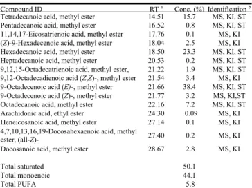
Sixteen fatty acids were identified. These are given in Table 1, where all compounds are listed in order of their elution from the DB-5HT column. The major fatty acid was 9-octadecenoic acid (E) (38.4% of total), followed by hexadecanoic acid (23.3%), tetradecanoic acid (15.7%), octadecanoic acid (7.2%), and 9-octadecenoic acid (Z) (3.2%). The composition of fatty acids in I. galbana found in this study differs from the results of
NPC Natural Product Communications
2010
Vol. 5
No. 12
1941 - 1944
1942 Natural Product Communications Vol. 5 (12) 2010 Yu et al.
Table 1: Chemical composition of I. galbana ethanol/n-hexane extractive. Compound ID RT a Conc. (%) Identificationb
Tetradecanoic acid, methyl ester 14.51 15.7 MS, KI, ST Pentadecanoic acid, methyl ester 16.52 0.8 MS, KI, ST 11,14,17-Eicosatrienoic acid, methyl ester 17.76 0.1 MS, KI (Z)-9-Hexadecenoic acid, methyl ester 18.04 2.5 MS, KI Hexadecanoic acid, methyl ester 18.50 23.3 MS, KI, ST Heptadecanoic acid, methyl ester 20.53 0.2 MS, KI, ST 9,12,15-Octadecatrienoic acid, methyl ester, 21.22 1.9 MS, KI, ST 9,12-Octadecadienoic acid (Z,Z)-, methyl ester 21.54 3.4 MS, KI 9-Octadecenoic acid (E)-, methyl ester 21.66 38.4 MS, KI, ST 9-Octadecenoic acid (Z)-, methyl ester 21.77 3.2 MS, KI,ST Octadecanoic acid, methyl ester 22.16 7.2 MS, KI, ST Arachidonic acid, ethyl ester 24.30 0.09 MS, KI Heneicosanoic acid, methyl ester 27.14 0.1 MS, KI 4,7,10,13,16,19-Docosahexaenoic acid, methyl
ester, (all-Z)- 27.40 0.2 MS, KI
Docosanoic acid, methyl ester 28.67 2.8 MS, KI
Total saturated 50.1
Total monoenoic 44.1
Total PUFA 5.8
a Retention time on a DB-5 HT column with reference to n-alkanes. b MS, NIST and Wiley library spectra, and the literature; RI, retention
index; ST, authentic standard compounds.
an earlier study, which showed higher DHA (22:6n-3) (7.91%) [5].
Also, some differences were found from the study by Lin et al., who determined DHA to be a major fatty acid at every growth phase [4]. It is interesting that we found a few fatty acids, such as pentadecanoic acid, heptadecanoic acid, and heneicosanoic acid, which had not been previously found [4]. This could be a result of different growth conditions.
Few existing studies have focused on the variety of pigments from I. galbana. Herein we have separated and identified the pigments from 1.65 g of an ethanolic extract of I. galbana. At least five chlorophyll compounds (Figure 1) were found including (132-R)-pheophytin-a (1.5 mg; RT = 8.03 min), pheophorbide-a (1 mg; RT = 15.82 min), 10R-10-hydroxypheophytin-a (3 mg; RT = 22.54 min), 10S-10-hydroxypheophytin-a (1 mg; RT = 23.51 min), and ethyl pheophorbide-a (4 mg; RT = 34.92 min). We also found brassicasterol (2 mg; RT = 15.10 min).
In 1981, Volkman et al. [6] were the first to report that 24-methyl-22-dehydrocholesterol is the dominant sterol in I. galbana [6]. Our experimental results were similar. Park et al. [7] demonstrated that autotrophically grown I. galbana contains three major sterols (24-oxocholesterol acetate, ergost-5-en-ol, and cholest-5-en-24-1, 3-(acetyloxy)-,3-ol), with 24-methylcholesta-5,22-dien- 3-ol as a minor sterol [7]. They found that the total sterol content clearly decreased during dark culture, with such decreases being particularly evident in two major sterols, 24-oxocholesterol acetate and ergost-5-en-3-ol.
Our study showed that the monosaccharide composition of a polysaccharide (IP) from I. galbana was 2.07%
Figure 1: The chemical structure of (1) pheophorbide-a; (2) ethyl pheophorbide-a; (3) (132-R)-pheophytin-a; (4)
10R-10-hydroxypheophytin-a; (5) 10S-10-hydroxypheophytin-10R-10-hydroxypheophytin-a; (6) brassicasterol.
Figure 2: Effect of Isochrysis polysaccharide (IP) on cell viability. J774A.1 macrophages (5 × 10³/well) were treated with IP or DMSO (control) for 24
h, followed by incubation with MTT reagent. Absorbance (A550-A690) was measured by spectrophotometry. Data are expressed as mean ± SE from three separate experiments.
fucose, 2.50% rhamnose, 2.72% arabinose, 8.49% xylose, 15.70% mannose, 32.73% galactose and 35.79% glucose. No cytotoxic effect was observed after J774A.1 cells were treated with various concentrations of IP for a period of 24 hours, as measured by MTT assay (Figure 2). It is well known that IL-1β is secreted mainly from activated macrophages; this could activate other immune cells and modulate immune responses. An IP-induced
IL-1β precursor, pro-IL-1β, was detected in whole cell lysates after IP stimulation by means of Western-blotting analysis. In this time course study, the expression of pro-IL-1β protein within IP-stimulated cells was detected at six hours post-stimulation. In addition, the expression of pro-IL-1β increased with increasing IP concentrations in a dose-dependent manner. Such results demonstrate that IP stimulates pro-IL-1β expression, a result that would appear to be similar to the ability of polysaccharides isolated from the algae Rhizoclonium riparium (Roth) Harvey and Chlorella pyrenoidosa Chick to stimulate pro-IL-1β expression within murine macrophages [8,9].
Chemical composition and bioactivities of the Isochrysis galbana Natural Product Communications Vol. 5 (12) 2010 1943 Fabregas et al. found that endocellular extracts of
I. galbana clearly inhibited viral hemorrhagic septicemia virus (VHSV) replication at a dose of 20 μg/mL, and that I. galbana contained sulfated soluble exopolysaccharides [10].
Experimental
Algal culture and collection: I. galbana used in this
study was obtained from the Tungkang Biotechnology Research Center, Fisheries Research Institute, Republic of China. Purified I. galbana CCMP 1324 (0.9 L, 680 nm, OD 1.10) was inoculated and cultured in Walne’s medium [9]. This included nutrient, vitamin and trace metal solutions in a 10 L PET tank containing 8.1 L seawater autoclaved at 120°C for 20 min, to which was added 9.0 mL of nutrient solution and 0.9 mL of vitamin solution. The culture medium was agitated gently by bubbling air with a flow rate of 4.7 L/min and a culture time of 7 d. Continuous illumination at an irradiance of 5900 lx was provided by fluorescent lamps. The culture medium was then centrifuged (Himac CR22-GII, Hitachi, Japan) continuously at 12000 rpm at 25ºC. After lyophilization the yield of alga was 0.1 g/L (dry weight) culture medium.
Total C, H, O, N, S: Total carbon, hydrogen, oxygen,
nitrogen and sulfur contents were determined by CHN elemental analysis. Freeze-dried samples (15 g each) were combusted in a 2400 CHN/O elemental analyzer (Perkin-Elmer, Waltham MA, USA) [11].
Extraction and composition of n-hexane/ethanol extracts of I. galbana: Ten grams of sample was
extracted in a Soxhlet apparatus with n-hexane: ethanol (95% v/v) (50:50) for 48 h. The solution was rotary-evaporated at 65 ºC to provide a hydro-ethanolic extractive (HAE) (2.175 g). The HAE (0.5 g) was trimethylsilylated with Sylon HTP (HMDS/TMCS/ pyridine, 3:1:9) trimethylsilylation reagent (Supelco, Bellefonte PA, USA). The final derivatives were kept in n-hexane for gas chromatography–mass spectrometric (GC-MS) analysis. A Hewlett-Packard HP 6890 gas chromatograph equipped with a DB-5HT fused silica capillary column (30 m x 0.25 mm x 0.25 μm film thickness; Agilent Technologies, Santa Clara CA, USA) and a FID detector were used for quantitative determination of the components. The oven temperature was programmed as follows: 100ºC for 2 min, rising to 275ºC at 5ºC/min; injector temperature, 270ºC; carrier gas, He with a flow rate of 1 mL/min; detector temperature, 250ºC; split ratio 50.1:1. One μL sample was injected. Identification of the oil components was based on their retention indices and MS results. The GC analysis parameters listed above and the MS were obtained (full scan mode; scan time, 0.3 s; mass range, MHz 30–500) in the electron impact (EI) mode at 70 eV.
Extraction, purification and
identification
of ethanolicextracts of I. galbana: Dry alga (5 g) was treated with
ethanol (95% v/v for 10 d, repeated 3 times) at room temperature. Then the extract was concentrated to provide the ethanolic extract (AE). AE (1.65 g) was applied to a silica gel column (Si 60) and eluted with acetone/n-hexane to give 43 sub-fractions. Each eluted fraction was 150 mL. The chlorophyll compounds were purified by preparative HPLC (KNAUER RI detector 2400, pump 100; KNAUER, Germany) on a Merck (Germany) Hibar Fertigsaule RT column Si 60 (25 cm length, 1 cm i.d., 5.0 μm). The separation conditions were as follows: flow rate 4 mL/min; mobile phase, acetone/n-hexane = 1/9. The sterol was separated by a Phenomenex Luna silica (2) column (25 cm length, 1 cm i.d., 5.0 μm) under the following conditions: flow rate 4 mL/min; mobile phase, acetone/n-hexane = 1/15. The structures of the compounds were confirmed by comparison of physical and spectral data (including optical rotation, EIMS, 1H NMR) with previously reported values.
Extraction of polysaccharides from I. galbana: Five
grams dry alga was extracted with n-hexane/ethanol. The extractive was ground into a fine powder, and then suspended in 100 mL distilled water. After autoclaving at 121°C for 30 min, the extract was filtered through a 0.2 μm membrane. The extract was then vacuum- concentrated at 50°C, giving a final volume of 30 mL to which 5 volumes of 95% ethanol was added slowly at 4°C. Then the mixture was centrifuged to produce a precipitate of ca. 1.910 g, dry wt. Sixty mg of the precipitate was treated further with 3 mg proteinase K for removal of the peptide part, and dialyzed against H2O
(Spectra/Por® membrane, molecular weight cutoff 1,000 Da), resulting in 17.5 mg polysaccharide [I. galbana (IP)].
Sugar composition analysis: Sugar composition was
determined by GC-MS. The polysaccharide content of I. galbana was determined by methanolysis with 0.5 M methanolic HCl at 80°C for 16 h, and trimethylsilylation with Sylon HTP. The final trimethylsilylated (TMS) derivatives were kept in n-hexane for GC-MS analysis [12]. Carbohydrate analysis was done with inositol as the internal standard; integrated peak area was used to establish the relative amounts of the constituents. Compounds were identified by comparing of their mass spectrometric fragmentation patterns with those of authentic standards, and the quantity of compounds was obtained by integrating the peak area of the spectra.
Microculture tetrazolium (MTT) assay for cell viability:
J774A.1 macrophages were seeded in 96-well plates at a density of 5 × 10³ cells/well. Cells were incubated with IP
for 24 h. Cell viability was determined using colorimetric MTT assays.
1944 Natural Product Communications Vol. 5 (12) 2010 Yu et al.
Cell cultures: Murine J774A.1 macrophages were
obtained from the American Type Culture Collection (ATCC) (Rockville MD, USA). All cells were propagated in RPMI-1640 medium supplemented with 10% heat-inactivated FCS and 2 mM L-glutamine (Life
Technologies, Carlsbad CA, USA), and cultured in a 37°C, 5% CO2 incubator [8,9].
Western blotting: Whole cell lysates were separated by
12% SDS-PAGE and electrotransferred to a PVDF membrane. The membrane was incubated in blocking solution (5% nonfat milk in PBS with 0.1% Tween 20) at room temperature for 1 h. The membrane was then incubated with anti-IL-1β antibody at room temperature for 2 h. After washing 3 times in PBS with 0.1% Tween 20, the membrane was incubated with an HRP-conjugated secondary antibody directed against the primary antibody. The membrane was developed by an enhanced chemiluminescence Western-blotting detection system
(DuPont NEN® Research Products, Boston MA, USA) according to the manufacturer’s instructions [8,9].
Statistical analysis: All values are given as mean ± SE.
Data analysis involved one-way ANOVA with subsequent Scheffé test.
Acknowledgment - This work was supported by a grant
from the Taiwan Power Research Institute (contract/grant numbers TPC-546-2517-9802, NSC 96-2116-M-039-001 -MY3 and NSC 98-3114-B-001-001) Taiwan Department of Health Clinical Trial and Research Center of Excellence (DOH99-TD-B-111-004) and Taiwan Department of Health Clinical Trial Research Center of Excellence (DOH 99-TD-B-111-004) and of Health Cancer Research Center of Excellence (DOH-99-TD-C-111-005) for financial support.
References
[1] Wikfors GH, Ferris GE, Smith BC. (1992) The relationship between gross biochemical composition of cultured algal foods and
growth of the hard clam, Mercenaria mercenaria (L.). Aquaculture, 108, 135-154.
[2] Sánchez S, Martínez M, Espinola F. (2000) Biomass production and biochemical variability of the marine microalga Isochrysis
galbana in relation to culture medium. Biochemical Engineering Journal, 6, 13-18.
[3] Pernet F, Tremblay R, Demers E, Roussy M. (2003) Variation of lipid class and fatty acid composition of Chaetoceros muelleri and
Isochrysis sp. grown in a semicontinuous system. Aquaculture, 221, 393–406.
[4] Lin YH, Chang FL, Tsao CY, Leu JY. (2007) Influence of growth phase and nutrient source on fatty acid composition of Isochrysis
galbana CCMP 1324 in a batch photoreactor. Biochemical Engineering Journal, 37, 166-176.
[5] Cho JY, Jin HJ, Lim HJ, Whyte JNC, Hong YK. (1999) Growth activation of the microalga Isochrysis galbana by the aqueous
extract of the seaweed Monostroma nitidum. Journal of Applied Phycology, 10, 561-567.
[6] Volkman JK, Smith DJ, Eglinton G, Forsberg TEV, Corner. EDS (1981) Sterol and fatty acid composition of four marine
Haptophycean algae. Journal of the Marine Biological Association of the United Kingdom, 61, 509-527.
[7] Park DW, Jo Q, Lim HJ, Véron B. (2002) Sterol composition of dark-grown Isochrysis galbana and its implication in the seed
production of Pacific oyster, Crassostrea gigas. Journal of Applied Phycology, 14, 351-355.
[8] Hsu HY, Hua KF, Su YC, Chu LC, Su SC, Chiu HW, Wong CH, Chen ST, Shieh CW, Yang SS, Chen YM, Chao LK. (2006)
Study on regulation of cytokine gene expression in macrophages with an alkali-soluble polysaccharide of Rhizoclonium riparium alga. Journal of Agricultural and Food Chemistry, 54, 3558-3565.
[9] Hsu HY, Jeyashoke N, Yeh CH, Song YJ, Hua KF, Chao LK. (2010) Immunostimulatory bioactivity of algal polysaccharides from
Chlorella pyrenoidosa activates macrophages via Toll-like receptor 4. Journal of Agricultural and Food Chemistry, 58, 927-936
[10] Fabegas J, García D, Fernandez-Alonso M, Rocha AI, Gómez-Puertas P, Escribano JM, Otero A, Coll JM. (1999) In vitro inhibition
of the replication of haemorrhagic septicaemia virus (VHSV) and African swine fever virus (ASFV) by extracts from marine microalgae. Antiviral Research, 44, 67-73.
[11] Barbarino E, Lourenço SO. (2005) An evaluation of methods for extraction and quantification of protein from marine macro- and
microalgae. Journal of Applied Phycology, 17, 447-460.
[12] Yang FL, Lu CP, Chen CS, Chen MY, Hsiao HL, Su Y, Tsay SS, Zou W, Wu SH. (2004) Structural determination of the polar
glycoglycerolipids from thermophilic bacteria Meiothermus taiwanensis. European Journal of Biochemistry/FEBS J, 271, 4545-4551.
Inhibition of Protein Tyrosine Phosphatase 1β by Hispidin Derivatives Isolated from the Fruiting Body of Phellinus linteus
Yeon Sil Lee, Il-Jun Kang, Moo Ho Won, Jae-Yong Lee, Jin Kyu Kim and Soon Sung Lim 1927 A New Azafluorenone from the Roots of Polyalthia cerasoides and its Biological Activity
Kanchana Pumsalid, Haruthai Thaisuchat, Chatchanok Loetchutinat, Narong Nuntasaen,
Puttinan Meepowpan and Wilart Pompimon 1931
Evaluation of Antiviral Activities of Curcumin Derivatives against HSV-1 in Vero Cell Line
Keivan Zandi, Elissa Ramedani, Khosro Mohammadi, Saeed Tajbakhsh, Iman Deilami, Zahra Rastian,
Moradali Fouladvand, Forough Yousefi and Fatemeh Farshadpour 1935
Hyloglyceride and Hylodiglyceride: Two New Glyceride Derivatives from Hylodendron gabunensis
Awazi Tengu Nyongha, Hidayat Hussain, Etienne Dongo, Ishtiaq Ahmed and Karsten Krohn 1939 Chemical Composition and Bioactivities of the Marine Alga Isochrysis galbana from Taiwan
Chi-Cheng Yu, Hsiao-Wei Chen, Mao-Jing Chen, Yu-Ching Chang, Shih-Chang Chien, Yueh-Hsiung Kuo,
Feng-Ling Yang, Shih-Hsiung Wu, Jie Chen, Hsiao-Hui Yu and Louis Kuop-Ping Chao 1941 An Efficient Protocol for High-frequency Direct Multiple Shoot Regeneration from Internodes of
Peppermint (Mentha x piperita)
Sanjog T. Thul and Arun K. Kukreja 1945
Essential Oil Yield and Chemical Composition Changes During Leaf Ontogeny of Palmarosa (Cymbopogon martinii var. motia)
Bhaskaruni R. Rajeswara Rao, Dharmendra K. Rajput, Rajendra P. Patel and Somasi Purnanand 1947 Essential Oil Composition of Four Endemic Ferulago Species Growing in Turkey
Ceyda Sibel Kılıç, Ayşe Mine Gençler Özkan, Betül Demirci, Maksut Coşkun and Kemal Hüsnü Can Başer 1951 Essential Oils of Daucus carota subsp. carota of Tunisia Obtained by Supercritical Carbon Dioxide
Extraction
Hanen Marzouki, Abdelhamid Khaldi, Danilo Falconieri, Alessandra Piras, Bruno Marongiu, Paola Molicotti
and Stefania Zanetti 1955
Oil Constituents of Artemisia nilagirica var. septentrionalis Growing at Different Altitudes
Flora Haider, Narendra Kumar, Ali Arif Naqvi and Guru Das Bagchi 1959 Volatile Oil Composition of Pogostemon heyneanus and Comparison of its Composition with Patchouli Oil
Ramar Murugan, Gopal Rao Mallavarapu, Kyathsandra Venkataramaiah Padmashree,
Ramachandra Raghavendra Raoand Christus Livingstone 1961
Chemical Composition of Volatile Oils of Aquilaria malaccensis (Thymelaeaceae) from Malaysia
Saiful Nizam Tajuddin and Mashitah M. Yusoff 1965
Chemical Composition and Phytotoxic Effects of Essential Oils from Four Teucrium Species
Laura De Martino, Carmen Formisano, Emilia Mancini, Vincenzo De Feo, Franco Piozzi, Daniela Rigano
and Felice Senatore 1969
Chemical Constituents and Larvicidal Activity of Hymenaea courbaril Fruit Peel
José Cláudio D. Aguiar, Gilvandete M. P. Santiago, Patrícia L. Lavor, Helenicy N. H. Veras, Yana S. Ferreira, Michele A. A. Lima, Ângela M. C. Arriaga, Telma L. G. Lemos, Jefferson Q. Lima, Hugo C. R. de Jesus,
Péricles B. Alves and Raimundo Braz-Filho 1977
CaryophylleneOxide-richEssentialOilsofLithuanianArtemisiacampestrisssp.campestrisandTheirToxicity
Asta Judzentiene, Jurga Budiene, Rita Butkiene, Eugenija Kupcinskiene, Isabelle Laffont-Schwoband
Véronique Masotti 1981
Comparison of Antibacterial Activity of Natural and Hydroformylated Essential Oil of Thymus capitatus Growing Wild in North Sardinia with Commercial Thymus Essential Oils
Marianna Usai, Marzia Foddai, Barbara Sechi, Claudia Juliano and Mauro Marchetti 1985 Composition and Chemical Variability of the Leaf Oil from Corsican Juniperus thurifera
Integrated Analysis by GC(RI), GC-MS and 13C NMR
Josephine Ottavioli, Joseph Casanova and Ange Bighelli 1991
CombinedAnalysisbyGC (RI),GC-MSand13C NMRof theSupercriticalFluid Extract of Abies alba Twigs
Emilie Duquesnoy, Bruno Marongiu, Vincent Castola, Alessandra Piras, Silvia Porcedda and Joseph Casanova 1995
Review/Account
Eugenol: A Natural Compound with Versatile Pharmacological Actions
Natural Product Communications
2010
Volume 5, Number 12
Contents
Original Paper
Page
Anticonvulsant Activity of the Linalool Enantiomers and Racemate: Investigation of Chiral Influence
Damião P. de Sousa, Franklin F. F. Nóbrega, Camila C. M. P. Santos and Reinaldo N. de Almeida 1847 Kinetic Analysis of Genipin Degradation in Aqueous Solution
Paul Slusarewicz, Keng Zhu and Tom Hedman 1853
Microbial Transformation of Marine Halogenated Sesquiterpenes
Aurelio San Martin, Juana Rovirosa, Alvaro Carrasco, Silvia Orejarena, Jorge Soto-Delgado,
Renato Contreras and M. Cristina Chamy 1859
Two New Guaianolides from Amberboa ramosa
Muhammad Ibrahim, Rehan Khan and Abdul Malik 1865
Antiplasmodial and Cytotoxic Activities of Drimane Sesquiterpenes from Canella winterana
Mary H. Grace, Carmen Lategan, Flaubert Mbeunkui, Rocky Graziose, Peter J. Smith, Ilya Raskin and
Mary Ann Lila 1869
Three New 18-Oxygenated ent-Kaurane Diterpenoids from Isodon leucophyllus
Hai Bo Zhang, Jian Xin Pu, Yong Zhao, Fei He, Wei Zhao, Li Guang Lou, Wei Lie Xiaoand Han Dong Sun 1873 Immunomodulatory Action of Monosulfated Triterpene Glycosides from the Sea Cucumber
Cucumaria okhotensis: Stimulation of Activity of Mouse Peritoneal Macrophages
Dmitry L. Aminin, Alexandra S. Silchenko, Sergey A. Avilov, Vadim G. Stepanov and Vladimir I. Kalinin 1877 Three New Aaptamines from the Marine Sponge Aaptos sp. and Their Proapoptotic Properties
Larisa K. Shubina, Tatyana N. Makarieva, Sergey A. Dyshlovoy, Sergey N. Fedorov, Pavel S. Dmitrenok and
Valentin A. Stonik 1881
Isolation and Characterization of Crotosparsamide, a New Cyclic Nonapeptide from Croton sparsiflorus
Rashad Mehmood and Abdul Malik 1885
Two New Lavandulyl Flavonoids from Sophora flavescens
Dan Liu, Xiulan Xin, Dong-hai Su, Junying Liu, Qing Wei, Bo Li and Jian Cui 1889 Biotransformation of Naringenin to Eriodictyol by Saccharomyces cerevisiea Functionally Expressing
Flavonoid 3’ Hydroxylase
Ilef Limem-Ben Amor, Alain Hehn, Emmanuel Guedon, Kamel Ghedira, Jean-Marc Engasser,
Leila Chekir-Ghedrira and Mohamed Ghoul 1893
Two New 3-C-Carboxylated Flavones from the Rhizomes of Caragana conferta
Rehan Khan, Abdul Malik, Shazia Yasmeen and Nighat Afza 1899
Kaempferol Glycosides in the Flowers of Carnation and their Contribution to the Creamy White Flower Color
Tsukasa Iwashina, Masa-atsu Yamaguchi, Masayoshi Nakayama, Takashi Onozaki, Hiroyuki Yoshida,
Shuji Kawanobu, Hiroshi Ono and Masachika Okamura 1903
Factors Influencing Glabridin Stability
Mingzhang Ao, Yue Shi, Yongming Cui, Wentao Guo, Jing Wang and Longjiang Yu 1907 Effect of Different Strains of Agrobacterium rhizogenes and Nature of Explants on Plumbago indica
Hairy Root Culture with Special Emphasis on Root Biomass and Plumbagin Production
Moumita Gangopadhyay, Saikat Dewanjee, Somnath Bhattacharyya and Sabita Bhattacharya 1913 Fujianmycin C, A Bioactive Angucyclinone from a Marine Derived Streptomyces sp. B6219
Muna Ali Abdalla, Elisabeth Helmke and Hartmut Laatsch 1917
Dioscorealide B from the Traditional Thai Medicine Hua-Khao-Yen Induces Apoptosis in MCF-7 Human Breast Cancer Cells via Modulation of Bax, Bak and Bcl-2 Protein Expression
Jiraporn Saekoo, Potchanapond Graidist, Wilairat Leeanansaksiri, Chavaboon Dechsukum and Arunporn Itharat 1921