Extraskeletal osteoma of the hand.
4
0
0
全文
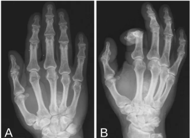
(2) Y ick-Fung Jim, et al.. 129. DISCUSSION. A. B. Fig. 1. A: Radiograph of hand in AP projection shows a mass lesion of high density at the soft tissue between the third and fourth fingers. Bone trabeculae inside is evident. B: Radiograph of the right hand in oblique projection shows the lesion in the soft tissue.. A. B. Fig. 2. A: T1WI of the right hand. B: T2WI of the right hand. They show high signal intensity of the lesion, similar to that of normal bone marrow.. The patient underwent a near-margin resection and the tumor was resected successfully. Pathology revealed that the tumor was about 2 centimeters in diameter and consisted of normal bone trabeculae and fatty marrow; an extraskeletal osteoma was diagnosed (Fig. 3). Radiograph of the patient's hand at follow-up confirmed that the tumor had been completely resected. He recovered well after the operation and had no complications at his most recent visit to our clinic.. Osteoma is a benign, often asymptomatic neoplasm consisting of well-differentiated mature bone [1]. Osteomas of the paranasal sinuses are slow-growing benign tumors most frequently found in the frontal sinus in 47% to 80% of cases [2]. Osteomas are rare bone tumors and occur in the following sites: mastoid [3], the mandibular condyle [4], ulna [5], thyroid cartilage [6] and larynx [7]. An osteoma is a dense protrusion of normal bone. The lesions are confined to areas of the bone that are normally produced by the periosteal membrane [8]. Osteomas usually occur in the skull and facial bones; however, they may be present in the pelvis or tubular bones of extremities. Diagnosis is based on imaging studies. Extra-skeletal osteoma has the same internal components as that of osteoma of bone; they show the same pattern and features on radiographs. High radiopaque density of the lesion is present; bone trabecular pattern may be demonstrated. Osteoma of soft tissue should be considered when mature bone tissue is detected in soft tissue [8]. Osteomas of soft tissue are extremely uncommon. They generally occur in the head, usually in the posterior portion of the tongue, or in the thigh [9]. Our patient's osteoma occurred in the soft tissue of the right hand. Some atypical sites of osteomas of soft tissue include the base of the tongue [8,10] and the ovary [11]. Recognition of this benign bone tumor is especially important to avoid misdiagnosis of other, potentially more aggressive types of malignancies such as osteosarcoma or chondrosarcoma. Dense calcification or bone marrow is characteristic of the tumor and may be relied upon to exclude alternative soft tissue lesions. Ma'luf et al suggested that trauma might be a factor in the pathogenesis of osteoma [12]. Soft tissue osteoma is generally regarded as a benign bone tumor, however, Kasper et al proposed that it may be a reactive lesion [13]. Metaplastic ossification due to tumor or inflammation is one of the differential diagnoses;.
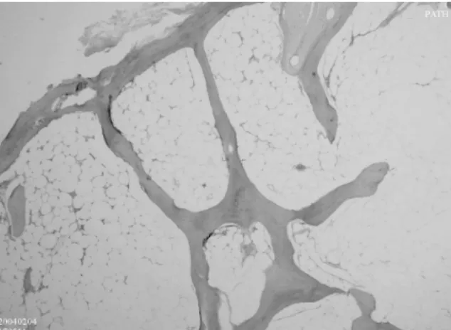
(3) 130. Extraskeletal Osteoma of the Hand. REFERENCES. Fig. 3. The mass consisted of normal bone trabeculae and fatty tissue.. detailed history of the patient can aid in the inclusion or exclusion of metaplastic ossification. Myositis ossifican is another differential diagnosis; the correct diagnosis can be made if we carefully examine any trauma history, and the sequential change of ossification inside the lesion. Once discovered, osteomas usually remain unchanged on series studies. Our patient had the lesion for more than 10 years. However, he felt that the mass had become larger recently. Surgical intervention was indicated to eliminate any possibility of malignancy since histopathologic confirmation of the tumor is the definitive method for diagnosis. MRI scan provides valuable information about soft tissue. The contour and margin of the lesion can be demonstrated in three dimensional planes. The nature of the lesion may be predicted if the lesion consists mainly of fatty tissue or bone marrow. The lesion in our patient consisted of bone marrow; therefore, extra-skeletal osteoma was included in our differential diagnosis.. 1. Johann AC, de Freitas JB, de Aguiar MC, et al. Peripheral osteoma of the mandible: case report and review of the literature. [Review] J Craniomaxillofac Surg 2005;33:276-81. 2. Dispenza C, Saraniti C, Ferrara S, et al. Frontal sinus osteoma and palpebral abscess: case report. Rev Laryngol Otol Rhinol (Bord) 2005;126:49-51. 3. Magliulo G, Pulice G, Mastoid osteoma. An Otorrinolaringol Ibero Am 2005;32:271-8. 4. Ortakoglu K, Gunaydin Y, Aydintug YS, et al. Osteoma of the mandibular condyle: report of a case with 5-year follow-up. Mil Med 2005;170:117-20. 5. Chikuda H, Goto T, Ishida T, et al. Juxtacortical osteoma of the ulna. J Orthop Sci 2002;7:721-3. 6. Redman AG, Hide IG, Zammit-Maempel I. Osteoma of the thyroid cartilage--an unusual cause of difficult intubation. Br J Radiol 2000;73:899-900. 7. Batti JS, Abramson A. First report of a case of osteoma of the larynx. Ear Nose Throat J 2000;79: 564-8. 8. Resnick D. Tumors and tumor-like lesions of soft tissues. In: Resnick D, 3rd eds. Bone and Joint Imaging. Eliser Inc. 2005:1225. 9. Lekas MD, Sayegh R, Finkelstein SD. Osteoma of the base of the tongue. Ear Nose Throat J 1997;76:827-8. 10. Yang SW, Chen CY, Lin CY. Lingual osteoma: case report. Chang Gung Med J 2000;23:498-502. 11. Celik C, Gungor S, Acar A, et al. Ovarian osteoma. Arch Gynecol Obstet 2003;268:222-3. 12.Ma'luf RN, Ghazi NG, Zein WM, et al. Orbital osteoma arising adjacent to a foreign body. Ophthal Plast Reconstr Surg 2003;19:327-30. 13.Kasper HU, Adermahr J, Dienes HP. Soft tissue osteoma: tumour entity or reactive lesion? Paraarticular soft tissue osteoma of the hip. Histopathology 2004;44:91-3..
(4) 131. 1. 1. 67 10. 2006;11:128-31. 404. 2. 2005. 10. 12. 2005. 11. 11. 2005. 11. 4.
(5)
數據
相關文件
Fig. 3 MR images of the mandible. a T1-weighted MR image showing a decrease in the signal intensity of the bone marrow in the left lower premolar and molar regions, except for
In an immunohistochemical and ultrastructural study of sialolipoma cases, Nagao et al [5] observed that the glandular components within the tumor consisted of regularly
Microscopically, FD comprises irregular trabeculae of woven bone, blending into the surrounding normal bone (figure 4) and lying within a cellular fibrous stroma with
As was the case in this study, several reports found that the best success rate in terms of pulp and periodontal healing was observed when the donor tooth was at the ½ to ¾ stage
The second mode was found in case 2, case 3, and case 4: the bony window on CT scans showed ossification lesions around the condyle, and the bone cortex was destroyed, MRI
Iatrogenic Delay in Diagnosis of Temporo-Mandibular Joint Ankylosis: A Cross Sectional Analysis of Thirty Four Trauma Patients from Central India.. Bailoor Durgesh, Gupta
Loss of vascular content, increase of fat in the bone marrow cavity, and fibrosis showed a linear relation with time after radiation and were considered the end stage of
This case report describes the diagnosis and management of a 55-year-old woman with a synovial sarcoma of the right lateral border of the tongue that was initially diagnosed as a
