Size dependence of photoluminescence and resonant Raman scattering from ZnO
quantum dots
Hsin-Ming Cheng, Kuo-Feng Lin, Hsu-Cheng Hsu, and Wen-Feng Hsieh
Citation: Applied Physics Letters 88, 261909 (2006); doi: 10.1063/1.2217925
View online: http://dx.doi.org/10.1063/1.2217925
View Table of Contents: http://scitation.aip.org/content/aip/journal/apl/88/26?ver=pdfcov Published by the AIP Publishing
Articles you may be interested in
Identification of visible emission from ZnO quantum dots: Excitation-dependence and size-dependence J. Appl. Phys. 111, 083521 (2012); 10.1063/1.4705395
Anomalous optical processes in photoluminescence from ultrasmall quantum dots of ZnO J. Vac. Sci. Technol. A 29, 03A120 (2011); 10.1116/1.3578344
Size dependence of the electronic structures and electron-phonon coupling in ZnO quantum dots Appl. Phys. Lett. 91, 262101 (2007); 10.1063/1.2824396
Study on the quantum confinement effect on ultraviolet photoluminescence of crystalline ZnO nanoparticles with nearly uniform size
Appl. Phys. Lett. 90, 263113 (2007); 10.1063/1.2750527
Raman scattering and photoluminescence of As ion-implanted ZnO single crystal J. Appl. Phys. 96, 175 (2004); 10.1063/1.1756220
This article is copyrighted as indicated in the article. Reuse of AIP content is subject to the terms at: http://scitation.aip.org/termsconditions. Downloaded to IP: 140.113.38.11 On: Thu, 01 May 2014 01:56:14
Size dependence of photoluminescence and resonant Raman scattering
from ZnO quantum dots
Hsin-Ming Cheng,a兲Kuo-Feng Lin, Hsu-Cheng Hsu, and Wen-Feng Hsiehb兲
Department of Photonics and Institute of Electro-Optical Engineering, National Chiao Tung University, 1001 Tahsueh Road, Hsinchu 30050, Taiwan, Republic of China
共Received 17 March 2006; accepted 24 May 2006; published online 28 June 2006兲
ZnO quantum dots共QDs兲 of controlled sizes have been fabricated by a simple sol-gel method. The blueshift of room-temperature photoluminescence measurement from free exciton transition are observed decreasing with the QD size that is ascribed to the quantum confinement effect. From the resonant Raman scattering, the coupling strength between electron and longitudinal optical phonon, deduced from the ratio of the second- to the first-order Raman scattering intensity, diminishes with reducing the ZnO QD diameter. The size dependence of electron-phonon coupling is principally a result of the Fröhlich interaction. © 2006 American Institute of Physics.关DOI:10.1063/1.2217925兴
ZnO is a promising material for short-wavelength pho-tonic devices since it has a large direct band gap of 3.37 eV, and a large exciton binding energy of 60 meV, all of which are advantageous for light-emitting diode and low-threshold optical pumped laser applications at room temperature. For the feasible requirement, low-dimensional ZnO nanostruc-tures, such as quantum dots共QDs兲,1,2nanoparticles共NPs兲,3,4 nanobelts,5 nanowires,6 and quantum wells,7 have been widely investigated. In particular, ZnO QDs and NPs are of great interest because of the three-dimensional confinement of carrier and phonon leads not only continuous tuning of the optoelectronic properties but also improvement in device performance. Recently, Demangeot et al.4have reported the resonant Raman scattering共RRS兲 and low-temperature pho-toluminescence 共PL兲 from ZnO NPs with different particle sizes which were synthesized by a room-temperature organo-metallic method. However, the study showed no size effects from the aspect of either carrier or phonon. The origin of weak size dependence of longitudinal optical 共LO兲 phonon frequency was explained by the ligands bonded to the par-ticle surface, and no shift from the low-temperature PL mea-surement indicated that UV emission was most likely domi-nated by weakly bound localized defects, which could come from the surface-bound ionized acceptor-exciton complexes, rather than the size-dependent quantum confinement effect. It is therefore important to note that the nanocrystals synthe-sized by chemical methods indeed occasionally cause the product suffering the active surround, such as ligands, which could intensely transform the intrinsic properties of the core. Accordingly, the demand for surface passivation of the NPs and the QDs is significant from both the fundamental scien-tific research and photonic application points of view. In this letter, we demonstrate the size-dependent PL and RRS of ZnO QDs which are fabricated by a simple sol-gel method without placing any ligands. The enlargement of free exciton transition energy is responsible for the blueshift of near-band-edge emission in ZnO QDs at room temperature. More-over, the size dependence of electron-phonon coupling, which can be addressed principally as a result of the Fröhlich
interaction, is confirmed by resonant Raman spectra. The ZnO QDs were synthesized via a simple sol-gel method without placing any ligands. The average size can be tailored by the concentration of precursor, zinc acetate dihy-drate关99.5% Zn共OAc兲2, Riedel-deHaen兴. A high speed
cen-trifuge was used to separate the final colloids into the upper suspension and the white bottom layer, which include the monodispersed single crystalline ZnO QDs and the second-ary ZnO clusters, respectively. In this present work, we con-sidered only the monodispersed ZnO QDs to avoid the intri-cately mutual interaction. Additional detail of the synthesis can be found elsewhere.8The microstructures were analyzed by JEOL JEM-2100F field emission transmission electron microscope共FETEM兲 operated at 200 KeV. The phase and average crystallite size were characterized using Bede D1 thin film x-ray diffractometer 共TFXRD兲. Resonant Raman spectroscopy was carried out by a He–Cd laser 共=325 nm兲, and a Jobin-Yvon T64000 microspectrometer with a 1800 grooves/ mm grating in the backscattering con-figuration. Room-temperature PL measurements were also performed using the 20 mW He–Cd laser共=325 nm兲.
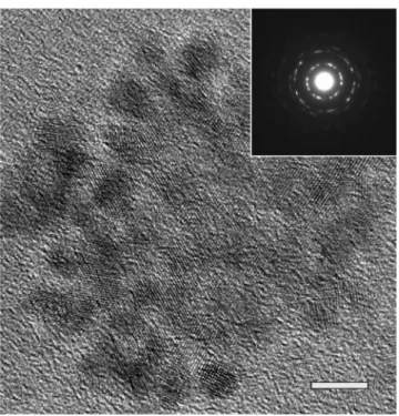
Figure 1 shows a TEM micrograph of the ZnO QDs formed using 0.06M zinc precursor. The ZnO QDs are little aggregated but still appear to be sphere and ellipsoid in shape. The mean-particle size is estimated to be ca. 4.2 nm. The electron and x-ray diffractions共not shown兲 all coincide with the presence of hexagonal wurzite crystallites with cell constants of a = 3.251 Å and c = 5.208 Å. No excess peaks are detected, which indicates that all the precursors have been completely decomposed. Blueshift of room-temperature ultraviolet共UV兲 emission was investigated in a series of ZnO QDs with different average sizes, as shown in Fig. 2共a兲. The broad shape of PL peaks is apparently due to the moderate size distribution of ZnO QDs. The nature of the UV-PL from ZnO QDs itself is still a matter of controversy.1 Aforemen-tioned Demangeot et al.4have reported that ligands coordi-nated at the surface of the nanoparticles may induce some changes in ZnO bonds, leading to some modifications of ei-ther their mechanical or dielectric properties. The UV-PL that came from surface-bound ionized acceptor-exciton com-plexes revealed no significant energy shift with varying size of ZnO NPs; furthermore, the weak bound emission vanished at the measured temperature higher than 15 K. In contrast, in
a兲Also at: Material and Chemical Research Laboratories, Industrial
Technol-ogy Research Institute, Hsinchu 310, Taiwan, Republic of China.
b兲Author to whom correspondence should be addressed; electronic mail:
APPLIED PHYSICS LETTERS 88, 261909共2006兲
0003-6951/2006/88共26兲/261909/3/$23.00 88, 261909-1 © 2006 American Institute of Physics
This article is copyrighted as indicated in the article. Reuse of AIP content is subject to the terms at: http://scitation.aip.org/termsconditions. Downloaded to IP: 140.113.38.11 On: Thu, 01 May 2014 01:56:14
the present work, the relaxed state near the band edge is attributed to the free exciton emission with high electronic density of states because no ligands were added during our synthesis process and the intense UV emission still exhibited even at room temperature. The UV emission shifts to the higher energy from 3.3 to 3.43 eV as the particle size re-duces from 12 to 3.5 nm. Moreover, the discernible broad green band共2.1–2.8 eV兲 was observed only when the size of ZnO QDs is smaller than 4.2 nm as shown in Fig. 2共b兲, the surface-located complex emission reasonably be enhanced while the surface volume of QDs were increased as decreas-ing the particle size. The relatively weak visible emission indicated that the ZnO QDs contain less intrinsic defects at the surface. Since the exciton Bohr radius aBof bulk ZnO is
2.34 nm,9 the carrier confinement in these ZnO QDs are in
the moderate to strong confinement regimes. The enlarged band gap from 3.41 to 3.60 eV as the ZnO QD size reduces from 12 to 3.5 nm has been estimated by effective mass model in our previous study.8 Although the energy differ-ences come from the relaxed state of the exciton and its binding energy, the parallel tendency consists with our as-sumption of quantum confinement effect. To circumvent other probabilities, we operated the PL measurements at dif-ferent power. The unchanged energies of the UV emission peaks, as shown in Fig. 2共c兲, exhibit no considerable local heating effect in ZnO QDs, and the obtained exponent value about 1.3 of power law confirmed our assignment that the observed UV emission bands are due to excitonic transition. The electron-phonon interaction could be straightly probed by the RRS when the exciting photon energy is reso-nant with the electronic interband transition energy of the wurtzite ZnO. The polar symmetry makes the A1共LO兲 and
E1共LO兲 modes the dominant ones, while the nonpolar E2
phonon is not visible. An intense multiphonon scattering of the ZnO QDs with various sizes was observed in the resonant Raman spectra of Fig. 3 with background subtracted, where three major bands were observed to result mainly from the polar symmetry modes A1共LO兲 and E1共LO兲 and their over-tones. Multiphonon scattering processes that have been pre-viously reported for one-dimensional共1D兲, two-dimensional 共2D兲, and three-dimensional 共3D兲 ZnO systems,10–14
in par-ticular, have been recently reported intensely for zero-dimensional共0D兲 systems.2,4,15
It is remarkable that the intensities of the first-order Ra-man modes and their overtones are enhanced while the size of ZnO QDs decreases. The reason can be explained using the total Raman cross section for an n-phonon process as a result of the energy of the incoming or the scattered photon that matches real electronic states in the material to enhance the Raman scattering cross section. The band gap of the present ZnO QDs certainly tends to approach the excitation laser energy as decreasing its size because of the quantum-confined effect mentioned above. Alim et al.2 have shown FIG. 1. TEM image of the ZnO QDs fabricated using 0.06M precursor with
the inset of its corresponding selected area electron diffraction共SAED兲 pat-tern. The scale bar has a length of 5 nm.
FIG. 2.共Color online兲 Room-temperature PL spectra 共a兲 and green emission 共b兲 of ZnO QDs with various sizes. 共c兲 PL spectra of ZnO QDs 共4.2 nm in diameter兲 as a function of excitation laser intensity 共from 1.1 to 23 mW兲, the exponent␥of power law I⬃L␥lies about 1.3.
FIG. 3.共Color online兲 Resonant Raman scatterings of ZnO QDs with vari-ous particle sizes measured at room temperature using a He–Cd laser共 = 325 nm兲.
261909-2 Cheng et al. Appl. Phys. Lett. 88, 261909共2006兲
This article is copyrighted as indicated in the article. Reuse of AIP content is subject to the terms at: http://scitation.aip.org/termsconditions. Downloaded to IP: 140.113.38.11 On: Thu, 01 May 2014 01:56:14
that the large redshifts in the RRS spectra from 20 nm ZnO QDs are most likely due to the local heating by UV laser excitation. In the present RRS spectra, the 1LO frequencies were all located at⬃575 cm−1 共within ±2 cm−1 fluctuation兲 for the ZnO QDs of different sizes. The heating effect com-ing from the inspection of micro-Raman seems to be negli-gible, because we used the laser power of only 0.8 mW at the spot size about 100m2.
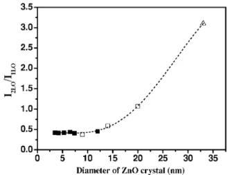
Beyond the phonon frequency shift, by observing the size dependence of intensity ratio between the second- and the first-order LO Raman scatterings, one can evaluate the coupling strength of the electron-phonon interaction. Within the Franck-Condon approximation,16 the electronic oscilla-tion strength distribuoscilla-tion over nth phonon mode is defined as
I⬃Sne−S/ n!, in which S is Huang-Rhys parameter, and also
can be used to express the coupling strength of the electron to the LO phonon. The ratio between the second- and the first-order Raman scattering cross sections was found to in-crease remarkably from 0.4 to 3.1 while an inin-crease of the ZnO crystallite size was from 3.5 to 33 nm, as shown in Fig. 4.
The electron-phonon coupling is generally determined by two mechanisms: the deformation potential and the Fröhlich potential. On the one hand, following Loudon17and Kaminow and Johnston,18the transverse optical共TO兲 Raman scattering cross section is determined by the deformation po-tential that involves the short-range interaction between the lattice displacement and the electrons, and on the other, the LO Raman scattering cross section includes contributions from both the deformation potential and Fröhlich potential that involves the long-range interaction generated by the
macroscopic electric field associated with the LO phonons. We found that under the resonant conditions the intensity of TO phonon in ZnO QDs is almost insensitive, while that of LO phonon is greatly enhanced. Therefore, we believe that the electron-phonon coupling as decreasing the nanocrystal size is mainly associated with the Fröhlich interaction. Al-though the complex origin of coupling is not well under-stood, the result in this study is extremely consistent with reports in other low-dimensional ZnO nanosystems.7,19,20
In summary, the enlarged free-exciton transition energy is responsible for the blueshift of near-band-edge PL spectra in ZnO QDs and gives significant evidence for the quantum confinement effect. Moreover, an increasing electron-phonon coupling was also discovered from RRS analysis while the diameter of ZnO crystal increases. The size dependence of electron-phonon coupling is principally as a result of the Fröhlich interaction.
Research supported by the National Science Council 共NSC兲 共Project No. NSC-94-2112-M-009-015兲 and the Nano Technology Research Center 共NTRC/ITRI兲 共Project No. A341XSYM91兲 in Taiwan.
1V. A. Fonoberov and A. Balandin, Appl. Phys. Lett. 85, 5971共2004兲. 2K. A. Alim, V. A. Fonoberov, and A. Balandin, Appl. Phys. Lett. 86,
053103 共2005兲; K. A. Alim, V. A. Fonoberov, M. Shamsa, and A. Balandin, J. Appl. Phys. 97, 124313共2005兲.
3L. Guo, S. Yang, C. Yang, P. Yu, J. Wang, W. Ge, and G. K. L. Wang,
Appl. Phys. Lett. 76, 2901共2000兲.
4F. Demangeot, V. Paillard, P. M. Chassaing, C. Pagès, M. L. Kahn, A.
Maisonnat, and B. Chaudret, Appl. Phys. Lett. 88, 071921共2006兲.
5Z. W. Pan, Z. R. Dai, and Z. L. Wang, Science 291, 1947共2001兲. 6M. H. Huang, S. Mao, H. Feick, H. Yan, Y. Wu, H. Kind, E. Weber, R.
Russo, and P. Yang, Science 292, 1897共2001兲.
7T. Makino, C. H. Chia, N. T. Tuan, H. D. Sun, Y. Segawa, M. Kawasaki,
A. Ohtomo, K. Tamura, and H. Koinuma, Appl. Phys. Lett. 77, 975 共2000兲.
8K. F. Lin, H. M. Cheng, H. C. Hsu, L. J. Lin, and W. F. Hsieh, Chem.
Phys. Lett. 409, 208共2005兲.
9R. T. Senger and K. K. Bajaj, Phys. Rev. B 68, 045313共2003兲. 10J. F. Scott, Phys. Rev. B 2, 1209共1970兲.
11X. T. Zhang, Y. C. Liu, Z. Z. Zhi, J. Y. Zhang, Y. M. Lu, D. Z. Shen, W.
Xu, G. Z. Zhong, X. W. Fan, and X. G. Kong, J. Phys. D 34, 3430共2001兲.
12V. V. Ursaki, I. M. Tiginyanu, V. V. Zalamai, V. M. Masalov, E. N.
Sama-rov, G. A. Emelchenko, and F. J. Briones, J. Appl. Phys. 96, 1001共2004兲.
13H. T. Ng, B. Chen, J. Li, J. Han, M. Meyyappan, J. Wu, S. X. Li, and
E. E. Haller, Appl. Phys. Lett. 82, 2023共2003兲.
14H. M. Cheng, H. C. Hsu, Y. K. Tseng, L. J. Lin, and W. F. Hsieh, J. Phys.
Chem. B 109, 8749共2005兲.
15H. M. Cheng, K. F. Lin, H. C. Hsu, C. J. Lin, L. J. Lin, and W. F. Hsieh,
J. Phys. Chem. B 109, 18385共2005兲.
16K. Huang and A. Rhys, Proc. R. Soc. London, Ser. A 204, 406共1950兲. 17R. Loudon, Adv. Phys. 13, 23共1964兲.
18I. P. Kaminow and W. D. Johnston, Phys. Rev. 160, 19共1967兲. 19T. Makino, K. Tamura, C. H. Chia, Y. Segawa, M. Kawasaki, A. Ohtomo,
and H. Koinuma, Phys. Rev. B 66, 233305共2002兲.
20R. P. Wang, G. Xu, and P. Jin, Phys. Rev. B 69, 113303共2004兲.
FIG. 4. Ratio between the second- and the first-order Raman scattering cross sections as a function of ZnO diameter. The experimental values of ZnO QDs in this work共squares兲 compared with ZnO NPs of Ref. 15 共empty squares兲 and nanocrystalline ZnO thin films of Ref. 11 共empty triangle兲. The dashed line joining the data points is just a guide for the eyes.
261909-3 Cheng et al. Appl. Phys. Lett. 88, 261909共2006兲
This article is copyrighted as indicated in the article. Reuse of AIP content is subject to the terms at: http://scitation.aip.org/termsconditions. Downloaded to IP: 140.113.38.11 On: Thu, 01 May 2014 01:56:14