Rapid and sensitive detection of rare cancer cells
by the coupling of immunomagnetic nanoparticle
separation with ELISA analysis
Hao-Yuan Cheng1
Lee-Jene Lai2
Fu-Hsiang Ko1
1Department of Materials Science and
Engineering, National Chiao Tung University, Taiwan, Republic of China;
2National Synchrotron Radiation
Research Center, Taiwan, Republic of China
Correspondence: Hao-Yuan Cheng National Chiao Tung University, 1001 University Road, Hsinchu, Taiwan, Republic of China Tel +88 635 780 281 Fax +88 635 789 816
Email [email protected]. edu.tw
Abstract: This study presents a rapid and sensitive method for detecting cancer cells occurring
at low concentration. The method involves the simultaneous detection of two biomarkers of T helper cancer cells. One biomarker conjugates with immunofunctionalized magnetic nano-particles (MNPs), enabling the separation of the T helper cells from a mixed population of cells. The other biomarker is used for detection during enzyme-linked immunosorbent assay (ELISA) analysis. The specific T helper cells can be quantified according to their ELISA absorbance values following magnetic separation. The experimental results demonstrate that immunofunctional-ized MNPs can function as magnetic sensors and separate specific T helper cells from a mixed population with high efficiency and high specificity. Coupled with the ELISA technique, the immunofunctionalized MNPs can simultaneously detect rare cells. Results indicated increasing absorbance with increasing T cell number (from 10 to 106). The total detection time was less than
15 minutes, even at a low T cell count. The advantages of the proposed method for detecting specific cells at low concentration include ease of preparation, low cost, rapid detection, and high sensitivity. The proposed system can be adopted to detect circulating tumor cells in early tumor stages for diagnostic or prognostic purposes.
Keywords: ELISA, magnetic nanoparticles, immunoassay, cancer cell
Introduction
In biotechnological research, the development of methods that enable the rapid and sensitive detection of rare cells and the early diagnosis, staging, and prognosis of viral infections or cancer has become a high priority.1,2 Systems to rapidly and sensitively detect low-frequency cancer cells have the potential to significantly improve cancer diagnosis and prognosis.3,4 In the human circulatory system, particularly in the blood, the number of circulating tumor cells is closely correlated with the recurrence of cancer and relapse. During the early stages of a tumor, cells circulate in the blood at extremely low concentrations, and their detection is thus a difficult task.5 The con-ventional methods for detecting trace cells are culture techniques and enzyme-linked immunospot assays. These require increasing cell numbers by culturing in vitro or by incubating with specific peptides.6,7 Studies have reported other approaches, based on flow cytometry, to detect low-frequency cells for the prognosis of minimal residual disease in childhood acute lymphoblastic leukemia.8–10 However, detecting numerous cells is a relatively labor-intensive and time-consuming process when using enzyme-linked immunospot or flow cytometric approaches. Developing highly sensitive and rapid methods of detecting specific cancer cells occurring at low frequency after appropriate treatment is, therefore, of significant prognostic value.
O R I g I N A L R E S E A R C H open access to scientific and medical research Open Access Full Text Article
Number of times this article has been viewed
This article was published in the following Dove Press journal: International Journal of Nanomedicine
Immunological methods provide powerful tools for the chemical detection of proteins in situ. The enzyme-linked immunosorbent assay (ELISA) is one of the most com-monly used techniques for the detection and quantification of antibodies, antigens, hormones, cytokines, and various other molecules, including synthetic peptides. Because of its quantitative, sensitive, and rapid response, the use of ELISA in research and clinical laboratories has become widespread.11,12 In addition, immunomagnetic separation has been shown to be a simple, fast, and efficient method of isolating specific micro-metastatic cells from colorectal cancer and stem cells.13,14
The present study demonstrates a high-throughput and sensitive method for detecting cells occurring in low numbers, using specific immunofunctionalized magnetite nanoparticles (MNPs) coupled with the ELISA technique. Human Jurkat cells (T helper lymphocytes) provided a model for circulating tumor cells, with antibodies and immu-nofunctionalized MNPs targeting CD3 and CD4 markers, respectively, on the T helper cell membrane. Human C1R cells (B lymphocytes) provided the other cells in the mixed population. Experimental results indicated that the detection of specific cells occurs within approximately 15 minutes, even at a low cell number, and that the sensitivity for distinction of specific T helper lymphocytes from B cells is approximately 0.001%. The proposed system thus provides a rapid and sensi-tive method for detecting and quantifying rare cells.
Materials and methods
Chemical materials
Chemicals such as iron (III) chloride hexahydrate (FeCl3 ⋅6H2O), 3-aminopropyltriethoxysilane (APTES), glutaraldehyde solution, 10 × concentrated phosphate buff-ered saline (10 × PBS) (diluted to 1 × PBS using ultrapure water for further use), and 3,3′,5,5′-tetramethylbenzidine were obtained from Sigma-Aldrich (St Louis, MO). Iron (II) chloride tetrahydrate (FeCl2 ⋅4H2O) was obtained from Alfa Aesar (Ward Hill, MA). The reagents ammonium hydroxide (NH4OH) and horseradish peroxidase-streptavidin (HRP-stv) were purchased from J.T. Baker (Covidien, Mansfield, MA) and Thermo Scientific (Thermo Fisher Scientific). Human T helper lymphocytes (ATCC TIB-152, Jurkat cell line) and human B lymphocytes (ATCC CRL-1993, C1R) were obtained from the American Type Culture Collection (Manassas, VA). Anti-human CD4 (14-0049) and biotinylated anti-human CD3 (13-0038) antibodies were purchased from eBioscience, Inc (San Diego, CA). Ultrapure water (18 MΩcm), obtained using a Milli-Q purification system (Millipore, MA), was used for the preparation of all solutions.
Cell culture
The Jurkat cells (T helper lymphocytes) were maintained in an RPMI-1640 medium (11875, Gibco®; Life Technologies, Carlsbad, CA) supplemented with 25 mm HEPES (SH30237, HyClone; Thermo Fisher Scientific, Waltham, MA), 10% (v/v) heat-inactivated fetal clone III serum (SH30109; Hyclone), 1% penicillin (P0781; Sigma-Aldrich), and 0.005 mM 2-Mercaptoethanol (M7522; Sigma-Aldrich) at 37°C and 5% CO2. The C1R cells (B lymphocytes) were cultured in an Iscove’s Modified Dulbecco’s medium (12200-028, Gibco; Life Technologies), supplemented with 10% (v/v) heat- inactivated fetal clone III serum and 1% penicillin at 37°C and 5% CO2 in a humidified atmosphere. A hemocytometer was used to count the number of cells in the cultured solutions. Cells were then concentrated by centrifuging or diluted and washed with 1 × phosphate buffered saline (PBS) for further use.
Transmission electron microscopy
For preparation of the samples for transmission electron microscopy, the particles were placed on a copper mesh surface for critical point drying using CO2 (CPD 030; Leica Microsystems [Bal-Tec], Wetzlar, Germany). The morphol-ogy of the sample was observed using a JEOL (Tokyo, Japan) 2010F transmission electron microscope.
Experimental protocols
MNPs were synthesized according to the following procedure. Iron (II) chloride tetrahydrate and FeCl3 ⋅6H2O were mixed in the ratio Fe+2:Fe+3= 1:2 and dissolved in ultrapure water. The mixtures were then placed in a water bath maintained at 80°C, and aqueous NH4OH solution (8 M) was added until a black precipitate appeared. The mixture was stirred continuously for 30 minutes in the 80°C water bath. When the reaction was complete, the MNPs in the solution were isolated using a magnet and washed repeatedly with hot water to remove any unreacted impurities. The synthesized MNPs were reacted with APTES overnight to create MNPs-APTES. To introduce functional immunoglobulin on the particles, glutaraldehyde was conjugated to the terminal NH2 group of MNPs-APTES. Excess anti-CD4 antibodies were added to create the immunofunctionalized MNPs.
For immunomagnetic separation, anti-CD4 MNPs were introduced to the mixture of T helper cells and B lympho-cytes for 15 minutes on ice, yielding MNP-bound T cells. The suspension of the mixed cell population containing these MNP-bound T cells was transferred into a new tube, and 1 × PBS solution was added to reach a total volume of 1.5 mL. The tube was loaded onto the magnet for separation
of the immunomagnetic particles from the mixed cells. The supernatant fraction was pipetted off, leaving the immunomag-netically labeled specific T helper cells remaining in the tube. The immunomagnetic separation procedure was conducted three times.
The specific lymphocytes were then detected using ELISA. Four µL of biotinylated anti-human CD3 antibody and HRP-stv were added to 100 µL of the separated T helper cells and incubated at room temperature for 15 minutes. The samples were washed with 1 × PBS to remove unreacted mat-ter, and 3,3′-,5,5′- tetramethylbenzidine was added for ELISA detection through measurement of absorbance at 650 nm (OD650). Detection was done using the EMax® endpoint ELISA microplate reader (Molecular Devices, Sunnyvale, CA), running SoftMax® Pro analysis software (Molecular Devices Corporation, Sunnyvale, CA). During detection, correction was done using B cells as blanks.
Results and discussion
Proof of concept
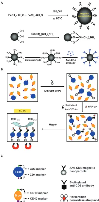
The present study demonstrates a method for rapid and sensitive detection of specific lymphocytes occurring at low frequency using immunofunctionalized MNPs and ELISA analysis. Figure 1A displays the methods employed for syn-thesis of MNPs using Fe+2- and Fe+3-containing materials. Synthesis of the MNPs occurred after treatment of the Fe+2/Fe+3 mixtures with alkaline solution in a water bath maintained at 80°C. Reaction of the MNPs with APTES then led to the formation of MNPs-APTES. To introduce functional immunoglobulin onto the particles, glutaraldehyde was conjugated to the terminal NH2 group of MNPs-APTES. The addition of excess anti-CD4 antibodies then created the immunofunctional MNPs. As shown in Figure 1B and C, the antigens on the membranes of the T helper cells differ from those on the membranes of the B cells. All T helper cell membranes contain the markers CD3 and CD4, whereas the membranes of B cells contain CD19 and CD40 mark-ers. The presented method takes advantage of these characteristics, using the CD3 and CD4 biomarkers to distin-guish the T helper cells (targets) from the B cells. The tight binding affinity of biotin-streptavidin15–17 enables linkage to the target T helper cells through biotinylated CD3 anti-body and HRP-stv. The membranes of the T helper cells thus conjugated with anti-human CD4-MNPs through the antigen-antibody reaction, yielding the MNP-bound T helper cells. After selection using a magnet, the membranes of the T helper cells were associated with biotin-conjugated anti-human CD3 antibodies and HRP-stv, creating the labeled T cells.
ELISA TMB(ox) TMB(red) Magnet Anti-CD4 MNPs Anti-CD4 antibody Si-(CH2)3NH2 Biotinylated Anti-CD3 Ab HRP-stv T cell CD3 marker CD4 marker B cell CD40 marker CD19 marker Anti-CD4 magnetic nanoparticle Biotinylated anti-CD3 antibody Horseradish peroxidase-streptavidin OH A B C OH OH OH OH OH OH OH OH OH OH OH OH OHC OHC OHC OHC CHO CHO CHO CHO Glutaraldehyde Si(OEt)3(CH2)3NH2 FeC12 • 4H2O + FeCI3 • 6H2O NH4OH ∆ 80°C NH2 NH2 NH2 NH2 NH2 H2N H2N H2N Magnetic nanoparticles O O O
Figure 1 (A) Synthesis of MNPs and immunofunctionalized MNPs. (B) The schema
illustrates the process for detection of specific T helper cells using two biomarkers: immunofunctionalized MNPs for immunoseparation and the ELISA technique for detection. Absorbance was recorded at a wavelength of 650 nm. (C) Diagrammatic
representations of the T helper cells, B cells, and other materials.
Abbreviations: MNPs, magnetite nanoparticles; ELISA, enzyme-linked
immunosorbent assay; HRP-stv, horseradish peroxidase-streptavidin; TMB, 3,3′,5,5′-tetramethylbenzidine.
Following the catalysis of 3,3′,5,5′-tetramethylbenzidine, the isolated T helper cells could be quantified according to their ELISA absorbance values.
Morphology evaluation
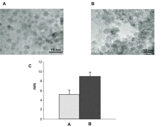
To verify the dimensions of the synthesized MNPs, transmission electron microscopy images were evaluated (Figure 2). The MNPs were spherical, with a uniform diameter (Figure 2A). The APTES-modified MNPs were also spherical
A B C B A 12 10 8 6 4 2 0 nm 15 nm 15 nm
Figure 2 Typical transmission electron microscopy images of (A) synthesized MNPs and (B) APTES-modified MNPs, with (C) a bar chart indicating their respective average
diameters.
Abbreviations: MNPs, magnetite nanoparticles; APTES, 3-aminopropyltriethoxysilane.
in transmission electron microscopy images (Figure 2B). Figure 2C shows the diameters of the MNPs (less than 6 nm) and the APTES-modified MNPs (less than 10 nm).
Phase identification
Previous studies have demonstrated that varying the prepara-tion condiprepara-tions of ferrites can create versatile magnetites.18,19 In the present study, the X-ray diffraction technique was selected for MNP identification. Phase identification of the samples was performed with a BL01C2 X-ray powder diffraction beam-line in the National Synchrotron Radiation Research Center, using an X-ray wavelength of 0.688807 Å. Figure 3A–C, and 3D represent the spectra of α-Fe2O3, Fe3O4, MNPs, and the APTES-modified MNPs, respectively. The diffraction peaks in Figure 3C are more consistent with those in Figure 3B than those in Figure 3A, indicating that the MNPs are more similar to Fe3O4 than to α-Fe2O3. The diffraction pattern of Figure 3D is identical to that of Figure 3C, indicating that APTES is amorphous. This suggests that APTES modifies MNPs yet has no apparent effects on their crystalline structures. Structural characterization
Fourier transform infrared spectra were obtained using a BL14A1 beamline at the National Synchrotron Radiation
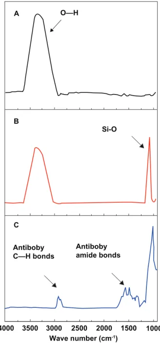
Research Center and used to characterize the APTES- modified MNPs and immunofunctionalized MNP composites. The spectra were compared with the MNP results. As shown in Figure 4A, the Fourier transform infrared spectrum of the MNPs displays a characteristic broad peak, from approximately 3000 cm−1 to approximately 3500 cm−1, assigned to the stretch-ing vibration of O–H groups on the MNPs. In Figure 4B, the spectrum of the APTES-modified MNPs displays a further peak at approximately 1020 cm−1, corresponding to the Si–O stretching vibration. Common biomolecules, such as nucleic acids, proteins, lipids, and carbohydrates, have characteristic and well-known vibration fingerprints, which can be identi-fied using infrared spectroscopy.20 As shown in Figure 4C, the spectrum of the anti-CD4 immunofunctionalized MNPs displays peaks that are frequently observed in biological samples, including lipids (at approximately 2900 cm-1), amide I (at approximately 1650 cm-1), and amide II (at approximately 1549 cm-1), representing stretching vibrations of the antibody signature. These results indicated the successful creation of anti-CD4 immunofunctionalized MNPs during multiple stages.
Specific binding and separation ability
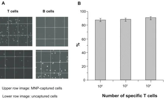
Specificity after conjugation was verified using the antigen-antibody recognition reaction occurring betweenthe CD3 markers of the T helper cells and biotinylated anti-human CD3, followed by a coupling reaction between biotinylated anti-human CD3 and HRP-stv and between the CD4 marker of the T helper cells and anti-human CD4-conjugated MNPs. Incubation of the T cells with first the anti-human CD4-conjugated MNPs, then biotinylated anti-human CD3, and finally, HRP-stv led to the formation of the HRP-tagged MNP-bound T cells. Figure 5A shows the specificity of the magnetic separation process for the T helper cells, following the sequential incubation of 106 T cells and 106 B cells with anti-human CD4-conjugated MNPs, bioti-nylated anti-human CD3, and HRP-stv. A high proportion of the target T helper cells were captured. In contrast, most of the B cells were not captured, despite the high cell count (106). This indicated that the anti-human CD4-conjugated
MNPs successfully attached to the membranes of T helper cell through the direct specificity of the antigen-antibody reaction and that cross-reaction with nontarget B cells did not occur. The separation ability was evaluated using dif-ferent numbers of T helper cells, from approximately 104 to 106, with results indicating an efficiency of almost 90% (Figure 5B). The experimental results further demonstrated that the anti-CD4 immunofunctionalized MNPs could func-tion as a magnetic sensor, separating T helper cells with high specificity and separation efficiency. When coupled with A (3.1.1) (4.4.0) (2.2.0) B C D 0 5 10 15 20 25 30 35 40 45 2θ (degree)
Figure 3 Synchrotron XRD spectra of (A) α-Fe2O3, (B) Fe3O4, (C) MNPs, and (D)
APTES-modified MNPs.
Abbreviations: XRD, X-ray diffraction; MNPs, magnetite nanoparticles; APTES,
3-aminopropyltriethoxysilane. A O—H Si-O B C Antiboby C—H bonds Antiboby amide bonds 1000 1500 2000 2500 3000 3500 4000 Wave number (cm-1)
Figure 4 Synchrotron Fourier transform infrared spectra of (A) MNPs, (B)
APTES-modified MNPs, and (C) immunofunctionalized MNPs.
T cells B cells
A B
Upper row image: MNP-captured cells
Lower row image: uncaptured cells Number of specific T cells
% 106 105 104 80 100 60 40 20 0
Figure 5 (A) Optical microscopic images indicating the specificity of immunofunctionalized MNPs for the T helper cells during magnetic separation and (B) a bar chart
indicating the separation efficiency of immunofunctionalized MNPs at various T cell counts.
Abbreviation: MNPs, magnetite nanoparticles.
1.4 1.2 1.0 0.8 0.6 0.4 0.2 0.0 Absorbance Number of T cells 0 101 102 103 104 105 106 y = 0.1779X + 0.0481 R2 = 0.9976
Figure 6 Semi-log plot of absorbance and the number of T helper cells detected by
ELISA in a mixed cell population.
Abbreviation: ELISA, enzyme-linked immunosorbent assay.
the ELISA technique, the anti-CD4 immunofunctionalized MNPs provide the further advantage of being able to detect rare cells.
Detection of T helper cells in a mixed cell population using ELISA
Following the sequential incubation of cells with anti-CD4 immunofunctionalized MNPs, biotinylated anti-human CD3, and the HRP-stv catalytic probe, a magnet was applied to separate the specific CD3- and MNP-labeled T helper cells. The separated T helper cell population was then quantified by measuring the absorbance according to CD3-labeling. Figure 6 displays the absorbance and T helper cell population
number (from 0 to approximately 106 cells) following magnetic separation from the B cells. Absorbance was recorded at a wavelength of 650 nm. Experimental results indicated increasing absorbance of the CD3-labeled T cells with increasing T helper cell quantity (from 10 to 106) in a mixed population with B cells. Detection time was less than 15 minutes, including the scanning period, even at a low T cell count. These results indicated that the system has high sensitivity for the detection of specific rare cells from a mixed cell population.
Conclusion
This study presents an efficient, specific, and sensitive method for detecting cancer cells occurring at low concentra-tion using ELISA and MNPs as probes. After funcconcentra-tionaliza- functionaliza-tion of the surfaces of the MNPs using APTES, the antibody is covalently immobilized on the MNPs. The immobilized molecules retain their specific binding activity. The immu-nofunctionalized MNPs display high ability to capture and separate target cancer cells. Coupled with the ELISA tech-nique, they can detect specific T cells from a mixed B cell-T cell population. Absorbance increases with increasing T cell population (from 10 to 106 cells). The immunofunctional-ized MNPs have approximately 90% separation efficiency and approximately 0.001% detection sensitivity for specific T helper lymphocytes from B cells, with detection occurring within 15 minutes. The anti-CD4-conjugated MNPs can, therefore, function as magnetic sensors and separate specific
International Journal of Nanomedicine
Publish your work in this journal
Submit your manuscript here: http://www.dovepress.com/international-journal-of-nanomedicine-journal
The International Journal of Nanomedicine is an international, peer-reviewed journal focusing on the application of nanotechnology in diagnostics, therapeutics, and drug delivery systems throughout the biomedical field. This journal is indexed on PubMed Central, MedLine, CAS, SciSearch®, Current Contents®/Clinical Medicine,
Journal Citation Reports/Science Edition, EMBase, Scopus and the Elsevier Bibliographic databases. The manuscript management system is completely online and includes a very quick and fair peer-review system, which is all easy to use. Visit http://www.dovepress.com/ testimonials.php to read real quotes from published authors.
T helper cells from a mixed cell population, with HRP-stv coupled with anti-CD3 biotin providing a reliable ELISA platform for T cell detection. The advantages of the system include ease of manipulation, low cost, rapid detection, and high sensitivity. In the future, the proposed method could be applied for the detection of Epstein Barr virus–specific memory T lymphocytes in nasopharyngeal carcinoma patients and could prove useful for the early diagnosis, stag-ing, and prognosis of cancer diseases.
Disclosure
The authors report no conflicts of interest in this work.
References
1. Gross HJ, Verwer B, Houck D, Hoffman RA, Recktenwald D. Model study detecting breast cancer cells in peripheral blood mononu-clear cells at frequencies as low as 10-17. Proc Natl Acad Sci U S A.
1995;92(2):537–541.
2. Sha MY, Xu H, Natan MJ, Cromer R. Surface-enhanced Raman scattering tags for rapid and homogeneous detection of circulating tumor cells in the presence of human whole blood. J Am Chem Soc. 2008;130:17214–17215.
3. Li G, Cuilleron M, Gentil-Perret A, et al. Rapid and sensitive detection of messenger RNA expression for molecular differential diagnosis of renal cell carcinoma. Clin Cancer Res. 2003;9:6441–6446.
4. Ishii T, Fujiwara Y, Ohnaka S, et al. Rapid genetic diagnosis with the transcription–reverse transcription concerted reaction system for cancer micrometastasis. Ann Surg Oncol. 2004;11:778–785.
5. Pantel K, Otte M. Occult micrometastasis: enrichment, identification and characterization of single disseminated tumour cells. Semin Cancer Biol. 2001;11:327–337.
6. Pahar B, Li J, Rourke T, Miller CJ, McChesney MB. Detection of antigen-specific T cell interferon γ expression by ELISPOT and cytokine flow cytometry assays in rhesus macaques. J Immunol Methods. 2003;282:103–115.
7. Alix-Panabières C, Rebillard X, Brouillet JP, et al. Detection of circulat-ing prostate-specific antigen-secretcirculat-ing cells in prostate cancer patients.
Clin Chem. 2005;51:1538–1541.
8. Fornas O, Garcia J, Petriz J. Flow cytometry counting of CD34+ cells in whole blood. J Nat Med. 2000;6:833–836.
9. Bajaj S, Welsh JB, Leif RC, Price JH. Ultra-rare-event detection performance of a custom scanning cytometer on a model preparation of fetal nRBCs. Cytometry. 2000;39:285–294.
10. Neale G, Coustan-Smith E, Stow P, et al. Comparative analysis of flow cytometry and polymerase chain reaction for the detection of minimal residual disease in childhood acute lymphoblastic leukemia. Leukemia. 2004;18:934–938.
11. Engvall E, Perlmann P. Enzyme-linked immunosorbent assay, ELISA. 3. Quantitation of specific antibodies by enzyme-labeled immunoglobulin in antigen-coated tubes. J Immunol. 1972;109(1): 129–135.
12. Engvall E. Enzyme immunoassay ELISA and EMIT. Methods Enzymol. 1980;70(A):419–439.
13. Flatmark K, Bjørnland K, Johannessen HO, et al. Immunomagnetic detection of micrometastatic cells in bone marrow of colorectal cancer patients. Clin Cancer Res. 2002;8:444–449.
14. Kekarainen T, Mannelin S, Laine J, Jaatinen T. Optimization of immu-nomagnetic separation for cord blood-derived hematopoietic stem cells.
BMC Cell Biol. 2006;7:30.
15. Ijiro K, Ringsdorf H, Birch-Hirschfeld E, Hoffmann S, Schilken U, Strube M. Protein-DNA double and triple layers: interaction of bioti-nylated DNA fragments with solid supported streptavidin layers.
Langmuir. 1998;14(10):2796–2800.
16. Kuberan B, Gunay NS, Dordick JS, Linhardt RJ. Preparation and isolation of neoglycoconjugates using biotin-streptavidin complexes.
Glycoconj J. 1999;16(6):271–281.
17. Aslan FM, Yu Y, Mohr SC, Cantor CR. Engineered single-chain dimeric streptavidins with an unexpected strong preference for biotin-4-fluorescein. Proc Natl Acad Sci U S A. 2005;102:8507–8512. 18. Tronc E, Belleville P, Jolivet JP, Livage J. Transformation of ferric
hydroxide into spinel by iron (II) adsorption. Langmuir. 1992;8: 313–319.
19. Yang J, Peng J, Liu K, Guo R, Xu D, Jia J. Synthesis of ferrites obtained from heavy metal solutions using wet method. J Hazard Mater. 2007;143(1–2):379–385.
20. Jamin N, Dumas P, Moncuit J, et al. Highly resolved chemical imaging of living cells by using synchrotron infrared microspectrometry. Proc
Natl Acad Sci U S A. 1998;95:4837–4840.