Moderate exercise training attenuates aging-induced cardiac
inflammation, hypertrophy and fibrosis injuries of rat hearts
Po-Hsiang Liao1,Dennis Jine-Yuan Hsieh2, Chia-Hua Kuo3, Cecilia-Hsuan Day4,
Chia-Yao Shen4, Chao-Hung Lai5, Ray-Jade Chen6, V. Vijaya Padma7, Wei- Wen
Kuo8*, Chih-Yang Huang 1,9,10*
1Graduate Institute of Basic Medical Science, China Medical University, Taichung,
Taiwan
2School of Medical Laboratory and Biotechnology, Chung Shan Medical University,
Taichung, Taiwan
3Laboratory of Exercise Biochemistry, Department of Sports Sciences, University of
Taipei, Taiwan
4Department of Nursing, Mei Ho University, Pingguang Road, Pingtung, Taiwan 5Division of Cardiology, Department of Internal Medicine, Armed Force Taichung
General Hospital, Taichung, Taiwan
6Department of Surgery, School of Medicine, College of Medicine, Taipei Medical
University, Taipei, Taiwan
7Department of Biotechnology, Bharathiar University, Coimbatore-641 046, India 8Department of Biological Science and Technology, China Medical University,
Taichung, Taiwan
9Graduate Institute of Chinese Medical Science, China Medical University,
Taichung, Taiwan
10Department of Health and Nutrition Biotechnology, Asia University, Taichung,
Taiwan
* These authors contributed equally to this paper.
Corresponding author: Chih-Yang Huang Ph.D., Graduate Institute of Chinese Medical Science, China Medical University, Taichung, Taiwan, No. 91, Hsueh-Shih Road, Taichung, 404, Taiwan. Tel: 22053366 ext 3313. Fax:
+886-4-22032295. E-mail address: [email protected] 1 2 3 4 5 6 7 8 9 10 11 12 13 14 15 16 17 18 19 20 21 22 23 24 25 26 27 28 29 30 31 32 33 34 35 36
Abstract
Aging is the most important risk factor in cardiovascular disease (CVD), which is the leading causes of death worldwide and the second major cause of death in Taiwan. The major factor in heart failure during aging is heart remodeling, including long-term stress-induced cardiac hypertrophy and fibrosis. Exercise is good for aging heart health, but the impact of exercise training on aging is not defined. This study used 3-, 12- and 18-month-old rats and randomly divided each age group into no exercise training control groups (C3, A12 and A18) and moderate gentle swimming exercise training groups (E3, AE12 and AE18). The protocol of exercise training was swimming five times weekly with gradual increases from the first week from 20 to 60 min for 12 weeks. Analyses of protein from rat heart tissues and sections revealed cardiac inflammation, hypertrophy and fibrosis pathway increases in aged rat groups (A12 and A18), which were improved in exercise training groups (AE12 and AE18). There were no heart injuries in young rat hearts in exercise group E3. These data suggest that moderate swimming exercise training attenuated aging-induced cardiac inflammation, hypertrophy and fibrosis injuries of rat hearts.
Key words: aging; cardiovascular disease; heart failure; moderate exercise training. 37 38 39 40 41 42 43 44 45 46 47 48 49 50 51 52 53 54 55 56 57 58 59 60 61 62
Introduction
Aging is an inevitable trend of the world's population, and it is accompanied with serious age-related health issues in modern society that must be investigated. The risks for high cholesterol, hypertension, ventricular hypertrophy, diabetes,
cerebrovascular disease, ischemic heart disease, myocardial infarction, obstructive sleep apnea (OSA), changes in tissue macrophages and related cancers increase with aging . According to World Health Organization statistics, heart disease is the leading causes of death in the world. Moreover, The Department of Health in Taiwan
published the top ten leading causes of death and reported that cardiovascular diseases (CVDs) are the second major cause of death and lead to atherosclerosis, hypertension and heart failure.
The process of heart failure includes stress-induced cardiac remodeling, cardiac apoptosis, cardiac fibrosis and heart failure. Cardiac remodeling in CVD is one of the most frequently occurring characteristics of aged hearts . The risk reasons for cardiac remodeling during aging include heredity, diabetes mellitus, obesity, hypertension, hyperlipidemia, and smoking . Previous studies indicated that aging induced an inflammatory response in the rat heart . Tumor necrosis factor α (TNF-α), nuclear factor-κB (NF-κB) and cyclooxygenase 2 (COX-2) are the major protein markers of the cardiac inflammation pathway . Inflammation, myocardial injury and hypertension results from pathological hypertrophy, which cannot be reversed . The interleukin 6 (IL-6) signaling pathway is a major pathway that regulates cardiac hypertrophy, IL-6 and IL-6 receptor-activated downstream genes, such as the mitogen-activated protein kinase 5 (MEK5), extracellular signal-regulated kinase (ERKs) and calcineurin, which promotes the translocation of transcription factor-signal transducer and activator of transcription 3 (STAT3) and GATA binding protein 4 to the nucleus . Previous 63 64 65 66 67 68 69 70 71 72 73 74 75 76 77 78 79 80 81 82 83 84 85 86 87
research on the concentric hypertrophy pathway demonstrated that mitogen-activated protein kinase (MEK1/2) activated ERK1/2 and c-Jun N-terminal kinase (JNK), which induced calcineurin expression and promoted the transcription factor nuclear factor of activated T-cells, cytoplasmic 3 (NFATc3) into the nucleus and translocated downstream gene b-type natraretic peptide (BNP), the levels of BNP which is a risk for heart disease . . Some studies indicated that activation of the MEK5/ERK5/STAT3 signaling pathway promoted eccentric hypertrophy, which is one type of pathological hypertrophy .
Fibrosis is the end-stage step after cardiac myocyte apoptosis . Fibroblasts result in fibrosis, which leads to reduced cardiac physiological functions . Heart injury and remodeling, such as inflammation and hypertrophy, induce cardiac fibrosis, which plays an important role in progression of heart disease, but the mechanisms and risk factors of fibrosis are not clear . Our recent paper demonstrated that transforming growth factor beta (TGF-β) activated downstream signaling pathways and increased connective tissue growth factor (CTGF) . CTGF expression in the fibrosis pathway upregulated and translocated to the cell nucleus, which leads to fibroblast proliferation . Our previous paper demonstrated that another fibrosis pathway of fibroblast growth factor 2 (FGF-2) activated urokinase/tissue-type plasminogen activator (uPA/tPA) and matrix metalloproteinase 2 (MMP-2) in cardiomyoblast cells . Moderation of exercise training prevents colonic inflammation via regulation of peroxisome proliferator-activated receptor gamma in obese mice , and exercise training reduced pathological cardiac hypertrophy, which was induced by pressure overload . Another paper indicated that exercise training reduced vascular fibrosis in obese rats . This research demonstrated that exercise training improved some protective function, but there was no evidence indicating that exercise training prevented heart injury in natural aging 88 89 90 91 92 93 94 95 96 97 98 99 100 101 102 103 104 105 106 107 108 109 110 111 112
rats.
Our previous study discovered that exercise training decreased aging-induced cardiomyocyte apoptosis, which is the end-step in heart failure progression, and long-term stress followed the age increase in cardiac inflammation, hypertrophy and fibrosis. . Therefore, suitable exercise training prevented heart injury by reducing heart failure progression caused by natural aging in rat heart, as expected .
Exercise training decreased the risk for heart failure and improved cardiac function . This study used moderate exercise training of open and free swimming in warm water at a suitable depth in a swimming pool to avoid excessive fear of drowning and struggle. The effects of this moderate swimming exercise training on aging-induced cardiac inflammation, hypertrophy and fibrosis injuries in rat hearts were investigated. 113 114 115 116 117 118 119 120 121 122 123 124 125 126 127 128 129 130 131 132 133 134 135 136 137 138 139
Materials and methods Animal models
Twenty-six Sprague-Dawley (SD) male rats at different age stages were used. Ten 3-month-old and 12-month-old rats and six rats at 18 month olds were used. Each age group was randomly divided into two groups: C3: 3-month-old control group; E3: 3-month-old exercise training aging rats; A12: 12-month-old control group; AE12: 12-month-old exercise training aging rats; A18:18-month-old control group; and AE18: 18-month-old exercise training aging rats. No more than 3 rats were housed per cage. Room temperature was maintained at 22-24°C, and rats were housed on a 12-hour light-dark cycle. Rats were supplemented with standard laboratory chow (Lab Diet 5001; PMI Nutrition International Inc., Brentwood, MO, USA), and water was available ad libitum. The Institutional Animal Care and Use Committee of China Medical University, Taichung, Taiwan approved all protocols, and the principles of laboratory animal care (NIH publication) were followed.
Exercise training protocol
The exercise training protocol was based on a previous protocol . Rats in the control group were soaked in the right amount of water, and the water temperature was 25±2oC. The depth of swimming pool (35 cm) could not lead to rats drowning,
and there were only two rats swimming at the same time. Exercise training began for rats in the exercise group (E3, AE12, AE18) swam for 20 min per day for the first 2 weeks. Swimming training time was extended to 30 min per day at the third week. The swimming training time was extended to 1 h per day in the remaining weeks, and all of the swimming times were recorded 5 times weekly. Animals were sacrificed after the experimental period and heart tissues were collected. The left ventricle tissues were isolated, and the tibia length was measured.
140 141 142 143 144 145 146 147 148 149 150 151 152 153 154 155 156 157 158 159 160 161 162 163 164 165
Tissue extraction
The entire heart weight was measured, and the left ventricle tissues from each group were isolated and washed 3 times in PBS buffer. Tissue samples (0.1 mg) were collected, and 1 mL lysis buffer (50 mM Tris-HCL, 1% NP-40, 0.5%
Na-deoxycholate, 0.1% SDS, 150 mM NaCl, 2 mM EDTA, 50 mM NaF) was added to the mixture. The tissue was homogenized for 5 min and centrifuged at 2,200 rpm at 4 °C. Samples were homogenized, centrifuged at 12,500 rpm, and a clean upper layer suspension was extracted. The centrifugation step was repeated, and a clean upper layer suspension was extracted.
Heart slices area tomography analysis
The heart tissue cross-section were made from identical location and stained by hematoxylin and eosin. Heart chamber area was measured by imageJ software.
Western blotting analysis
Tissue protein samples were mixed with 5× loading dye and placed on 95ºC for 5 min. The samples were loaded onto sodium dodecyl sulfate polyacrylamide gel electrophoresis (SDS-PAGE). The upper SDS-PAGE was a 5 % gel, and the underlying gel was an 8, 10 or 12 % separating gel. The prepared SDS-gels were placed in a vertical electrophoresis system. The prepared protein samples were loaded into U-shaped wells. The gel electrophoresis system worked at 100 volts (V). The gel was removed after the electrophoresis was completed and covered with a
polyvinylidene difluoride (PVDF) membrane. The gel and PVDF membrane were placed into a transfer tank at 90 V for 70 min at 4 °C. The PVDF membrane was soaked in a 5 % blocking buffer/Tris-buffered saline (TBS)-fat-free milk solution on a 166 167 168 169 170 171 172 173 174 175 176 177 178 179 180 181 182 183 184 185 186 187 188 189 190 191
shaker for 40 min at room temperature. The PVDF membrane was washed with TBS buffer 3 times for 10 min each and incubated with the primary antibody overnight at 4 °C. The PVDF membrane was washed with TBS buffer 3 times for 10 min each and incubated with the secondary antibody for 1 hr at room temperature. Membrane data was collected using an LAS-4000 mini (GE Healthcare Life Sciences), and data were quantified using Image J software.
Hematoxylin and eosin staining
Tissue sections were stained using hematoxylin and eosin (H&E). Sections were deparaffinized by immersion in xylene and stained with hematoxylin for 5 min. Sections were washed in double-distilled water (DDW) three times and placed in 85 % alcohol for 2 min. The sections were stained with eosin for 5 min and incubated in an ascending alcohol concentration gradient (70, 80, and 90 %) for 5 min. Sections were placed in 100 % alcohol for 5 min and twice in xylene for 1 min.
Photomicrographs were detected using a microscope (OLYMPUS Microscope).
Masson’s trichrome staining
Rat heart tissue from each group was stored in 10% formalin for 2 weeks, dehydrated in an ascending series of alcohols (75%, 85%, 90%, and 100% alcohol, 5 min each) and embedded in paraffin wax. The 2 μm-thick paraffin sections were sliced from these paraffin-embedded tissue blocks. Tissue sections were
de-paraffinized via immersion in xylene (3 times, 5 min each) and rehydrated using an descending series of alcohols (100%, 90%, 85%, and 75% alcohol, 5 min each). Biopsy samples were stained using Masson’s trichrome stain to investigate heart morphological and fibrotic changes. Blue staining represented collagen accumulation. The results were visualized using an OLYMPUS microscope.
192 193 194 195 196 197 198 199 200 201 202 203 204 205 206 207 208 209 210 211 212 213 214 215 216 217
Statistics
The experimental data are provided as the means ± SEM. Student ’ s t test was used to compared two differences groups. One-way ANOVA was used for
comparisons between multiple groups. P <0.05 was considered significant.
Results 218 219 220 221 222 223 224 225 226 227 228 229 230 231 232 233 234 235 236 237 238 239 240 241 242 243 244
Exercise training improve inflammation in aging rats
We measured TNFα, p-IKKα/β, p-Iκβa, pNFκB, COX2 and iNOS to investigate the major pathways of cardiac inflammation in aging rats and aging rats after exercise training (Fig. 1A). The aging groups (A12 and A18) exhibited myocardial
inflammatory compared with the younger group (C3). The cardiac inflammation was age-dependent because inflammation in the A18 group was more severe than the A12 group. Cardiac inflammation was down-regulated in the E3, AE12 and AE18 group after exercise training. Notably, exercise training reduced the expression of members of the inflammation signaling pathway in aging groups (AE12 and AE18) better than the young group (E3), especially in the AE18 group.
Body weight, cardiac histopathological analysis and heart weight change
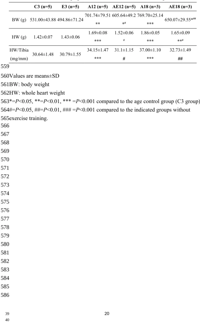
Rat body weights (BW) were measured and analyzed. Table 1 shows that the BW of rats increased with increasing age, and BW decreased after exercise training, especially in the aging groups (AE12 and AE18). Rat heart tissues were analyzed to compare differences between groups. Residual adipose tissue and any remaining blood was removed, and paper was used to dry excess water. Whole heart (HW) weight and tibia length were determined in each group.
We investigated the correlation between HW weight and tibia length. This value avoids external factors and more closely displays changes in HW weight for comparisons between groups. Comparisons of this ratio between each groups demonstrated that the ratio in experimental rats changed with HW weight. Table 1 indicates that the HW weight and heart chamber wall thickness (Fig. 2) increased proportionally with age. HW weight increased with age in the aging without exercise rats (A12, and A18). The HW weights of the exercise training groups (E3, AE12, and AE18) were reduced (P< 0.05). The exercise training effect was demonstrated in the 245 246 247 248 249 250 251 252 253 254 255 256 257 258 259 260 261 262 263 264 265 266 267 268 269 270
young group (E3) and aging groups (AE12, AE18), and it exhibited a very significant difference.
Histopathological tomography section analyses of cardiac tissue
Histopathological tomography analyses of whole hear tissues were performed using H&E staining (Fig. 2A). Images were viewed under a microscope and the control group C3 exhibited normal cardiac myocyte architecture and volume.
However, after measured by imageJ (Fig. 2B), the heart sizes in aging rats (A12, and A18) were found to be increased, and heart walls were thickened. Heart chambers (left ventricular and right ventricle) narrowed with age, but this phenomenon was rescued in both aging with exercise training groups (AE12, and AE18).
Exercise training prevented pathological hypertrophy in aged rat hearts.
Our previous data demonstrated that aging induced heart weight gain (Table 1 and Fig. 2). We investigated whether aging affected the pathological hypertrophy pathway in cardiomyocytes. We determined the genes that are involved in pathological
hypertrophy (Fig. 3A). The data demonstrated that upstream genes (p-JNK, p-p38 and p-ERK1/2) were up-regulated in aging groups (A12 and A18) compared with the C3 group, and the pathological hypertrophy transcription factors, NFATc3 and
p-GATA4, were increased in aging rats hearts (A18). The cardiac hypertrophy marker B-type natriuretic peptide (BNP), which is regulated by NFATc3, was
up-regulated compared to the control group. Aging with exercise training reversed the up-regulated expression of members of the pathological hypertrophy pathway. The protective effect in the A18 group was more significant (P<0.01). These results indicated that exercise training reduced aging-induced cardiac pathological hypertrophy. 271 272 273 274 275 276 277 278 279 280 281 282 283 284 285 286 287 288 289 290 291 292 293 294 295 296
Exercise training retarded cardiac fibrosis in aged rat hearts
Aging is a risk factor for heart failure. This study demonstrated that aging induced cardiac inflammation and pathological cardiac hypertrophy (Fig. 1-3), which follows the process of heart failure. We detected cardiac fibrosis using Masson’s trichrome staining of heart tissue sections (Fig. 4). Collagen accumulation occurred with aging in the aging groups (A12 and A18). The collagen accumulation in the A18 group was more serious than the A12 and C3 groups. Exercise training reduced collagen
accumulation in aging rats groups (AE12 and AE18). We also investigated the
fibrosis molecular signaling pathways. Fig. 5A shows that TGFb1, which is the ligand in the TGF-dependent fibrosis pathway, was up-regulated in the aging groups (A12 and A18) and resulted in CTGF overexpression compared to the control group (C3). The aging with exercise training groups (AE12 and AE18) exhibited a reversal of aging-activated TGF-dependent fibrosis pathway expression (Fig. 5A). We
investigated FGF2/uPA/MMP2 in the cardiac fibrosis pathway. FGF2 expression was up-regulated in aged rats (A18), and aging also induced the expression of the down-stream genes uPA and MMP2 (Fig. 6). Notably, exercise training prevented aging-induced cardiac fibrosis in the AE18 group, after exercise training reduced FGF2, uPA and MMP2 expression.
Discussion 297 298 299 300 301 302 303 304 305 306 307 308 309 310 311 312 313 314 315 316 317 318 319 320 321 322
It is well known that reactive oxygen species (ROS) also play a role during aging, previous studies revealed that aging-associated cardiovascular disease involve the role of nicotinamide adenine dinucleotide phosphate-oxidase induced ROS accumulation resulting in cardiac inflammation and fibrosis . Many researches have shown that drug or herbal extract mediated amelioration of cardiac injury is correlated with the extent of ROS scavenging . In this paper we revealed that moderate exercise can attenuate the aging-induced cardiac inflammation, hypertrophy and fibrosis injuries of rat hearts, however we need more experiments to verify whether
correlation of ROS regulation and moderate exercise effect on cardiovascular disease in aged rats.
Our previous studies demonstrated that diabetes, high blood pressure, obesity and aging were CVD risk factors . Apoptosis of cardiomyocytes always appeared in aging rat hearts . Long-term inflammation is one of the major risk factors that leads to cardiac hypertrophy . Our data demonstrated that cardiac inflammation existed in aging hearts, and this inflammation was induced by the activated NFκB signaling pathway. Figure 1 show that p-IKKα/β and p-Iκβ were up-regulated, which activated NFκB and up-regulated COX2 and iNOS expression. COX2 and iNOS are
downstream genes of NFκB and key markers of inflammation. These data indicated aging resulted in cardiac inflammation, and exercise training prevented aging-induced cardiac inflammation, especially in elderly rats (A18 and AE18 groups) (Fig. 1). Other studies demonstrated an effect of exercise training on inflammation in adipose tissue, muscle and brain . The present study demonstrated that the inflammatory pathway was up-regulated in aged rats hearts, and training with moderate exercise decreased inflammation in aging rats hearts (A18 and AE18) (Fig. 1).
Cardiac hypertrophy, or remodeling, includes changes in cell size and 323 324 325 326 327 328 329 330 331 332 333 334 335 336 337 338 339 340 341 342 343 344 345 346 347
morphology . Changes in pre-load, after-load or any pressure (e.g., inflammation and hypertension) trigger cardiomyocyte remodeling, which involves hypertrophy and fibrosis. The overloading pressure promotes cardiomyocyte transformation from physiological hypertrophy to pathological hypertrophy, and pathological hypertrophy is an irreversible process in the heart . No studies indicated whether moderate exercise training affected aging rat heart remodeling. Table 1, figure 2 and figure 4 show that aging induced cardiac remodeling. Whole heart weight and left ventricular wall thickness increased in the aging groups. Pathological hypertrophy can be divided into two types, concentric hypertrophy and eccentric hypertrophy. The heart wall thickens in concentric hypertrophy and the chamber size is reduced. The heart wall becomes thin or does not change in eccentric hypertrophy, and the chamber size is enlarged . Our findings demonstrated that aging induced cardiac pathological concentric hypertrophy via ERK1/2/JNK and NFATc3 expression (Fig. 3). Notably, training with moderate exercise decreased the risk for cardiac hypertrophy via down-regulation of the concentric hypertrophy pathway.
We examined the mechanism of aging-induced cardiomyocyte apoptosis, which may occur through inflammation and cardiac hypertrophy, and whether exercise training rescued the injury. Aging induced collagen accumulation (Fig. 4) and cardiac fibrosis via the TGF-β and FGF2 pathways (Fig. 5 and Fig. 6). Notably, we clarified that aging-induced cardiac fibrosis primarily occurred through the FGF2/uPA /MMP2 pathway, and fibrosis in aged rats was more severe (Fig. 6). Exercise training
decreased cardiac fibrosis in the aged rats group (AE18).
In conclusion, aging caused heart injury and activated the process of heart failure. Moderate exercise training decreased the heart injury via the down-regulation of inflammation, hypertrophy and fibrosis (Fig. 7). No significant injury in rat hearts 348 349 350 351 352 353 354 355 356 357 358 359 360 361 362 363 364 365 366 367 368 369 370 371 372
was found in the young rats in the exercise training group (E3). These findings demonstrated that moderate exercise training provided cardio-protection in aged rat hearts via the down-regulation of cardiomyocyte inflammation and remodeling. Notably, exercise training is neither a treatment nor therapy, but it decreased aging injuries in rat hearts. These results demonstrate that moderate exercise training provide aging rat hearts a rest environment with increased cardiorespiratory function and enhanced heart function and structure to reduce the possibility of heart failure in aging hearts.
Acknowledgment
This study was supported in part by the Taiwan Ministry of Health and Welfare Clinical Trial and Research Center of Excellence (MOHW104-TDU-B-212-113002).
References
1. Merkin SS, Karlamangla A, Roux AV, Shrager S and Seeman TE. Life course socioeconomic status and longitudinal accumulation of allostatic load in adulthood: multi-ethnic study of atherosclerosis. American journal of public health. 2014; 104(4):e48-55.
2. Seeman TE, Singer BH, Rowe JW, Horwitz RI and McEwen BS. Price of adaptation--allostatic load and its health consequences. MacArthur studies of successful aging. Archives of internal medicine. 1997; 157(19):2259-2268. 3. Lasry A and Ben-Neriah Y. Senescence-associated inflammatory responses: aging and cancer perspectives. Trends in immunology. 2015.
4. Cheitlin MD. Cardiovascular physiology-changes with aging. The American journal of geriatric cardiology. 2003; 12(1):9-13.
5. Khang D, Lu J, Yao C, Haberstroh KM and Webster TJ. The role of nanometer and sub-micron surface features on vascular and bone cell adhesion on titanium. Biomaterials. 2008; 29(8):970-983.
6. Pinto AR, Godwin JW, Chandran A, Hersey L, Ilinykh A, Debuque R, Wang L and Rosenthal NA. Age-related changes in tissue macrophages precede cardiac functional impairment. Aging. 2014; 6(5):399-413.
7. Lai CH, Ho TJ, Kuo WW, Day CH, Pai PY, Chung LC, Liao PH, Lin FH, Wu 373 374 375 376 377 378 379 380 381 382 383 384 385 386 387 388 389 390 391 392 393 394 395 396 397 398 399 400 401 402 403 404
ET and Huang CY. Exercise training enhanced SIRT1 longevity signaling replaces the IGF1 survival pathway to attenuate aging-induced rat heart apoptosis. Age. 2014; 36(5):9706.
8. Dai X, Hummel SL, Salazar JB, Taffet GE, Zieman S and Schwartz JB.
Cardiovascular physiology in the older adults. Journal of geriatric cardiology : JGC. 2015; 12(3):196-201.
9. Wang M and Shah AM. Age-associated pro-inflammatory remodeling and functional phenotype in the heart and large arteries. Journal of molecular and cellular cardiology. 2015; 83:101-111.
10. Hobi A, Roy S, Vuille C, Perdrix J and Darioli R. [Evolution of cardiac risk factors management among patients aged 65 years and older with coronary artery disease]. Revue medicale suisse. 2006; 2(56):658-663.
11. Linge HM, Lee JY, Ochani K, Koga K, Kohn N, Ojamaa K, Powell SR and Miller EJ. Age influences inflammatory responses, hemodynamics, and cardiac proteasome activation during acute lung injury. Experimental lung research. 2015:1-12.
12. Wang M and Shah AM. Age-associated pro-inflammatory remodeling and functional phenotype in the heart and large arteries. J Mol Cell Cardiol. 2015. 13. Xu W, Chen J, Lin J, Liu D, Mo L, Pan W, Feng J, Wu W and Zheng D. Exogenous H2S protects H9c2 cardiac cells against high glucose-induced injury and inflammation by inhibiting the activation of the NF-kappaB and IL-1beta pathways. International journal of molecular medicine. 2015; 35(1):177-186.
14. Heinemann JC, Duerr GD, Keppel K, Breitbach M, Fleischmann BK, Zimmer A, Wehner S, Welz A and Dewald O. CB2 receptor-mediated effects of
pro-inflammatory macrophages influence survival of cardiomyocytes. Life sciences. 2014. 15. Hill JA and Olson EN. Cardiac plasticity. The New England journal of medicine. 2008; 358(13):1370-1380.
16. Hirota H, Yoshida K, Kishimoto T and Taga T. Continuous activation of gp130, a signal-transducing receptor component for interleukin 6-related cytokines, causes myocardial hypertrophy in mice. Proceedings of the National Academy of Sciences of the United States of America. 1995; 92(11):4862-4866.
17. Leenders JJ, Pinto YM and Creemers EE. Tapping the brake on cardiac growth-endogenous repressors of hypertrophic signaling. Journal of molecular and cellular cardiology. 2011; 51(2):156-167.
18. Tonne JM, Holditch SJ, Oehler EA, Schreiber CA, Ikeda Y and Cataliotti A. Cardiac BNP gene delivery prolongs survival in aged spontaneously hypertensive rats with overt hypertensive heart disease. Aging. 2014; 6(4):311-319.
19. Kanda T and Takahashi T. Interleukin-6 and cardiovascular diseases. Japanese 405 406 407 408 409 410 411 412 413 414 415 416 417 418 419 420 421 422 423 424 425 426 427 428 429 430 431 432 433 434 435 436 437 438 439 440 441 442
heart journal. 2004; 45(2):183-193.
20. Kodama H, Fukuda K, Pan J, Makino S, Baba A, Hori S and Ogawa S. Leukemia inhibitory factor, a potent cardiac hypertrophic cytokine, activates the JAK/STAT pathway in rat cardiomyocytes. Circulation research. 1997; 81(5):656-663.
21. Bleumink GS, Knetsch AM, Sturkenboom MC, Straus SM, Hofman A, Deckers JW, Witteman JC and Stricker BH. Quantifying the heart failure epidemic:
prevalence, incidence rate, lifetime risk and prognosis of heart failure The Rotterdam Study. European heart journal. 2004; 25(18):1614-1619.
22. Lok SI, Nous FM, van Kuik J, van der Weide P, Winkens B, Kemperman H, Huisman A, Lahpor JR, de Weger RA and de Jonge N. Myocardial fibrosis and pro-fibrotic markers in end-stage heart failure patients during continuous-flow left
ventricular assist device support. European journal of cardio-thoracic surgery : official journal of the European Association for Cardio-thoracic Surgery. 2015.
23. Lai CH, Han CK, Shibu MA, Pai PY, Ho TJ, Day CH, Tsai FJ, Tsai CH, Yao CH and Huang CY. Lumbrokinase from earthworm extract ameliorates second-hand smoke-induced cardiac fibrosis. Environmental toxicology. 2014.
24. Hwang JM, Wu CH, Kuo WW, Jong GP, Lai CH, Tsai CH, Tsai FJ, Chang MH, Wu JP and Huang CY. Pro-inflammation and pro-fibrosis factors were highly
induction in heart tissues of carotid arteries balloon-injured animal model. Cell biochemistry and function. 2012; 30(5):390-394.
25. Yang CH, Ting WJ, Day CH, Ju DT, Yeh YL, Chung LC, Tsai FJ, Tsai CH, Tsai Y and Huang CY. SHSST cyclodextrin complex prevents the fibrosis effect on CCl(4)-induced cirrhotic cardiomyopathy in rats through TGF-beta pathway
inhibition effects. International journal of molecular sciences. 2014; 15(5):8037-8048. 26. Cheng YC, Kuo WW, Wu HC, Lai TY, Wu CH, Hwang JM, Wang WH, Tsai FJ, Yang JJ, Huang CY and Chu CH. ZAK induces MMP-2 activity via JNK/p38 signals and reduces MMP-9 activity by increasing TIMP-1/2 expression in H9c2
cardiomyoblast cells. Molecular and cellular biochemistry. 2009; 325(1-2):69-77. 27. Liu WX, Wang T, Zhou F, Wang Y, Xing JW, Zhang S, Gu SZ, Sang LX, Dai C and Wang HL. Voluntary exercise prevents colonic inflammation in high-fat diet-induced obese mice by up-regulating PPAR-gamma activity. Biochemical and biophysical research communications. 2015; 459(3):475-480.
28. Xu T, Tang H, Zhang B, Cai C, Liu X, Han Q and Zou L. Exercise preconditioning attenuates pressure overload-induced pathological cardiac hypertrophy. International journal of clinical and experimental pathology. 2015; 8(1):530-540.
29. Kim SY and Lee J. Exercise Training suppresses vascular fibrosis in aging 443 444 445 446 447 448 449 450 451 452 453 454 455 456 457 458 459 460 461 462 463 464 465 466 467 468 469 470 471 472 473 474 475 476 477 478 479 480
obesity induced rats. Journal of exercise nutrition & biochemistry. 2014; 18(2):175-180.
30. Liao SY, Liu Y, Zuo M, Zhang Y, Yue W, Au KW, Lai WH, Wu Y, Shuto C, Chen P, Siu CW, Schwartz PJ and Tse HF. Remodelling of cardiac sympathetic re-innervation with thoracic spinal cord stimulation improves left ventricular function in a porcine model of heart failure. Europace : European pacing, arrhythmias, and cardiac electrophysiology : journal of the working groups on cardiac pacing, arrhythmias, and cardiac cellular electrophysiology of the European Society of Cardiology. 2015.
31. Smith RN, Agharkar AS and Gonzales EB. A review of creatine supplementation in age-related diseases: more than a supplement for athletes. F1000Research. 2014; 3:222.
32. Whellan DJ, Kraus WE, Kitzman DW, Rooney B, Keteyian SJ, Pina IL, Ellis SJ, Ghali JK, Lee KL, Cooper LS and O'Connor CM. Authorship in a multicenter clinical trial: The Heart Failure-A Controlled Trial Investigating Outcomes of Exercise Training (HF-ACTION) Authorship and Publication (HAP) scoring system results. American heart journal. 2015; 169(4):457-463 e456.
33. Asrar Ul Haq M, Goh CY, Levinger I, Wong C and Hare DL. Clinical utility of exercise training in heart failure with reduced and preserved ejection fraction. Clinical Medicine Insights Cardiology. 2015; 9:1-9.
34. Chen Hi H, Chiang IP and Jen CJ. Exercise Training Increases Acetylcholine-Stimulated Endothelium-Derived Nitric Oxide Release in Spontaneously
Hypertensive Rats. Journal of biomedical science. 1996; 3(6):454-460.
35. Vendrov AE, Vendrov KC, Smith A, Yuan J, Sumida A, Robidoux J, Runge MS and Madamanchi NR. NOX4 NADPH Oxidase-Dependent Mitochondrial Oxidative Stress in Aging-Associated Cardiovascular Disease. Antioxidants & redox signaling. 2015.
36. Yin Q, Lu H, Bai Y, Tian A, Yang Q, Wu J, Yang C, Fan TP, Zhang Y, Zheng X, Zheng X and Li Z. A metabolite of Danshen formulae attenuates cardiac fibrosis induced by isoprenaline, via a NOX2/ROS/p38 pathway. British journal of
pharmacology. 2015.
37. Kwon MJ, Lee KY, Lee HW, Kim JH and Kim TY. SOD3 variant R213G altered SOD3 function, leading to ROS mediated inflammation and damage in multiple organs of premature aging mice. Antioxidants & redox signaling. 2015. 38. Escobales N, Nunez RE, Jang S, Parodi-Rullan R, Ayala-Pena S, Sacher JR, Skoda EM, Wipf P, Frontera W and Javadov S. Mitochondria-targeted ROS scavenger improves post-ischemic recovery of cardiac function and attenuates mitochondrial abnormalities in aged rats. Journal of molecular and cellular 481 482 483 484 485 486 487 488 489 490 491 492 493 494 495 496 497 498 499 500 501 502 503 504 505 506 507 508 509 510 511 512 513 514 515 516 517 518
cardiology. 2014; 77:136-146.
39. Drapkina OM and Shepel RN. [Telomeres and chronic heart failure]. Kardiologiia. 2014; 54(4):60-67.
40. Cencioni C, Spallotta F, Mai A, Martelli F, Farsetti A, Zeiher AM and Gaetano C. Sirtuin function in aging heart and vessels. Journal of molecular and cellular cardiology. 2015; 83:55-61.
41. Guzzardi MA and Iozzo P. Fatty heart, cardiac damage, and inflammation. The review of diabetic studies : RDS. 2011; 8(3):403-417.
42. Masson GS, Costa TS, Yshii L, Fernandes DC, Soares PP, Laurindo FR, Scavone C and Michelini LC. Time-dependent effects of training on cardiovascular control in spontaneously hypertensive rats: role for brain oxidative stress and inflammation and baroreflex sensitivity. PloS one. 2014; 9(5):e94927.
43. Knudsen JG, Murholm M, Carey AL, Bienso RS, Basse AL, Allen TL, Hidalgo J, Kingwell BA, Febbraio MA, Hansen JB and Pilegaard H. Role of IL-6 in exercise training- and cold-induced UCP1 expression in subcutaneous white adipose tissue. PloS one. 2014; 9(1):e84910.
44. Gliemann L, Olesen J, Bienso RS, Schmidt JF, Akerstrom T, Nyberg M, Lindqvist A, Bangsbo J and Hellsten Y. Resveratrol modulates the angiogenic response to exercise training in skeletal muscles of aged men. American journal of physiology Heart and circulatory physiology. 2014; 307(8):H1111-1119.
45. Aiello VD and Binotto MA. Myocardial remodeling in congenital heart disease. Arquivos brasileiros de cardiologia. 2007; 88(6):e185-186.
46. Tezuka F and Takahashi T. Pathology of cardiac hypertrophy in pressure overload. Japanese circulation journal. 1976; 40(10):1111-1118.
47. Bernardo BC, Weeks KL, Pretorius L and McMullen JR. Molecular distinction between physiological and pathological cardiac hypertrophy: experimental findings and therapeutic strategies. Pharmacology & therapeutics. 2010; 128(1):191-227. 48. Kanitz AC, Delevatti RS, Reichert T, Liedtke GV, Ferrari R, Almada BP, Pinto SS, Alberton CL and Kruel LF. Effects of two deep water training programs on cardiorespiratory and muscular strength responses in older adults. Experimental gerontology. 2015; 64:55-61.
49. Levine BD. Response to the Letters Regarding Article, "Can Intensive Exercise Harm the Heart? The Benefits of Competitive Endurance Training for Cardiovascular Structure and Function". Circulation. 2015; 131(23):e525.
519 520 521 522 523 524 525 526 527 528 529 530 531 532 533 534 535 536 537 538 539 540 541 542 543 544 545 546 547 548 549 550 551 552 553 554 555 556
Table 1 Characteristics of aging rats with or without exercise training (A and AE)
C3 (n=5) E3 (n=5) A12 (n=5) AE12 (n=5) A18 (n=3) AE18 (n=3)
BW (g) 531.00±43.88 494.86±71.24701.74±79.51 605.64±49.2 769.70±25.14650.07±29.55*## ** *# *** HW (g) 1.42±0.07 1.43±0.06 1.69±0.08 1.52±0.06 1.86±0.05 1.65±0.09 *** # *** **# HW/Tibia (mg/mm) 30.64±1.48 30.79±1.55 34.15±1.47 31.1±1.15 37.00±1.10 32.73±1.49 *** # *** ##
Values are means±SD BW: body weight HW: whole heart weight
*=P<0.05, **=P<0.01, *** =P<0.001 compared to the age control group (C3 group); #=P<0.05, ##=P<0.01, ### =P<0.001 compared to the indicated groups without exercise training. 557 558 559 560 561 562 563 564 565 566 567 568 569 570 571 572 573 574 575 576 577 578 579 580 581 582 583 584 585 586
Figures Figure 1 (A) (B) 587 588 589 590 591 592 593 594 595 596 597 598 599 600 601 602 603 604 605 606 607 608 609 610 611 612 613 614 615
Figure 2 (A) (B) 616 617 618 619 620 621 622 623 624 625 626 627 628 629 630 631 632
Figure 3 (A) (B) 633 634 635 636 637 638 639 640 641 642 643 644 645 646 647 648 649 650 651 652 653 654 655 656 657 658 659 660
Figure 4 661 662 663 664 665 666 667 668 669 670 671 672 673 674 675 676 677 678 679 680 681 682 683 684 685
Figure 5 (A) (B) 686 687 688 689 690 691 692 693 694 695 696 697 698 699 700 701 702 703 704 705 706 707 708 709 710 711 712 713 714 715
Figure 6 (A) (B) 716 717 718 719 720 721 722 723 724 725 726 727 728 729 730 731 732 733 734 735 736 737 738 739 740 741 742 743 744 745 746
Figure 7 747 748 749 750 751 752 753 754 755 756 757 758 759 760 761 762 763 764 765 766 767 768 769
Figure Captions
Fig. 1 The representative inflammatory protein products extracted from the left ventricles of two rats in each group: control rats (C3), aging rats (A12, A18), and aging, exercise-trained rats (E3, AE12, AE18) were measured using Western blotting. β-actin was used as an internal control.
*=P<0.05, **=P<0.01, *** =P<0.001 compared to the age control group (C3 group); #=P<0.05, ##=P<0.01, ### =P<0.001 compared to the indicated groups without exercise training.
Fig. 2 Hematoxylin and eosin staining. Cardiac tissue sections stained with
hematoxylin and eosin. (A) The myocardial architecture images were magnified ×10. The scale bar is 1 cm. RV: right ventricle, LV: left ventricle. (B) Quantization of cardiac ventricular area.
*=P<0.05, **=P<0.01, *** =P<0.001 compared with the age control group (C3 group); #=P<0.05, ##=P<0.01, ### =P<0.001 compared with the indicated groups without exercise training.
Fig. 3 Representative hypertrophy protein products extracted from the left ventricles of two rats in each group: control rats (C3), aging rats (A12, A18), and aging, exercise-trained rats (E3, AE12, AE18) were measured using Western blotting. β-actin was used as an internal control.
*=P<0.05, **=P<0.01, *** =P<0.001 compared with the age control group (C3 group); #=P<0.05, ##=P<0.01, ### =P<0.001 compared with the indicated groups without exercise training.
Fig. 4 Masson’s trichrome staining. Cardiac tissue sections stained with Masson’s trichrome staining buffer. Collagen accumulation is shown in blue color (black arrow). The myocardial architecture images were magnified ×400. The scale bar is 10 μm.
Fig. 5 Representative protein products of the TGFβ-dependent fibrosis pathway extracted from the left ventricles of two rats in each group: control rats (C3), aging rats (A12, A18), and aging, exercise-trained rats (E3, AE12, AE18) were measured using Western blotting. β-actin was used as an internal control.
770 771 772 773 774 775 776 777 778 779 780 781 782 783 784 785 786 787 788 789 790 791 792 793 794 795 796 797 798 799 800 801 802 803 804 805 806 807
*=P<0.05, **=P<0.01, *** =P<0.001 compared with the age control group (C3 group); #=P<0.05, ##=P<0.01, ### =P<0.001 compared with the indicated groups without exercise training.
Fig. 6 Representative protein products of the FGF2-dependent fibrosis pathway extracted from the left ventricles of two rats in each group: control rats (C3), aging rats (A12, A18), and aging, exercise-trained rats (E3, AE12, AE18) were measured using Western blotting. β-actin was used as an internal control.
*=P<0.05, **=P<0.01, *** =P<0.001 compared with the age control group (C3 group); #=P<0.05, ##=P<0.01, ### =P<0.001 compared with the indicated groups without exercise training.
Fig. 7 Moderate exercise training prevents aging-induced heart injury via down-regulated inflammation, hypertrophy and fibrosis.
808 809 810 811 812 813 814 815 816 817 818 819 820 821 822