ANALGESIC AND ANTI-INFLAMMATORY ACTIVITIES OF AN ETHANOL EXTRACT OF DUNALIELLA SALINA TEOD.
(CHLOROPHYCEAE)
PEI-YU CHOU1*, GUAN-JHONG HUANG2*, HSU-CHEN CHENG1, CHIEH-HSI WU3, YI-CHUNG CHIEN1, JWO-SHENG CHEN4, MING-HSING HUANG5, KAI-JENG HSU3and MING-JYH SHEU3,6
1Department of Life Science, National Chung Hsing University Taichung 402, Taiwan
2Institute of Chinese Pharmaceutical Sciences, College of Pharmacy China Medical University
Taichug 404 Taiwan
3School of Pharmacy, College of Pharmacy China Medical University
Taichung 404, Taiwan
4Department of Sports Medicine, China Medical University Taichung 404, Taiwan
5Department of Cosmetic Science Chia Nan University of Pharmacy and Science 60, Erh-Jen Road, Sec.1, Jen-Te, Tainan, 707 Taiwan
Accepted for Publication May 23, 2009
ABSTRACT
This study investigated the analgesic and anti-inflammatory effects of an ethanol extract of Dunaliella salina Teod. (Chlorophyceae) (EDS) in Imprint-ing Control Region mice. Standard all-trans-b-carotene and the amount of all-trans-b-carotene in an EDS were analyzed by high-performance liquid chromatography (HPLC). In HPLC analysis, the fingerprint chromatogram of EDS was established. Both all-trans-b-carotene and EDS showed similar peaks at the retention time of 24 min. This implied that EDS contained the active ingredient all-trans-b-carotene.
Treatment of animals with EDS significantly inhibited the numbers of acetic acid-induced writhing responses at doses of 0.5 g/kg (P< 0.01), * Both authors equally contributed this paper.
6 Corresponding author. TEL:+011-886-4-2205-3366-5158; FAX: +011-886-4-2207-3709; EMAIL: [email protected]
DOI: 10.1111/j.1745-4514.2010.00389.x
Journal of Food Biochemistry •• (2010) ••–••.
© 2010 Wiley Periodicals, Inc.
1.0 g/kg (P< 0.001) and 2.0 g/kg (P < 0.001). This inhibitory effect of EDS (1.0 and 2.0 g/kg) on acetic acid-induced writhings was similar to that of the positive control indomethacin (10 mg/kg) (P< 0.001). EDS did not signifi-cantly inhibit the formalin-induced pain in the early phase; however, at doses of 0.1 g/kg (P< 0.01), 0.5, 1.0 and 2.0 g/kg, EDS significantly inhibited the formalin-induced pain in the late phase (P< 0.001). Finally, EDS at doses of 1.0 and 2.0 g/kg also inhibited the development of paw edema induced by l-carrageenan (carrageenan). EDS (1.0 and 2.0 mg/kg) decreased the level of nitiric oxide (NO) in edematous paw tissue and in serum level, and diminished the level of serum tumor necrosis factor-a (TNF-a) at the fifth hour after carrageenan injection. Based on these findings, EDS probably exerts anti-inflammatory effects by suppressing TNF-a and NO. These results suggest that EDS might be a potential pharmacological analgesic and anti-inflammatory agent.
PRACTICAL APPLICATIONS
Dunaliella salina (Dunal) Teod. is a valuableb-carotene source. D. salina is cultivated in a great quantity in the southern part of Taiwan. In order to find its potential application for this product, the analgesic and anti-inflammatory effects were investigated. It was found that D. salina could attenuate the acetic acid-induced writhings and formalin-induced analgesia. We also demonstrate that D. salina could lessen the carrageenan-induced paw edema. Also, the anti-inflammatory effects verified that nitric oxide and TNF-a could play an import role.
INTRODUCTION
Dunaliella salina, a unicellular halophilic green microalgae, is a
well-known source of b-carotene. It is widely consumed in China, Japan and
Taiwan. Several reports indicate that Dunaliella has anti-oxidative effects (Lavy et al. 2003; Chidambara Murthy et al. 2005; Murthy et al. 2005; Vanitha et al. 2007). D. salina exhibits potent hepatoprotective effects on
CCl4-induced liver damage in mice, and it is believed that these effects may be
due to both the increase of anti-oxidant enzymes activities and inhibition of lipid peroxidation (Hsu et al. 2008). In vivo, it has been shown that anti-oxidative effect of algal carotenoid is similar to that of synthetic carotene
(Chidambara Murthy et al. 2005).b-carotene-rich algae D. bardawil markedly
inhibited spontaneous mammary tumorigenesis in mice by increasing the
bardawil promotes the growth of normal mammary gland cells, but inhibits neoplastic cells (Fujii et al. 1993). It has also been shown that an extract of D. salina could significantly inhibit NSAR-induced carcinogenesis (Xue 1993). Levin et al. reported that the antiperoxidative effect of 9-cis-b-carotene was more potent than that of all-trans-isomer in preventing malignant and cardio-vascular diseases (Levin et al. 1997). In a recent study, D. salina exerted a protective effect against experimentally induced fibrosarcoma in Wistar rats (Raja et al. 2007). In our previous studies, we found that the ethanol extract of D. salina (EDS) inhibits proliferation and induces apoptosis in the human lung cancer cell line A549 (Sheu et al. 2008). However, little information is avail-able on the analgesic and anti-inflammatory effects of D. salina. Therefore, we examined the analgesic effects of D. salina on acetic acid- and formalin-induced nociception. We also evaluated the anti-inflammatory effects of D. salina on paw edema induced by carrageenan in mice and investigated its related mechanisms.
MATERIALS AND METHODS
Materials
Acetic acid and formalin were purchased from Merck (Darmstadt, Germany). All-trans-b-carotene, carrageenan and indomethacin were obtained from Sigma-Aldrich (St. Louis, MO).
Preparation of D. Salina
Spray-dried algae material from D. salina cultured in outdoor cultivation pools were prepared by GONG BIH Enterprise Co., Ltd (Doo-Liu City, Taiwan). The composition of D. salina was analyzed and is shown in Table 1. D. salina (1.5 kg) was soaked in 70% ethanol (each 10 L) at 100C for 20 min.
TABLE 1.
COMPOSITIONS OF DS DRY POWDER
Compositions mg/g a-carotene 120/100 b-carotene 6,882/100 Xanthohyll 194/100 Zeaxanthin 275/100 Lycopene 7.18/100 Chlorophyll 1.124/100
Atiob-carotene ratio(9-cis/all-trans) 47:53.
The supernatants were collected, concentrated with a vacuum evaporator (Eyela N-N Serials, Rikakikai Co. Ltd., Tokyo, Japan) until the volume was
reduced to 5 mL and stored at-20C in a refrigerator (Herrero et al. 2006).
Samples were filtered with filter paper (Advantec No.1, Advantec MFS Inc., Tokyo, Japan) while the residue was further extracted under the same condi-tions thrice. The filtrates collected from these separate extraccondi-tions were com-bined and evaporated to dryness under vacuum at 50C. The yield obtained was 4% (60 g) from the ethanol extract of D. salina.
Compositional analysis ofb-carotene and EDS by High-Performance Liquid Chromatography (HPLC)
HPLC was conducted to analyze both the standard (all-trans-b-carotene) and EDS. The purity of the standard was more than 95% based on reverse phase HPLC analysis (Instrument: Jasco system; column: C18 reversed-phase
column with particle size 5mm, Vydac 201 TP54 stainless column
[25 cm¥ 4.6 mm i.d.]; Mobile phase : methanol : acetonitrile [9:1, v/v];
mobile phase flow rate:1.0 mL/min). Animals
Male ICR mice (18 g to 25 g) were obtained from BioLASCO Taiwan Co., Ltd. (Nankang, Taiwan). The animals were kept in plexiglass cages at a
constant temperature of 22⫾ 1C, relative humidity 55 ⫾ 5% with 12-h
dark–light cycle for at least 2 weeks before the experiment. They were given food and water ad libitum. All experimental procedures were performed according to the NIH Guide for the Care and Use of Laboratory Animals. The control groups were given 0.1 mL/10 g saline intraperitoneally (i.p.) using a bent blunted 27-gauge needle connected to a 1-mL syringe. All tests were conducted under the guidelines of the International Association for the Study of Pain (Zimmermann 1983). This study was approved by the ethics committee of the Institutional Animal Care and Use Committee of China Medical University.
Acetic Acid-Induced Writhing Response
After a 2-week adaptation period, male ICR mice (18–25 g) were
ran-domly assigned to six groups (n= 8) including a normal control, an
indomethacin (Indo) positive control and four EDS-treated groups. Control group received normal saline and the positive control group received indomethacin (10 mg/kg, i.p.) 25 min before i.p. injection of 1.0% acetic acid (10 mL/kg body weight). EDS-treated groups received EDS (0.1, 0.5, 1.0, and 2.0 g/kg, p.o.) 55 min before i.p. injection of 1.0% acetic acid (10 mL/kg body
weight). Five minutes after the i.p. injection of acetic acid, the number of writhings during the following 10 min was recorded. Control mice received normal saline (Taber et al. 1969).
Formalin Test
The antinociceptive activity of the drugs was determined using the for-malin test described by Dubuisson and Dennis (1977). Twenty microliters of 5% formalin was injected into the dorsal surface of the right hind paw 60 min after administration of EDS (0.1, 0.5, 1.0 and 2.0 g/kg, p.o.) and 30 min after administration of indomethacin (10 mg/kg, i.p.). The mice were observed for 30 min after the injection of formalin, and the amount of time spent licking the injected hind paw was recorded. The first 5 min post-formalin injection is referred to as the early phase and the period between 15 min and 40 min as the late phase. The total time spent licking or biting the injured paw (pain behav-ior) was measured with a stop watch. The activity was recorded in 5 min intervals.
l-Carrageenan (Carrageenan)-Induced Edema
Carrageenan-induced hind paw edema model was used for determination of anti-inflammatory activity (Winter et al. 1962). After a 2-week adaptation period, male ICR mice (18–25 g) were randomly assigned to five groups
(n= 8) including control, carrageenan, positive indomethacin control and three
EDS-treated groups. The control group only received normal saline. The
carrageenan group received 1% carrageenan (50mL). EDS at doses of 0.5, 1.0
and 2.0 g/kg were orally administered 2 h before the injection with 1%
carra-geenan (50mL) in the plantar side of right hind paws of the mice.
Indometha-cin (10 mg/kg) was intraperitoneally administered 90 min before the injection
with 1% carrageenan (50mL) in the plantar side of right hind paws of the mice.
Paw volume was measured immediately after carrageenan injection at 1, 2, 3, 4 and 5 h intervals using a plethysmometer (model 7159, Ugo Basile, Varese, Italy). The degree of swelling induced was evaluated by the ratio a/b, where a is the volume of the right hind paw after carrageenan treatment, and b is the volume of the right hind paw before carrageenan treatment. Indo was used as a positive control (Mascolo et al. 1989). After 5 h, the animals were sacrificed and the carrageenan-induced edema paw was dissected. The right hind paw tissue was rinsed in ice-cold normal saline, and immediately placed in cold normal saline four times their volume and homogenized at 4C. Then, the
homogenate was centrifuged at 12,074¥ g for 5 min. The supernatant was
obtained and stored at-20C refrigerator for the NO assays. Also, blood was
Total Protein Assay
The protein concentration of the sample was determined by the Bradford dye-binding assay (Bio-Rad, Hercules, CA).
Determination of Nitric Oxide (NO)
Nitrite, a stable end product of NO, was then measured using the Griess
reaction (Liao et al. 2007). Samples of 100mL aliquots mixed with 100 mL of
Griess reagent (0.1% N-(1-naphthyl) ethylenediamide dihydrochloride, 1% sulfanilamide in 5% phosphoric acid), followed by spectrophotometric mea-surement at 550 nm. Nitrite concentrations in the supernatants were deter-mined by comparison with a sodium nitrite standard curve.
Measurement of Serum TNF-a by Enzyme Linked Immunosorbent Assay (ELISA)
Serum levels of TNF-a were determined using a commercially available ELISA kit according to the manufacturer’s instructions. TNF-a was deter-mined from a standard curve for the cytokine. The concentrations were expressed as pg/mL (Chun et al. 2007).
Statistical Analysis
Data are expressed as mean⫾ SEM Statistical evaluation was
carragee-nanied out by one-way analysis of variance (ANOVA followed by Scheffe’s
multiple range tests). Statistical significance is expressed as *P< 0.05,
**P< 0.01 and ***P < 0.001.
RESULTS
Compositional Analyses ofb-carotene and EDS by HPLC
D. salina contains a-carotene, b-carotene, lutein, cryptoxanthin and zeoxanthin (Table 1). The composition of all-trans-b-carotene (standard) and that of EDS were analyzed by HPLC. The HPLC fingerprint demonstrated that EDS and the standard (all-trans-b-carotene) had similar peaks at the retention time of 24 min. The chromatogram indicated that EDS contained the active ingredient all-trans-b-carotene.
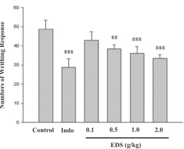
Acetic Acid-Induced Writhing Response
The cumulative amount of abdominal stretching correlated with the level of acetic acid-induced pain (Fig. 1). EDS treatment (1.0 and 2.0 g/kg)
signifi-cantly inhibited the number of writhings in comparison with the normal
controls (P< 0.001). Also, 0.5 g/kg EDS inhibited the number of writhings
in comparison with the normal (P< 0.01). This inhibiting effect of acetic
acid-induced writhings by EDS (1 and 2 g/kg) was similar to that produced by a positive control indomethacin (10 mg/kg).
Formalin Test
EDS 0.1 g/kg (P< 0.01) and EDS (0.5, 1 and 2 g/kg) significantly
(P< 0.001) inhibited formalin-induced pain in the late phase (Fig. 2);
however, there was no inhibition in the early phase (data not shown). The
positive control indomethacin (10 mg/kg) also significantly (P< 0.001)
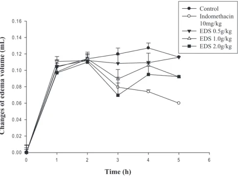
inhib-ited the formalin-induced pain in the late phase. Carrageenan-Induced Edema
EDS (1.0 g/kg) (P< 0.05) and EDS (2.0 g/kg) (P < 0.01) significantly
inhibited the development of carrageenan-induced paw edema after 3 h. of treatment; however, it only showed inhibition in the early phase. Indomethacin (10 mg/kg) significantly decreased the carrageenan-induced paw edema after 3
(P< 0.01), 4 (P < 0.01) and 5 h of treatment (P < 0.001) (Fig. 3).
EDS (g/kg) Numbers of W r ithing Response Control Indo 0.1 0.5 1.0 2.0
FIG. 1. The effect of EDS on 1% acetic acid-induced writhing response in mice. 1% acetic acid (10 mL/kg) was intraperitoneally injected to mice 55 min after administration of the EDS (0.1, 0.5,
1 and 2 g/kg, p.o.) and 25 min of indomethacin (Indo, 10 mg/kg, i.p.). Data are represented as mean⫾ SEM (n = 8). **P < 0.01, ***P < 0.001 compared with the control group. (One-way
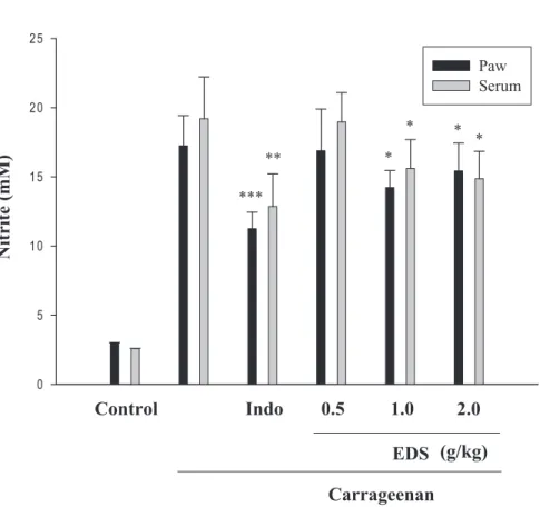
Effects of EDS on NO Measurement
EDS (1.0 g/kg and 2.0 g/kg) significantly decreased the NO level in
edematous paw tissue (P< 0.05) and in serum (P < 0.05) (Fig. 4).
Indometha-cin (10 mg/kg) significantly decreased the NO level in the edematous tissue
and in serum at the fifth hour after carrageenan injection (P< 0.001).
Effects of EDS on TNF-a Level
EDS (1.0 g/kg) and EDS (2.0 g/kg) decreased the TNF-a level in serum
at the fifth hour after carrageenan injection (P< 0.01) (Fig. 5). Indomethacin
(10 mg/kg) significantly decreased the TNF-a level in serum at the fifth hour
after carrageenan injection (P< 0.01).
DISCUSSION
The anti-nociceptive effects of test samples were assayed using the acetic acid- and formalin-induced analgesic models. Our results indicate that EDS treatment significantly inhibited the number of writhings in comparison with
Control Indo 0.1 0.5 1.0 2.0
Licking T
ime
(sec)
EDS (g/kg)
FIG. 2. The effects of EDS on the late phase B (15–40 min) on 1% formalin-induced inflammation in mice. 20mL of 5% formalin was injected into the dorsal surface of the right hind-paw paw of
mice 60 min after administration of the EDS (0.1, 0.5, 1 and 2 g/kg, p.o.) and 30 min of indomethacin (Indo, 10 mg/kg, i.p.). Data are represented as mean⫾ SEM (n = 8). **P < 0.01,
***P< 0.001 compared with the control group. (One-way ANOVA followed by Scheffe’s multiple range test).
the normal controls (P< 0.001) (Fig. 1). Also, EDS significantly inhibited formalin-induced pain in the late phase (Fig. 2). The acetic writhing test is commonly used to study the peripheral analgesic effects of drugs and widely used for analgesic screening (Shibata et al. 1989). We found that EDS (0.5, 1 and 2 g/kg) exhibited an antinociceptive effect in acetic acid-induced writhing response.
The formalin test is a valid and reliable model of nociception and is sensitive for various classes of analgesic drugs. The formalin test produces a distinct biphasic response, and different analgesics may act differently in the early and late phases of this test. Therefore, the test can be used to clarify the possible mechanism of an antinociceptive effect of a proposed analgesic (Tjolsen et al. 1992). Centrally acting drugs such as opioids inhibit both phases equally (Shibata et al. 1989), but peripherally acting drugs such as aspirin, indomethacin and dexamethasone only inhibit the late phase. The inhibitory effect of EDS on the nociceptive response in the late phase of the
Time (h)
0 1 2 3 4 5 6
Changes of edema volume (mL)
0.00 0.02 0.04 0.06 0.08 0.10 0.12 0.14 0.16 Control Indomethacin 10mg/kg EDS 0.5g/kg EDS 1.0g/kg EDS 2.0g/kg
FIG. 3. The effects of EDS on mice hind-paw edema induced by carrageenan. 50mL of 1% carrageenan was intraperitoneally injected into the plantar side of right hind paws of the mice 120 min after administration of the EDS (0.1, 0.5, 1 and 2 g/kg, p.o.) and 90 min of indomethacin (Indo, 10 mg/kg, i.p.). Data are represented as mean⫾ SEM (n = 8). **P < 0.01, ***P < 0.001 as compared with the control group. (One-way ANOVA followed by Scheffe’s multiple range test).
formalin test suggested that the anti-nociceptive effect of the EDS could be due to its peripheral action.
The carrageenan-induced edema test is highly sensitive to nonsteroidal anti-inflammatory drugs, and has long been accepted as a useful phlogistic tool for investigating new anti-inflammatory drugs (Just et al. 1998). The degree of swelling of the carrageenan-injected paws was maximal 3 h after injection and the mean increase in volume at that time was about 100% in the control group.
Statistical analysis revealed that EDS (1.0 g/kg) (P< 0.05) and EDS (2.0 g/kg)
(P< 0.01) significantly inhibited the development of carrageenan-induced
paw edema after 3 h of treatment (Fig. 3).
The L-arginine-NO pathway has been proposed to play an important role in the carrageenan-induced inflammatory response (Salvemini et al. 1996).
Nitrite (mM)
0 5 10 15 20 25 Paw SerumControl
0.5
1.0
2.0
(g/kg)
Indo
EDS
Carrageenan
*** ** * * * *FIG. 4. Effects of the EDS and indomethacin on carrageenan-induced NO concentration of edematous paw tissue and serum at the fifth hour in mice. Each value represents as mean⫾ SEM
###P< 0.001 as compared with the control group. *P < 0.05, **P < 0.01 and ***P < 0.001 as compared with the carrageenan group (one-way ANOVA followed by Scheffe’s multiple range test).
The expression of the inducible isoform of NO synthase has been proposed as an important mediator of inflammation (Cuzzocrea et al. 1997). In our study, EDS at 1.0 and 2.0 g/kg significantly decreased the levels of NO in edematous paw tissue and in serum, indicating that EDS elicits an anti-inflammatory response via the L-arginine-NO pathway (Fig. 4).
TNF-a is a major mediator in inflammatory responses. It induces innate immune responses by activating T cells and macrophages, and stimulates secretion of other inflammatory cytokines (Beutler and Cerami 1989). Also, TNF-a is a mediator of carrageenan-induced inflammatory incapacitation, and is able to induce the further release of kinins and leukotrienes, which are suggested to play an important role in the maintenance of long-lasting noci-ceptive response (Tonussi and Ferreira 1999). In this study, we found that EDS decreased the TNF-a level in serum after carrageenan injection (Fig. 5).
Serum
TNF-α
(pg/mL)
0 200 400 600 800Control
Indo
0.5
1.0
2.0
EDS (g/kg)
Carrageenan
**
***
**
FIG. 5. Effects of the EDS and indomethacin (Indo) on carrageenan-induced TNF-a concentration of serum at the fifth hour in mice. Each value represents as mean⫾ SEM ###P < 0.001 as compared with the control group. *P< 0.05 and **P < 0.01 as compared with the carrageenan
Although algae is a source of protein in certain human foods and animal feeds, the effects of the algae are not clear. Certain algae are believed to possess anti-inflammatory activity (Price et al. 2002). For example, Spirulina has been shown to modulate the Th profile in patients with allergic rhinitis by inhibiting the production of IL-4, thereby suppressing the differentiation of Th2 cells (Mao et al. 2005). A pharmacological study of hydrosoluble and liposoluble extracts of the marine microalgae Chlorella stigmatophora and Phaeodactylum tricornutum indicated that hydrosoluble components of both species show significant anti-inflammatory, analgesic and free radical scav-enging activity. These activities were not detected in the liposoluble fractions (Guzman et al. 2001).
b-carotene is a bioactive molecule with anti-inflammatory activities (Bai et al. 2005). Our results show that D. salina contain 6% ofb-carotene, 0.12%
ofa-carotene, 0.3% of zeaxanthin and scarce amount of lycopene and
chlo-rophyll (Table 1). Study also showed that D. salina extract, a significant source
of b-carotene, had significantly higher anti-oxidant activity than all-trans
forms of a-carotene, b-carotene, lutein and zeaxanthin in all anti-oxidant
assays (Hu et al. 2008). D. salina could protect rats from tetrachloride-induced hepatotoxicity (Hsu et al. 2008). It has been proposed that the antihepatotxic activity of D. salina is mediated by the isomeric forms of beta-carotene. We propose that D. salina elicits an analgesic effect and anti-inflammatory activi-ties. We suggest that the mechanisms of D. salina may be associated with the inhibition of inflammatory mediator overproduction, including NO and TNF-a. These findings suggest that D. salina may be therapeutically useful for mitigating inflammatory pain.
ACKNOWLEDGEMENTS
We are very grateful to GONG BIH Enterprise Co., Ltd (Doo-Liu City, Taiwan). for providing dry Dunaliella salina powder. And also appreciate technical support from Ms. Shih Chen-I. This study was partially supported by CMC91-M-37, CMU92-M-12, CMU94-127, CMU95-247 and CMU97-141.
REFERENCES
BAI, S.K., LEE, S.J., NA, H.J., HA, K.S., HAN, J.A., LEE, H., KWON, Y.G., CHUNG, C.K. and KIM, Y.M. 2005. Beta-Carotene inhibits inflamma-tory gene expression in lipopolysaccharide-stimulated macrophages by suppressing redox-based NF-kappa-B activation. Exp. Mol. Med. 37(4), 323–334.
BEUTLER, B. and CERAMI, A. 1989. The biology of cachectin/TNF—A primary mediator of the host response. Annu. Rev. Immunol. 7, 625–655.
CHIDAMBARA MURTHY, K.N., VANITHA, A., RAJESHA, J.,
MAHADEVA SWAMY, M., SOWMYA, P.R. and RAVISHANKAR, G.A. 2005. In vivo antioxidant activity of carotenoids from Dunaliella salina – a green microalga. Life Sci. 76(12), 1381–1390.
CHUN, S.C., JEE, S.Y., LEE, S.G., PARK, S.J., LEE, J.R. and KIM, S.C. 2007. Anti-inflammatory activity of the methanol extract of moutan cortex in LPS-activated Raw264.7 cells. Evid. Based. Complement. Altern. Med. 4(3), 327–333.
CUZZOCREA, S., ZINGARELLI, B., CALAPAI, G., NAVA, F. and CAPUTI, A.P. 1997. Zymosanactivated plasma induces paw oedema by nitric oxide and prostaglandin production. Life Sci. 60(3), 215–220. DUBUISSON, D. and DENNIS, S.G. 1977. The formalin test: A quantitative
study of the analgesic effects of morphine, meperidine, and brain stem stimulation in rats and cats. Pain 4(2), 161–174.
FUJII, Y., SAKAMOTO, S., BEN-AMOTZ, A. and NAGASAWA, H. 1993. Effects of beta-carotene-rich algae Dunaliella bardawil on the dynamic changes of normal and neoplastic mammary cells and general metabo-lism in mice. Anticancer. Res. 13(2), 389–393.
GUZMAN, S., GATO, A. and CALLEJA, J.M. 2001. Antiinflammatory, anal-gesic and free radical scavenging activities of the marine microalgae Chlorella stigmatophora and Phaeodactylum tricornutum. Phytother. Res. 15(3), 224–230.
HERRERO, M., JAIME, L., MARTIN-ALVAREZ, P.J., CIFUENTES, A. and IBANEZ, E. 2006. Optimization of the extraction of antioxidants from Dunaliella salina microalga by pressurized liquids. J. Agric. Food Chem. 54(15), 5597–5603.
HSU, Y.W., TSAI, C.F., CHANG, W.H., HO, Y.C., CHEN, W.K. and LU, F.J. 2008. Protective effects of Dunaliella salina – a carotenoids-rich alga, against carbon tetrachloride-induced hepatotoxicity in mice. Food Chem. Toxicol. 46(10), 3311–3317.
HU, C.C., LIN, J.T., LU, F.J., CHOU, F.P. and YANG, D.J. 2008. Determina-tion of carotenoids in Dunaliella salina cultivated in Taiwan and antioxi-dant capacity of the algal carotenoid extract. Food Chem. 109(2), 439– 446.
JUST, M.J., RECIO, M.C., GINER, R.M., CUELLAR, M.J., MANEZ, S., BILIA, A.R. and RIOS, J.L. 1998. Anti-inflammatory activity of unusual lupane saponins from Bupleurum fruticescens. Planta. Med 64(5), 404– 407.
LAVY, A., NAVEH, Y., COLEMAN, R., MOKADY, S. and WERMAN, M.J. 2003. Dietary Dunaliella bardawil, a beta-carotene-rich alga, protects
against acetic acid-induced small bowel inflammation in rats. Inflamm. Bowel. Dis. 9(6), 372–379.
LEVIN, G., YESHURUN, M. and MOKADY, S. 1997. In vivo antiperoxida-tive effect of 9-cis beta-carotene compared with that of the all-trans isomer. Nutr. Cancer 27(3), 293–297.
LIAO, H., BANBURY, L.K. and LEACH, D.N. 2007. Elucidation of danzhix-iaoyao wan and its constituent herbs on antioxidant activity and inhibition of nitric oxide production. Evid. Based. Complement. Altern. Med 4(4), 425–430.
MAO, T.K., VAN DE WATER, J. and GERSHWIN, M.E. 2005. Effects of a Spirulina-based dietary supplement on cytokine production from allergic rhinitis patients. J. Med Food 8(1), 27–30.
MASCOLO, N., JAIN, R., JAIN, S.C. and CAPASSO, F. 1989. Ethnophar-macologic investigation of ginger (Zingiber officinale). J. Ethnopharma-col. 27(1-2), 129–140.
MURTHY, K.N., RAJESHA, J., SWAMY, M.M. and RAVISHANKAR, G.A. 2005. Comparative evaluation of hepatoprotective activity of carotenoids of microalgae. J. Med Food 8(4), 523–528.
NAGASAWA, H., FUJII, Y., KAGEYAMA, Y., SEGAWA, T. and BEN-AMOTZ, A. 1991. Suppression by beta-carotene-rich algae Dunaliella bardawil of the progression, but not the development, of spontaneous mammary tumours in SHN virgin mice. Anticancer. Res. 11(2), 713– 717.
PRICE, J.A., SANNY, C. and SHEVLIN, D. 2002. Inhibition of mast cells by algae. J. Med Food 5(4), 205–210.
RAJA, R., HEMAISWARYA, S., BALASUBRAMANYAM, D. and REN-GASAMY, R. 2007. Protective effect of Dunaliella salina (Volvocales, Chlorophyta) against experimentally induced fibrosarcoma on Wistar rats. Microbiol. Res. 162(2), 177–184.
SALVEMINI, D., WANG, Z., BOURDON, D.M., STERN, M.K., CURNE, M.G. and MANNING, P.T. 1996. Evidence of peroxynitrite involvement in the carrageenanageenan-induced rat paw edema. Eur. J. Clin. Pharma-col. 303(3), 217–220.
SHEU, M.J., HUANG, G.J., WU, C.H., CHEN, J.S., CHANG, H.Y., CHANG, S.J. and CHUNG, J.G. 2008. Ethanol extract of Dunaliella salina induces cell cycle arrest and apoptosis in A549 human non-small cell lung cancer cells. In Vivo 22(3), 369–378.
SHIBATA, M., OHKUBO, T., TAKAHASHI, H. and INOKI, R. 1989. Modi-fied formalin test: Characteristic biphasic pain response. Pain 38(3), 347–352.
TABER, R.I., GREENHOUSE, D.D., RENDELL, J.K. and IRWIN, S. 1969. Agonist and antagonist interactions of opioids on acetic acid-induced
abdominal stretching in mice. J. Pharmacol. Exp. Ther. 169(1), 29– 38.
TJOLSEN, A., BERGE, O.G., HUNSKAAR, S., ROSLAND, J.H. and HOLE, K. 1992. The formalin test: An evaluation of the method. Pain 51(1), 5–17.
TONUSSI, C.R. and FERREIRA, S.H. 1999. Tumor necrosis factor-alpha mediates carrageenin-induced knee-joint incapacitation and also triggers overt nociception in previously inflamed rat knee-joints. Pain 82(1), 81–87.
VANITHA, A., MURTHY, K.N., KUMAR, V., SAKTHIVELU, G., VEIGAS, J.M., SAIBABA, P. and RAVISHANKAR, G.A. 2007. Effect of the
carotenoid-producing alga, Dunaliella bardawil, on CCl4-induced
toxic-ity in rats. Int. J. Toxicol. 26(2), 159–167.
WINTER, C.A., RISELY, E.A. and NUSS, C.W. 1962. Carrageenan-induced oedema in hind paw of the rats as an assay for anti-inflammatory drugs. Proc. Soc. Exp. Biol. Med 111, 544–547.
XUE, L.X. 1993. Experimental study on extract of Dunaliella salina in pre-venting NSAR-induced cancer of proventriculus in mice. Zhonghua Yu Fang Yi Xue Za Zhi 27(6), 350–353.
ZIMMERMANN, M. 1983. Ethical guidelines for investigations of experi-mental pain in conscious animals. Pain 16(2), 109–110.